What Is the Best Initial Imaging for a Child with Suspected Sacroiliitis?
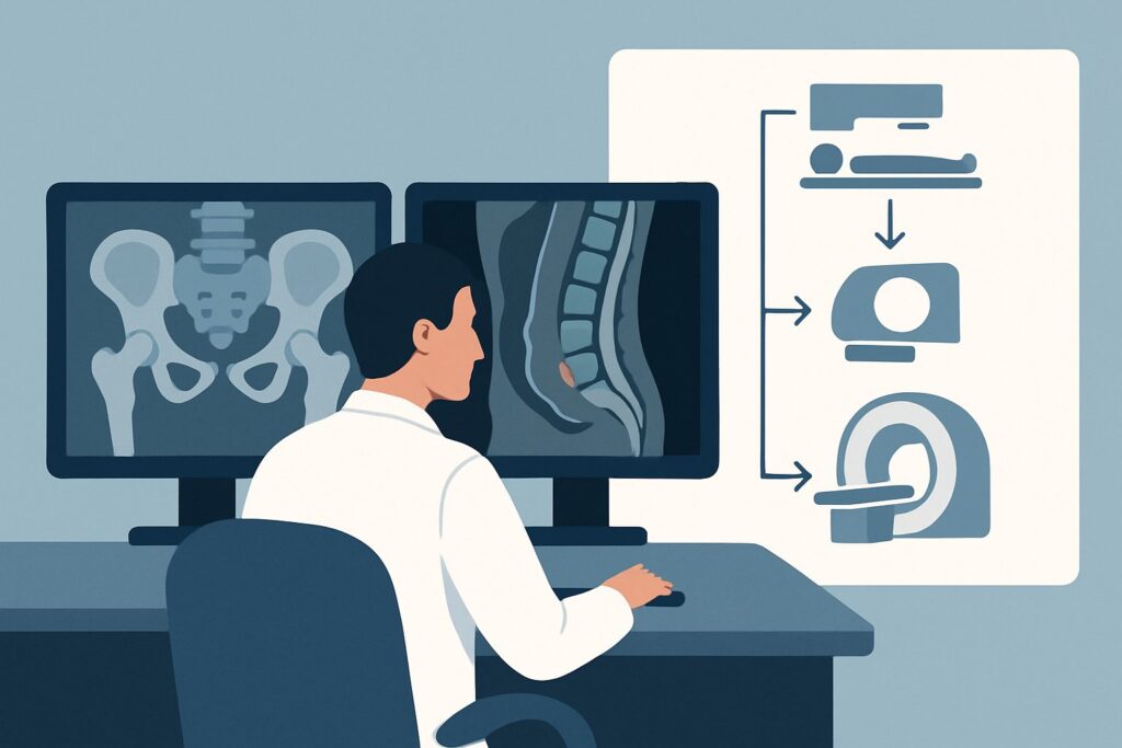
An 11-year-old boy presents to your clinic with a two-month history of insidious-onset buttock pain and low back stiffness, most pronounced in the morning. Physical examination elicits tenderness over both sacroiliac (SI) joints, and you suspect an inflammatory process like juvenile idiopathic arthritis (JIA). You need to confirm the diagnosis and assess for active inflammation, but which imaging study provides the clearest answer with the least risk? This article details the clinical workflow for a child with suspected sacroiliitis, focusing on the initial imaging decision. According to the American College of Radiology (ACR) Appropriateness Criteria, the most effective first step is MRI of the sacroiliac joints without IV contrast, which is rated Usually Appropriate.
Who Fits This Clinical Scenario?
This guidance applies specifically to the initial imaging workup of a child or adolescent with a clinical suspicion of idiopathic arthritis localized to the sacroiliac joints. The typical presentation involves insidious, non-traumatic pain in the buttock, hip, or low back region, often accompanied by inflammatory features like morning stiffness that improves with activity. This scenario is most common in children being evaluated for spondyloarthropathies, particularly enthesitis-related arthritis (ERA), a subtype of JIA.
This workflow is not intended for patients with:
- Acute, high-grade fever or significantly elevated inflammatory markers: This presentation raises the suspicion of infection (septic arthritis or osteomyelitis), which may alter the imaging approach, potentially requiring contrast-enhanced MRI.
- A clear history of acute, significant trauma: In cases of injury, the primary concern is fracture or ligamentous damage, and initial imaging often starts with radiographs.
- Predominantly appendicular joint symptoms: If the primary complaint is pain or swelling in the knees, ankles, or other peripheral joints, the workup follows a different ACR variant. See our guide on initial imaging for appendicular joint pain in children.
- Primary spinal pain without SI joint localization: If the pain is clearly localized to the thoracic or lumbar spine, the imaging protocol should be tailored accordingly.
What Diagnoses Are You Working Up in This Scenario?
When ordering initial imaging for suspected pediatric sacroiliitis, the primary goal is to identify active inflammation and differentiate it from other causes of low back and pelvic pain. The differential diagnosis guides the choice of imaging modality.
Sacroiliitis from Juvenile Idiopathic Arthritis (JIA): This is the leading consideration. Sacroiliitis is a hallmark of enthesitis-related arthritis (ERA) and can also be seen in psoriatic JIA. The underlying process is inflammation of the SI joint, particularly bone marrow edema (osteitis) in the adjacent sacrum and ilium. Early detection is crucial for initiating appropriate therapy to prevent long-term joint damage, such as erosions and ankylosis.
Infection (Osteomyelitis or Septic Arthritis): While less common, infection of the SI joint or adjacent bone is a critical diagnosis to exclude. It typically presents more acutely with fever and higher inflammatory markers (ESR, CRP) than JIA, but the initial symptoms can overlap. Imaging is essential to identify abscesses or widespread inflammation that would require urgent antibiotic therapy and possible surgical intervention.
Stress Injury or Apophysitis: In physically active adolescents, repetitive mechanical stress can cause stress fractures or inflammation at apophyses (growth plates), such as the iliac crest. The pain can mimic sacroiliitis, but the underlying pathology is mechanical rather than autoimmune. Imaging helps differentiate stress-related bone marrow edema from the inflammatory edema of sacroiliitis.
Neoplasm: Though rare, benign (e.g., osteoid osteoma) and malignant (e.g., Ewing sarcoma, leukemia, lymphoma) tumors can present with pain in the sacropelvic region. Red flags such as unrelenting night pain, weight loss, or constitutional symptoms should increase suspicion. Imaging is vital for identifying and characterizing any suspicious lesions.
Why Is MRI of the Sacroiliac Joints Without IV Contrast the Recommended Study?
The ACR designates MRI of the sacroiliac joints without IV contrast as Usually Appropriate for this clinical scenario because it directly visualizes the earliest sign of active sacroiliitis—bone marrow edema—without exposing the child to ionizing radiation.
The key advantage of MRI is its superior soft tissue and bone marrow contrast. Fluid-sensitive sequences, such as Short Tau Inversion Recovery (STIR), are exceptionally sensitive for detecting the increased water content associated with active inflammation (osteitis). This finding can be present long before any structural changes are visible on other imaging modalities, allowing for a definitive and early diagnosis.
For the initial evaluation of suspected JIA-related sacroiliitis, intravenous contrast is typically unnecessary. The primary diagnostic finding, bone marrow edema, is conspicuous on non-contrast STIR sequences. Adding contrast does not significantly increase sensitivity for this specific indication and adds potential risks (e.g., allergic reaction, nephrogenic systemic fibrosis in renal impairment) and costs.
How do alternative studies compare?
- Radiography of the sacroiliac joints is rated May be appropriate. While simple and widely available, radiographs are insensitive for early disease. They can only detect chronic changes like sclerosis, erosions, and joint space narrowing or fusion, which may take years to develop. A normal radiograph cannot rule out active sacroiliitis. Furthermore, it involves a low but non-zero dose of ionizing radiation (Pediatric RRL: ☢☢ 0.03-0.3 mSv).
- CT of the pelvis without contrast is rated Usually not appropriate. CT provides excellent detail of cortical bone and is superior to radiographs for detecting chronic structural changes. However, it is insensitive for detecting bone marrow edema and exposes the child to a significant radiation dose (Pediatric RRL: ☢☢☢☢ 3-10 mSv), making it unsuitable for an initial inflammatory workup in a pediatric patient.
The choice of a radiation-free modality is a critical consideration in pediatric imaging. By starting with a non-contrast MRI, you directly answer the clinical question—is there active inflammation?—with the highest sensitivity and no radiation burden.
What’s Next After MRI? Downstream Workflow
The results of the sacroiliac joint MRI will guide your next steps and determine the subsequent clinical pathway. The workflow branches based on whether the findings are positive, negative, or indeterminate for active sacroiliitis.
If the MRI is positive for active sacroiliitis: Findings of subchondral bone marrow edema consistent with osteitis confirm the diagnosis. This result warrants a prompt referral to a pediatric rheumatologist to initiate treatment. Management typically involves nonsteroidal anti-inflammatory drugs (NSAIDs), disease-modifying antirheumatic drugs (DMARDs), or biologic agents (e.g., TNF inhibitors) to control inflammation and prevent joint damage. Follow-up imaging may be considered later to monitor treatment response, which would fall under the ACR variant for follow-up of sacroiliac joint pain.
If the MRI is negative: A normal MRI makes active sacroiliitis highly unlikely. The next step is to reconsider the differential diagnosis. The pain may be mechanical, related to a stress injury not yet apparent, or referred from another source like the hip or lumbar spine. A thorough clinical re-evaluation is necessary. If symptoms persist, further investigation for other causes may be warranted, potentially including imaging of the lumbar spine or pelvis.
If the MRI is indeterminate or shows atypical features: Sometimes, findings may not be classic for JIA. For example, extensive soft tissue involvement, a destructive appearance, or a well-defined lesion might raise suspicion for infection or a tumor. In these cases, the next step is often a contrast-enhanced MRI to better characterize the abnormality. Consultation with a pediatric radiologist is crucial, and referrals to pediatric orthopedics or oncology may be necessary for further workup, which could include a biopsy.
Pitfalls to Avoid (and When to Get Help)
Navigating the workup for pediatric sacroiliac pain requires careful attention to avoid common diagnostic errors.
- Over-reliance on radiographs: The most common pitfall is concluding that a child does not have sacroiliitis based on a normal X-ray. Radiographs are insensitive to early inflammation; a negative result should not delay an MRI if clinical suspicion remains high.
- Ordering the wrong MRI protocol: A generic “MRI of the pelvis” or “MRI of the lumbar spine” may not include the specific thin-section, oblique coronal STIR sequences necessary to properly evaluate the SI joints. Be specific in your order: “MRI sacroiliac joints without contrast, with STIR sequences.”
- Misinterpreting normal developmental changes: The pediatric skeleton is still maturing, and normal red marrow, physeal plates, and developmental variations in the SI joints can mimic pathology. Interpretation by a radiologist with pediatric expertise is essential.
If clinical red flags are present—such as severe, unrelenting night pain, fever, weight loss, or a palpable mass—escalate immediately. This warrants urgent specialist consultation (rheumatology, orthopedics, or oncology) and may require expedited, and potentially different, imaging.
Related ACR Topics and Tools
This article focuses on a single clinical scenario. For a comprehensive overview of imaging for all presentations of suspected juvenile idiopathic arthritis, please consult our parent topic guide. You can also use the tools below to explore adjacent scenarios, review imaging techniques, and discuss radiation dose with families.
- Parent Topic Hub: For breadth across all scenarios in Joint Pain: Idiopathic Arthritis-Child, see our parent guide: Joint Pain: Idiopathic Arthritis-Child: ACR Appropriateness Decoded.
- ACR Appropriateness Criteria Lookup: https://gighz.com/acr-appropriateness-criteria/
- Imaging Protocol Library: https://gighz.com/imaging-protocol-library/
- Radiation Dose Calculator: https://gighz.com/radiation-dose-calculator/
Frequently Asked Questions
Why is a non-contrast MRI recommended over a contrast-enhanced MRI for the initial workup?
For the initial diagnosis of active sacroiliitis in suspected JIA, the key finding is bone marrow edema (osteitis). This is best visualized on non-contrast, fluid-sensitive MRI sequences like STIR. Adding gadolinium-based contrast does not significantly improve the detection of this primary finding and introduces unnecessary risks and costs. Contrast is typically reserved for cases where the diagnosis is unclear or there is a suspicion of an alternative diagnosis like an abscess or tumor.
If the MRI is negative, does that completely rule out JIA-related sacroiliitis?
A technically adequate MRI of the SI joints that shows no bone marrow edema makes active sacroiliitis very unlikely at that moment in time. However, JIA is a dynamic disease. If a patient has highly suggestive clinical symptoms that persist or evolve, a repeat MRI after a period of observation (e.g., 3-6 months) or a referral to pediatric rheumatology for further evaluation may still be appropriate.
Can ultrasound be used to evaluate the sacroiliac joints in a child?
According to the ACR Appropriateness Criteria, ultrasound of the sacroiliac joints is rated *Usually not appropriate* for this scenario. The SI joints are deep structures, and their visualization with ultrasound is limited by the overlying bone and soft tissues, especially in older or larger children. While ultrasound can sometimes detect increased blood flow with Doppler, it cannot visualize bone marrow edema, which is the key diagnostic feature. MRI remains the gold standard.
My patient also has back pain. Should I order an MRI of the lumbar spine at the same time?
The ACR lists ‘MRI sacroiliac joints and lumbar spine without IV contrast’ as *May be appropriate*. If the clinical symptoms are clearly localized to the SI joints, a dedicated SI joint MRI is sufficient and more targeted. If there is also significant, distinct lumbar spine tenderness or neurologic symptoms (e.g., radiculopathy), then a combined study may be reasonable. However, this expands the scan time and should be based on specific clinical indications beyond just suspected sacroiliitis.
What if my institution’s standard protocol for SI joints includes contrast?
It is best to communicate directly with the radiology department. You can specify ‘without contrast’ on the order and briefly state the indication is ‘initial evaluation for suspected JIA-related sacroiliitis.’ Citing the ACR Appropriateness Criteria can be helpful. Most pediatric radiologists are familiar with this indication and will agree that a non-contrast study is the most appropriate first step.
Reviewed by Pouyan Golshani, MD, Interventional Radiologist — June 27, 2026