What Imaging Should You Order for a Suspected Fracture in an Ankylosed Spine?
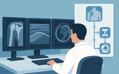
A 55-year-old man with a long-standing diagnosis of ankylosing spondylitis presents to the emergency department after a minor fall from a standing height. He reports new, sharp, and localized mid-thoracic back pain, distinctly different from his chronic, diffuse inflammatory discomfort. Given the biomechanical fragility of a fused spine, your suspicion for an occult fracture is high. What is the most appropriate initial imaging study to order in this specific clinical scenario? This article provides a step-by-step workflow based on the American College of Radiology (ACR) Appropriateness Criteria, explaining why the initial recommended study is rated as Usually Appropriate and what to do with the results.
Who Fits This Clinical Scenario for Suspected Fracture in an Ankylosed Spine?
This guidance is specifically for patients with a known diagnosis of axial spondyloarthritis (axSpA) who have progressed to spinal ankylosis, often described as a “bamboo spine” on imaging. The key elements of the presentation are:
- Established Disease: The patient has a pre-existing diagnosis of axSpA with radiographic evidence of spinal fusion.
- New or Worsening Pain: There is an acute change in the character or location of their back pain.
- History of Trauma (Even Minor): The new symptoms are often preceded by a fall or injury. Critically, the threshold for fracture in an ankylosed spine is very low; even seemingly trivial mechanisms like stepping off a curb or a sudden turn can be sufficient.
This workflow is distinct from other related clinical questions. This article does not apply to:
- Initial Diagnosis of axSpA: Patients with inflammatory back pain but no known diagnosis are covered in a different scenario focused on identifying sacroiliitis and early spinal changes.
- Monitoring Disease Activity: Imaging to assess treatment response or progression of inflammation in axSpA follows a separate pathway, often prioritizing MRI to detect active inflammation.
- Patients Without Ankylosis: The fracture risk and imaging approach are fundamentally different in patients with non-radiographic axSpA or early disease before significant fusion has occurred.
The unique biomechanics of the rigid, fused spine—which behaves like a long bone—is the central factor driving this specific imaging algorithm.
What Diagnoses Are You Working Up in This Scenario?
When a patient with an ankylosed spine presents with acute pain after trauma, the differential is narrow but the stakes are high. The primary goal of imaging is to identify or exclude conditions that require immediate stabilization or a change in management.
Vertebral Fracture
This is the most urgent and important diagnosis to exclude. The fused spine is brittle and cannot dissipate energy effectively, making it highly susceptible to fracture. These fractures are often transverse, extending through all three columns of the spine, and are inherently unstable. They carry a significant risk of neurologic injury, either at the time of injury or with subsequent displacement. Common locations include the cervicothoracic and thoracolumbar junctions.
Andersson Lesion (Spondylodiscitis)
This is a non-infectious inflammatory lesion or pseudoarthrosis that occurs at the discovertebral junction in patients with ankylosing spondylitis. It can present with acute, localized pain and may be initiated by minor trauma, mimicking a fracture. Radiographically, it appears as a localized area of destruction and sclerosis, which can be difficult to distinguish from a fracture-dislocation.
Acute Flare of Spondyloarthritis
While less likely to be precipitated by a distinct traumatic event, a severe inflammatory flare can cause intense, localized pain. However, in the setting of trauma, it is a diagnosis of exclusion that can only be made after a fracture has been confidently ruled out with appropriate imaging.
Epidural Hematoma
Fractures in the ankylosed spine can be associated with epidural hematoma, which can cause or worsen neurologic deficits. This is a key consideration when there is any evidence of spinal cord or nerve root compression on clinical exam.
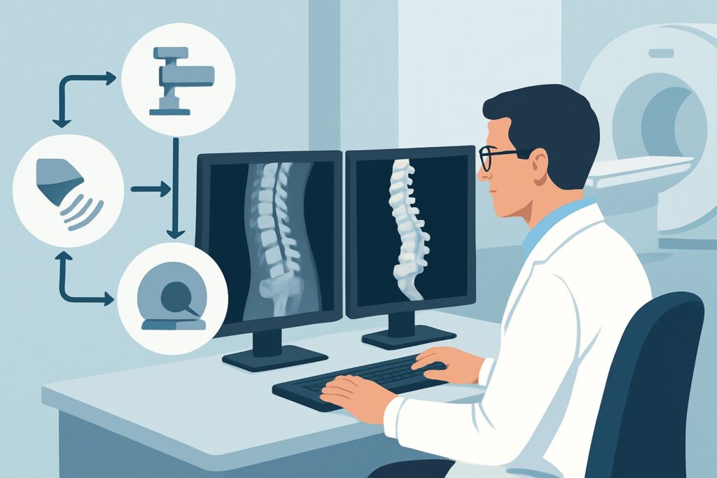
Why Is Radiography of the Spine the Recommended First Step for a Suspected Fracture?
The ACR Appropriateness Criteria provide clear guidance, rating three different modalities as Usually Appropriate for this scenario, each with a distinct role. The choice among them often depends on clinical stability, the severity of trauma, and local imaging availability.
Radiography spine area of interest is rated Usually Appropriate and is often the logical first step. It is fast, readily available, and uses a relatively low amount of ionizing radiation (RRL Varies). For a patient with a suspected fracture, obtaining anteroposterior (AP) and lateral views of the specific spinal region of maximal tenderness can quickly identify a displaced fracture, which would prompt immediate spinal immobilization and consultation with a spine surgeon. However, the sensitivity of plain radiographs for non-displaced or minimally displaced fractures in the ankylosed spine is notoriously low due to overlying structures and distorted anatomy.
Two other modalities are also rated Usually Appropriate and are critical to the workup:
- CT spine area of interest without IV contrast: CT is substantially more sensitive than radiography for detecting occult fractures. Given the high risk and subtle nature of these injuries, many institutions proceed directly to non-contrast CT, especially if the initial radiographs are negative but clinical suspicion remains high. CT provides exquisite bony detail, clearly delineates fracture lines, and is essential for surgical planning. It involves a higher radiation dose than radiography (RRL Varies) but is often necessary to make a definitive diagnosis.
- MRI spine area of interest without IV contrast: MRI is the best modality for evaluating the soft tissues, including the spinal cord, ligaments, and intervertebral discs. It is invaluable for detecting spinal cord compression, epidural hematoma, or ligamentous injury that may accompany a fracture. MRI can also help differentiate an acute fracture (which will show bone marrow edema) from an Andersson lesion. It uses no ionizing radiation (RRL O 0 mSv).
Why are other studies less appropriate?
Alternative studies are rated lower for good reason. CT with IV contrast is rated Usually not appropriate because contrast adds little value for the primary question of a bony fracture and increases risk and cost. Similarly, a Bone scan with SPECT/CT is Usually not appropriate; while sensitive to bony turnover, it is non-specific, provides poor anatomical detail compared to CT, and carries a significant radiation burden (☢☢☢ 1-10 mSv).
What’s Next After Radiography? Downstream Workflow
The results of the initial radiographs will guide your next steps in a critical decision tree. A high index of suspicion should be maintained throughout.
- If Radiographs Are Clearly Positive for Fracture: The patient requires immediate spinal immobilization (e.g., with a rigid collar or brace) and an urgent consultation with spine surgery or neurosurgery. Further characterization with a non-contrast CT is almost always required for surgical planning. An MRI should also be strongly considered to assess for spinal cord injury or epidural hematoma, especially if any neurologic deficits are present.
- If Radiographs Are Negative or Equivocal: Do not stop here. Given the low sensitivity of plain films in this setting, a negative radiograph does not rule out a fracture. If clinical suspicion persists (i.e., the patient has point tenderness and a history of trauma), the next step is a CT spine area of interest without IV contrast. This is the definitive study for excluding an occult bony injury.
- If CT Is Negative but Symptoms Persist: If the non-contrast CT is also negative for fracture but the patient continues to have significant pain or develops neurologic symptoms, an MRI spine area of interest without IV contrast is the next appropriate step. MRI can reveal bone marrow edema from a radiographically occult fracture, ligamentous injury, an inflammatory Andersson lesion, or an epidural hematoma that was missed on CT.
This stepwise approach balances speed and availability with diagnostic accuracy, ensuring that these high-risk, unstable injuries are not missed.
Pitfalls to Avoid (and When to Get Help)
Managing a suspected fracture in an ankylosed spine is fraught with potential pitfalls. Awareness of these common errors is key to preventing poor outcomes.
- Stopping the Workup After Negative Radiographs: This is the most common and dangerous pitfall. The threshold to proceed to CT should be extremely low. A negative plain film in a symptomatic patient with an ankylosed spine is an insufficient workup.
- Underestimating the Trauma Mechanism: Remember that significant, unstable fractures can occur with minimal or even no recalled trauma. Maintain a high index of suspicion regardless of the reported mechanism of injury.
- Inadequate Spinal Immobilization: Any patient with a confirmed or highly suspected fracture in an ankylosed spine should be handled with extreme care and placed in rigid spinal immobilization until the spine is cleared by a specialist to prevent iatrogenic neurologic injury.
- Failing to Image the Entire Spine: While imaging should be centered on the area of maximal tenderness, be aware that fractures can occur at a distance from the primary site of pain. Have a low threshold to extend imaging if the clinical picture is unclear.
If a fracture is identified or if the patient has any neurologic signs or symptoms, immediate escalation to a spine surgeon or neurosurgeon is mandatory.
Related ACR Topics and Tools
For a comprehensive overview of imaging for all related presentations, and for tools to help in your clinical practice, please see the following resources.
For breadth across all scenarios in Inflammatory Back Pain: Known or Suspected Axial Spondyloarthropathy, see our parent guide: Inflammatory Back Pain: Known or Suspected Axial Spondyloarthropathy: ACR Appropriateness Decoded.
- Imaging Appropriateness Selector — for adjacent scenarios
- Imaging Protocol Library — for technique on the recommended study
- Radiation Dose Calculator — for cumulative dose conversations
Frequently Asked Questions
Why is a CT scan often preferred over radiographs as the first test in a high-suspicion case?
While radiographs are a reasonable first step, CT is significantly more sensitive for detecting non-displaced fractures in an ankylosed spine. The complex, fused anatomy and overlying structures can easily obscure a subtle fracture line on a plain film. In a patient with high-risk features (e.g., significant trauma, neurologic symptoms, severe point tenderness), proceeding directly to a non-contrast CT is often the safest and most efficient diagnostic strategy.
If the patient has no neurologic symptoms, is an MRI still necessary if the CT shows a fracture?
Often, yes. Even without overt neurologic deficits, an MRI is valuable for assessing the stability of the fracture. It can reveal ligamentous injury, disc disruption, or an epidural hematoma that could predispose the patient to future neurologic decline. The spine surgery team will typically use information from both CT (for bone) and MRI (for soft tissues) to guide their management decisions.
Can an Andersson lesion be distinguished from an acute fracture on imaging?
It can be challenging. Both can present with localized pain and destructive changes on radiographs and CT. MRI is the most helpful modality for differentiation. An acute fracture will typically demonstrate intense bone marrow edema on T2-weighted sequences that conforms to the fracture line. An Andersson lesion may also show edema but often has features of chronic inflammation, sclerosis, and pseudoarthrosis formation.
What are the most common locations for fractures in a ‘bamboo spine’?
Fractures in an ankylosed spine most commonly occur at the junctions between the more mobile and more rigid segments of the spine. The cervicothoracic junction (C6-T1) and the thoracolumbar junction (T11-L2) are the most frequent sites. The subaxial cervical spine is also a common location.
Does the presence of diffuse idiopathic skeletal hyperostosis (DISH) carry the same fracture risk as ankylosing spondylitis?
Yes, patients with advanced DISH also develop a fused, rigid spine that is highly susceptible to unstable fractures from minor trauma. The clinical presentation and imaging workup for a suspected fracture in a patient with DISH are virtually identical to the workflow described here for axial spondyloarthritis.
Reviewed by Pouyan Golshani, MD, Interventional Radiologist — May 26, 2026