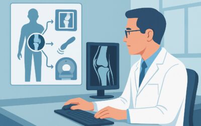
What Imaging Should You Order for Ankle Trauma with Positive Ottawa Rules?
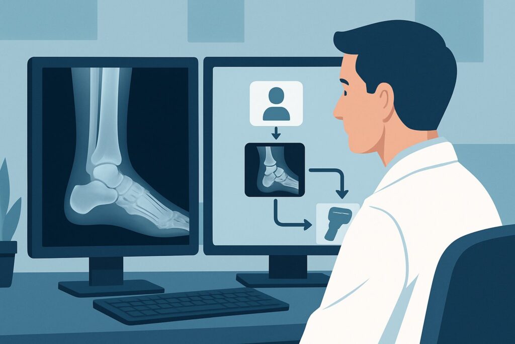
A 16-year-old presents to urgent care after twisting their ankle during a soccer match. They were unable to bear weight on the field and cannot walk four steps in the clinic without significant pain. On examination, you elicit point tenderness over the posterior edge of the lateral malleolus. The patient clearly meets the criteria for imaging based on the Ottawa Ankle Rules, but which study is the most direct and appropriate first step to rule out a clinically significant fracture? This is a common scenario where choosing the right initial imaging test prevents unnecessary radiation, cost, and delays in care.
According to the American College of Radiology (ACR) Appropriateness Criteria, for a patient with acute ankle trauma and positive Ottawa Ankle Rules, **Radiography ankle** is rated *Usually appropriate*.
Who Fits This Clinical Scenario for Ankle Trauma Imaging?
This guidance applies to a specific and very common clinical presentation: an adult or child aged 5 years or older who has experienced acute trauma to the ankle. It also includes patients with persistent pain for more than one week but less than three weeks following the injury.
The critical inclusion criterion is a positive finding on the Ottawa Ankle Rules, which are met if the patient has any of the following:
* Inability to bear weight both immediately after the injury and in the emergency department or clinic (for 4 steps).
* Bone tenderness at the posterior edge or tip of the lateral malleolus.
* Bone tenderness at the posterior edge or tip of the medial malleolus.
* Bone tenderness at the navicular bone (for foot injuries, part of the combined rule).
* Bone tenderness at the base of the fifth metatarsal (for foot injuries, part of the combined rule).
This workflow is intended for initial imaging and assumes no exclusionary criteria are present. It does *not* apply to patients with significantly altered sensation, intoxication, distracting injuries, or an open fracture. Similarly, patients who do *not* meet the Ottawa Ankle Rules fall under a different ACR variant where imaging is typically not recommended.
What Diagnoses Are You Working Up with a Positive Ottawa Ankle Rule?
When the Ottawa Ankle Rules are positive, the primary clinical question is whether a clinically significant fracture is present that would alter management from that of a simple sprain. The differential diagnosis is focused on identifying or excluding specific osseous injuries.
**Malleolar Fracture**
This is the most common fracture type around the ankle and the primary target of the Ottawa Ankle Rules. Fractures can involve the lateral malleolus (fibula), medial malleolus (tibia), or posterior malleolus (tibia). Identifying the number of malleoli involved (e.g., bimalleolar, trimalleolar) is critical for determining stability and the need for orthopedic consultation and potential surgical fixation.
**Talar or Calcaneal Fracture**
While less common from a simple twisting injury, fractures of the talus or calcaneus can occur and are specifically assessed by the tenderness criteria of the Ottawa rules. These injuries often require advanced imaging for characterization and carry different prognostic and management implications than malleolar fractures.
**Severe Ligamentous Injury (Ankle Sprain)**
If radiographs are negative, a severe ankle sprain is the presumed diagnosis. While radiographs do not directly visualize ligaments, they are essential for excluding a fracture, which is the key branch point in the management algorithm. The absence of a fracture confirms that conservative management (rest, ice, compression, elevation, and bracing) is the appropriate path forward.
**Maisonneuve Fracture**
This is a less common but consequential injury involving a proximal fibular fracture associated with an unstable ankle injury (medial malleolar fracture or deltoid ligament tear). Ankle radiographs may show a widened medial clear space, a sign of syndesmotic instability, which should prompt the clinician to examine the proximal fibula and obtain full-length tibia-fibula radiographs if tenderness is present.
Why Is Ankle Radiography the Recommended First Step for This Presentation?
The ACR rates **Radiography ankle** as *Usually appropriate* because it is a highly effective, low-risk, and resource-efficient tool for answering the primary clinical question in this scenario: is there a fracture?
The Ottawa Ankle Rules were developed and validated to identify patients with a very low probability of a clinically significant fracture, for whom radiography can be safely omitted. Conversely, for patients who are positive by these rules, radiography is the definitive next step. Standard ankle radiographs (including anteroposterior, lateral, and mortise views) have a very high sensitivity for detecting malleolar fractures and other significant osseous injuries of the ankle and hindfoot. The examination is fast, widely available, and provides the necessary information to guide immediate management.
In contrast, more advanced imaging modalities are rated *Usually not appropriate* for this initial evaluation:
* **CT ankle (without or with IV contrast):** While excellent for characterizing complex fractures, CT is not indicated as a first-line study. It delivers a higher radiation dose and is unnecessary if radiographs are negative or show a simple, non-displaced fracture. Its role is in pre-operative planning *after* a complex fracture has been identified on radiographs.
* **MRI ankle (without or with IV contrast):** MRI provides superior detail of soft tissues, cartilage, and bone marrow but is not the appropriate initial test for suspected fracture. It is more costly, time-consuming, and less available in an acute setting. MRI is reserved for cases where radiographs are negative but there is a high clinical suspicion for an occult fracture, osteochondral injury, or significant ligamentous disruption (e.g., syndesmotic injury) that would change management.
The radiation exposure from an ankle radiographic series is minimal. For an adult, the relative radiation level (RRL) is ☢ (<0.1 mSv), and for a child, it is even lower at ☢ (<0.03 mSv [ped]). This dose is negligible compared to the clinical value of diagnosing or excluding a fracture that requires specific orthopedic intervention.
What Is the Downstream Workflow After Ankle Radiography?
The results of the ankle radiographs create a clear decision tree for subsequent management.
* **If the study is POSITIVE for a fracture:** The next step is to determine ankle stability and arrange for appropriate orthopedic follow-up. Displaced or unstable fractures (e.g., bimalleolar fractures, any fracture with talar shift) often require urgent orthopedic consultation for reduction and possible surgical fixation. Non-displaced, stable fractures can typically be managed with immobilization (splint, cast, or boot) and non-urgent orthopedic follow-up. For complex fracture patterns, such as a pilon or comminuted intra-articular fracture, a CT scan may be ordered for pre-operative planning.
* **If the study is NEGATIVE for a fracture:** The diagnosis is an ankle sprain. Management focuses on conservative measures: rest, ice, compression, elevation (RICE), functional bracing, and a gradual return to activity with physical therapy. If, despite negative radiographs, there is high clinical suspicion for a syndesmotic injury (a “high ankle sprain”), the patient may fit a different ACR scenario where MRI becomes an appropriate next step to evaluate the ligaments connecting the tibia and fibula.
* **If the study is INDETERMINATE:** In rare cases, a fracture line may be suspected but not definitive. Consultation with the interpreting radiologist is recommended. Depending on the clinical suspicion, management may involve treating the injury as a fracture with immobilization and follow-up radiographs in 7-14 days, or proceeding to advanced imaging like CT or MRI to clarify the finding.
Common Pitfalls to Avoid in Evaluating Acute Ankle Trauma
When managing patients who meet this scenario’s criteria, several common errors can compromise care.
* **Misapplying the Ottawa Ankle Rules:** The rules are not validated for children under 5, patients with decreased sensation (e.g., diabetic neuropathy), or those with altered mental status or distracting injuries. Applying them outside of the validated population can lead to missed fractures.
* **Ordering an Incomplete Radiographic Series:** A complete ankle series requires three views: AP, lateral, and a mortise view (AP with 15-20 degrees of internal rotation). Omitting the mortise view can obscure talar shift or subtle fractures of the malleolar tips.
* **Forgetting to Examine the Proximal Fibula:** In any patient with medial ankle pain or signs of deltoid ligament injury, it is crucial to palpate the entire length of the fibula. Missing the tenderness associated with a Maisonneuve fracture can lead to mismanagement of an unstable ankle injury.
* **Overlooking Pediatric Fracture Patterns:** In skeletally immature patients, be vigilant for Salter-Harris fractures, which involve the physis (growth plate). These can be subtle on radiographs and are sometimes mistaken for sprains, but improper management can lead to growth arrest.
Related ACR Topics and Tools
For a comprehensive overview of imaging recommendations across all common presentations of ankle trauma, and for tools to help you apply these guidelines in practice, please see the following resources.
- For breadth across all scenarios in Acute Trauma to the Ankle, see our parent guide: Acute Trauma to the Ankle: ACR Appropriateness Decoded.
- To search for other clinical scenarios, visit the ACR Appropriateness Criteria Lookup.
- For details on imaging techniques, explore the Imaging Protocol Library.
- To discuss radiation exposure with patients, use the Radiation Dose Calculator.
Frequently Asked Questions
Why not get an MRI first to see the ligaments in a severe ankle injury?
While an MRI is excellent for visualizing ligaments, its primary role is not for initial evaluation after acute trauma when a fracture is suspected. The first and most critical step is to rule out a fracture, as this fundamentally changes management. Radiography is faster, cheaper, more available, and highly accurate for this purpose. An MRI is considered only after radiographs are negative and a complex ligamentous injury that would alter therapy (like a complete syndesmotic tear) is still suspected.
Are the Ottawa Ankle Rules 100% sensitive for all fractures?
The Ottawa Ankle Rules have a very high sensitivity (over 97%) for ‘clinically significant’ fractures, meaning those that typically require casting or surgery. However, they can miss some minor chip fractures or avulsion fractures that are managed like sprains anyway. Their primary strength is their high negative predictive value, allowing clinicians to safely rule out the need for radiography in patients who do not meet the criteria.
What if the patient has persistent pain two weeks after the injury and negative initial X-rays?
This scenario is still covered by this guideline (‘persistent pain for more than 1 week but less than 3 weeks’). If the Ottawa Ankle Rules were initially positive, obtaining radiographs is still the correct first step. If initial radiographs were negative and pain persists, the decision for further imaging (like MRI) depends on the specific physical exam findings, such as continued inability to bear weight or suspicion for an occult fracture, osteochondral lesion, or syndesmotic injury.
Should I order a foot series in addition to the ankle series?
You should order radiographs based on the location of the patient’s point tenderness. The Ottawa Ankle Rules include criteria for both the ankle (malleolar zones) and the midfoot (navicular and base of 5th metatarsal). If the patient has tenderness only in the malleolar zones, an ankle series is sufficient. If they also have tenderness in the midfoot zones, then both an ankle and a foot series should be ordered.
Is a CT scan ever the right first test for an acute ankle injury?
Almost never. CT is considered a secondary imaging modality in ankle trauma. Its main uses are for pre-operative planning of complex, intra-articular fractures already identified on radiographs, or for evaluating a suspected subtle fracture that is not visible on plain films but where MRI is contraindicated or unavailable. For the initial workup based on positive Ottawa Ankle Rules, radiography is the standard of care.
Reviewed by Pouyan Golshani, MD, Interventional Radiologist — May 21, 2026