

What Initial Imaging Is Best for High-Risk Placenta Accreta Spectrum Disorder?
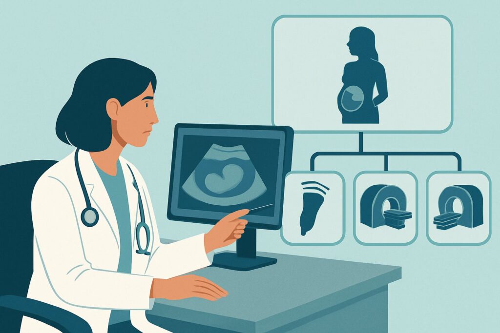
A 34-year-old G3P2 patient with a history of two prior cesarean sections presents for a routine 28-week prenatal visit. Her 20-week anatomy scan revealed a complete anterior placenta previa, placing her squarely in a high-risk category for Placenta Accreta Spectrum (PAS) disorder. As her obstetrician, you recognize the critical need for early and accurate diagnosis to plan for a complex delivery. The immediate question is which imaging study to order first to evaluate for signs of abnormal placental invasion. This article provides a detailed clinical workflow for this specific scenario, guiding the initial imaging decision for a patient at high risk for PAS. According to the American College of Radiology (ACR) Appropriateness Criteria, a `US duplex Doppler pregnant uterus` is Usually Appropriate as the initial imaging study.
Who Fits This Clinical Scenario for Placenta Accreta Spectrum Imaging?
This guidance is specifically for pregnant patients who have been identified as having a high risk for developing Placenta Accreta Spectrum disorder. The primary inclusion criteria are the presence of one or more significant clinical risk factors. The most common and highest-risk scenario is a patient with placenta previa in the setting of a prior cesarean delivery. The risk increases with the number of prior cesarean sections.
Other major risk factors include:
- History of other uterine surgeries, such as myomectomy that breached the endometrial cavity.
- Prior uterine artery embolization.
- Asherman syndrome (intrauterine adhesions).
- Advanced maternal age.
This workflow is distinct from other clinical situations. It does not apply to patients with a low pre-test probability for PAS, such as a primigravid patient with no history of uterine surgery and no placenta previa. That situation is covered in the ACR variant for low-risk initial imaging. Furthermore, this article addresses the initial diagnostic imaging step. It does not cover the workflow for a patient with an already-confirmed diagnosis who requires follow-up imaging to monitor progression or for detailed surgical planning, which is a separate clinical scenario.
What Diagnoses Are You Working Up in This Scenario?
When ordering initial imaging for a high-risk patient, the primary goal is to identify or exclude the presence and extent of Placenta Accreta Spectrum disorder. This is not a single diagnosis but a range of pathologic adherence of the placenta to the uterine wall.
Placenta Accreta: This is the most common and least invasive form within the spectrum. Here, the placental villi attach directly to the myometrium (the uterine muscle) without the normal intervening decidua basalis layer. While still a high-risk condition, it involves adherence without frank invasion into the muscle.
Placenta Increta: A more severe form where the placental villi invade directly into the myometrium. This deeper invasion significantly increases the risk of uterine rupture and catastrophic hemorrhage at the time of delivery, as the placenta cannot be separated from the uterine wall without causing severe damage.
Placenta Percreta: This is the most severe and dangerous form of the disorder. The placental villi invade completely through the myometrium and serosa (the outer layer of the uterus). The placenta may then invade adjacent organs, most commonly the urinary bladder. This condition carries the highest rates of morbidity and mortality and requires extensive multidisciplinary surgical planning.
Placenta Previa: While not a form of accreta itself, placenta previa (where the placenta covers the cervical os) is a critical co-diagnosis to establish. Its presence is a major risk factor for PAS, especially with a prior uterine scar, and its location dictates the surgical approach. The imaging study must confirm the relationship of the placenta to the cervix in addition to evaluating for invasion.
Why Is Ultrasound the Recommended Initial Study for This Presentation?
For the initial evaluation of a patient at high risk for Placenta Accreta Spectrum, the ACR designates `US duplex Doppler pregnant uterus`, `US pregnant uterus transabdominal`, and `US pregnant uterus transvaginal` as Usually Appropriate. Ultrasound is the frontline modality due to its safety, accessibility, and high diagnostic accuracy in experienced hands.
The combination of grayscale and color Doppler ultrasound allows for a detailed assessment of the key features suggestive of PAS. Grayscale imaging can identify the loss of the normal retroplacental “clear space,” thinning of the myometrium overlying the placenta, and the presence of placental lacunae (irregular vascular spaces within the placenta), which often have a “moth-eaten” or “Swiss cheese” appearance. Color Doppler is crucial for visualizing turbulent flow within these lacunae and identifying bridging vessels that cross from the placenta into the myometrium or beyond the uterine serosa.
A comprehensive examination typically involves both transabdominal and transvaginal approaches. The transabdominal view provides a broad overview of the uterus and placenta, while the transvaginal approach offers higher resolution imaging of the lower uterine segment and cervix—the most common site for PAS in patients with placenta previa and a prior cesarean scar.
In contrast, other imaging modalities are rated lower for this initial step. `MRI abdomen and pelvis without IV contrast` is considered May be appropriate. MRI can be a valuable problem-solving tool, particularly when ultrasound findings are equivocal, the placenta is posterior, or there is suspicion of posterior or parametrial invasion that is difficult to visualize with ultrasound. However, it is less available, more expensive, and generally reserved as a second-line or adjunct study. `MRI abdomen and pelvis without and with IV contrast` is rated Usually not appropriate. The use of gadolinium-based contrast agents during pregnancy is generally avoided due to theoretical concerns about fetal safety, and it typically does not add sufficient diagnostic information for PAS evaluation to justify the potential risk.
All recommended and appropriate studies for this scenario, including ultrasound and non-contrast MRI, involve no ionizing radiation (0 mSv), making them safe for both the mother and fetus.
What’s Next After Ultrasound? Downstream Workflow
The results of the initial ultrasound will guide the subsequent management and potential need for further imaging. The workflow branches based on whether the findings are positive, negative, or indeterminate for Placenta Accreta Spectrum.
If the ultrasound is positive for PAS: A definitive diagnosis on ultrasound prompts a cascade of actions. The patient should be referred to a tertiary care center with a multidisciplinary team experienced in managing PAS. This team typically includes maternal-fetal medicine specialists, gynecologic oncologists or experienced pelvic surgeons, anesthesiologists, neonatologists, and interventional radiologists. Further imaging with MRI may be considered to delineate the extent of invasion, especially if there is suspicion of percreta with bladder involvement, as this is critical for surgical planning. The primary goal shifts from diagnosis to preparation for a planned preterm cesarean hysterectomy.
If the ultrasound is negative: A high-quality ultrasound that shows no signs of PAS in a high-risk patient is reassuring but may not be completely exclusionary. Depending on the degree of clinical suspicion and the specific risk factors, serial ultrasounds may be performed later in the third trimester to monitor for developing signs. If the initial study was technically limited (e.g., due to maternal body habitus or a posterior placenta), and clinical suspicion remains high, proceeding to non-contrast MRI may be reasonable.
If the ultrasound is indeterminate or equivocal: This is the most common indication for the next step in imaging. When ultrasound findings are unclear, `MRI abdomen and pelvis without IV contrast` becomes the primary problem-solving tool. MRI can provide excellent soft-tissue contrast and a larger field of view, helping to clarify ambiguous findings like uterine bulging, placental heterogeneity, or the extent of deep myometrial invasion.
Pitfalls to Avoid (and When to Get Help)
Navigating the diagnosis of Placenta Accreta Spectrum requires careful attention to detail to avoid common pitfalls. A primary error is underestimation of the condition based on a non-specialized ultrasound. The sonographic signs of PAS can be subtle, and the accuracy of the diagnosis is highly operator-dependent. Ensure the study is performed at a center with expertise in high-risk obstetric imaging.
Another pitfall is relying solely on a transabdominal scan, which may miss signs of invasion in the lower uterine segment. A transvaginal component is often essential for a complete evaluation. Do not delay imaging; early identification in the second or early third trimester is key to allowing adequate time for multidisciplinary planning. Finally, avoid the use of gadolinium-based contrast agents in MRI unless there is a compelling, non-obstetric maternal indication where the benefits clearly outweigh the potential fetal risks.
If ultrasound results are equivocal or if there is any suspicion for placenta percreta, escalate care immediately by consulting with a maternal-fetal medicine specialist and arranging for evaluation at a center of excellence for placental disorders.
Related ACR Topics and Tools
For a comprehensive overview of all clinical scenarios related to Placenta Accreta Spectrum disorder, including low-risk and follow-up imaging, please see our parent guide. Additional GigHz tools can help you navigate imaging appropriateness, protocols, and radiation safety in your daily practice.
- For breadth across all scenarios in Placenta Accreta Spectrum Disorder, see our parent guide: Placenta Accreta Spectrum Disorder: ACR Appropriateness Decoded.
- For adjacent scenarios not covered here, use the ACR Appropriateness Criteria Lookup.
- To review technical details for various imaging studies, consult the Imaging Protocol Library.
- For discussions about cumulative exposure from other studies, use the Radiation Dose Calculator.
Frequently Asked Questions
At what gestational age should the initial ultrasound for high-risk PAS be performed?
While signs of Placenta Accreta Spectrum can sometimes be seen on the mid-trimester anatomy scan (around 18-22 weeks), a dedicated evaluation is often performed in the third trimester, typically between 28 and 32 weeks. This timing allows for better visualization of the developed placenta while still providing adequate time for multidisciplinary planning before delivery.
Is a transvaginal ultrasound always necessary in this scenario?
Yes, in most cases, a transvaginal ultrasound is a critical component of the evaluation. For patients with placenta previa and a prior cesarean section, the area of highest risk is the lower uterine segment overlying the old scar. The transvaginal approach provides superior resolution of this specific area compared to a transabdominal scan alone.
If the ultrasound is negative, can I definitively rule out Placenta Accreta Spectrum?
A high-quality negative ultrasound from an experienced center is very reassuring and has a high negative predictive value. However, it cannot completely exclude the diagnosis in 100% of cases, especially milder forms of placenta accreta. The final diagnosis is pathological. A high index of clinical suspicion should be maintained at delivery even with negative imaging.
Why is MRI only ‘May be appropriate’ and not the first-line test?
Ultrasound is the recommended initial test because it is highly accurate, widely available, less expensive, and does not require the patient to lie in a scanner for an extended period. MRI is reserved as a second-line, problem-solving tool for cases where ultrasound is inconclusive, technically difficult (e.g., posterior placenta), or when there is high suspicion for deep invasion into surrounding organs like the bladder.
What specific ultrasound findings should I look for in the report?
Key findings to look for in the radiology report include: presence of multiple large placental lacunae (irregular vascular spaces), loss of the normal retroplacental clear space, thinning of the myometrium to less than 1 mm, abnormalities of the uterine serosa-bladder interface, and turbulent flow within lacunae or vessels crossing the myometrium on color Doppler imaging.
Reviewed by Pouyan Golshani, MD, Interventional Radiologist — May 29, 2026


