What Is the ACR-Recommended Initial Imaging for Suspected Abuse in an Infant?
An 18-month-old is brought to the emergency department for irritability. The caregiver’s history is inconsistent, and on examination, you note scattered bruising in different stages of healing but no obvious deformity, focal weakness, or signs of acute distress. Your concern for non-accidental trauma is high, but the child’s examination doesn’t point to a specific skeletal, neurological, or abdominal injury. You are now faced with a critical decision: what is the appropriate imaging workup to screen for occult injuries that could confirm or refute your suspicion? This is a high-stakes scenario where choosing the correct initial studies is paramount for the child’s safety. For a child 24 months of age or younger with suspected physical abuse but no clinically apparent injuries, the American College of Radiology (ACR) Appropriateness Criteria rates a Radiography skeletal survey as ‘Usually appropriate’.
Who Fits This Clinical Scenario for Suspected Physical Abuse?
This guidance applies to a very specific and vulnerable patient population: a child 24 months of age or younger for whom there is a clinical concern for physical abuse. The suspicion may arise from an inconsistent or implausible history provided by a caregiver, the presence of sentinel injuries like bruising in a non-mobile infant, or other concerning social or clinical factors.
A key inclusion criterion for this workflow is the absence of clinically apparent major injuries. The purpose of imaging in this context is not to evaluate a known fracture or a clear neurological deficit, but rather to perform a comprehensive screen for occult (hidden) injuries.
This workflow is not appropriate for several similar-looking presentations that route to different ACR guidelines:
- Children older than 24 months: The patterns of injury, bone physiology, and differential diagnosis change in toddlers and older children, requiring a different imaging approach.
- Children with clinically apparent Central Nervous System (CNS) injury: If a child presents with seizures, altered mental status, a bulging fontanelle, or focal neurological deficits, the workup is driven by dedicated neuroimaging protocols under a different ACR variant.
- Children with suspected visceral injury: If the examination reveals abdominal tenderness, distension, a palpable mass, or a “seatbelt sign,” the focus shifts to abdominal imaging, again following a separate clinical pathway.
Applying this workflow correctly means using it for infants and young toddlers who appear relatively well on gross examination but for whom the clinical suspicion of abuse remains.
What Occult Injuries Are You Working Up in an Infant with Suspected Abuse?
The primary goal of initial imaging in this scenario is to identify injuries that are difficult or impossible to detect on physical examination alone. The differential diagnosis for the imaging findings is focused on identifying a pattern of trauma that is highly suggestive of physical abuse.
Occult Skeletal Fractures: This is the most critical diagnosis being investigated. Infants are unable to report pain or localize injury, and subtle fractures may have no external signs. Imaging seeks to identify high-specificity injuries that are rarely accidental. These include classic metaphyseal lesions (CMLs), also known as “corner” or “bucket-handle” fractures, which are caused by shearing forces from shaking or pulling on a limb. Posterior rib fractures, caused by forceful squeezing of the chest, are another hallmark finding. Fractures of the sternum, scapula, and spinous processes are also highly indicative of inflicted trauma.
Occult Intracranial Injury: Abusive head trauma is a leading cause of morbidity and mortality in this population. An infant can sustain significant intracranial injury, such as a subdural hematoma, from violent shaking without showing focal neurological signs on initial presentation. Irritability or lethargy may be the only symptoms. Therefore, screening for occult head injury is a co-primary goal of the initial workup.
Evidence of Repeated Trauma: A key role of the skeletal survey is its ability to reveal fractures in different stages of healing. The presence of both acute and healing or chronic fractures is powerful evidence of ongoing abuse and is a finding that a physical exam cannot provide. While other conditions like osteogenesis imperfecta can cause multiple fractures, the specific patterns and radiographic appearance of healing often help differentiate these mimics from non-accidental trauma.
Why Is a Radiography Skeletal Survey the Recommended Initial Study?
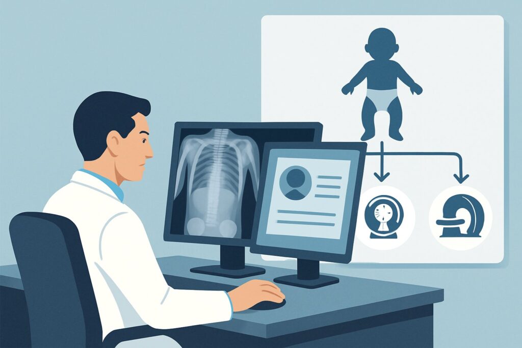
For a child under 24 months with suspected abuse and no localizing signs, a Radiography skeletal survey is rated ‘Usually appropriate’ and serves as the cornerstone of the evaluation. This is not a single “babygram” but a series of precisely positioned, high-detail radiographs of the entire skeleton, typically including 21 separate images. The rationale is its superior ability to detect the full spectrum of skeletal injuries, especially the high-specificity fractures like classic metaphyseal lesions and posterior rib fractures that are pathognomonic for abuse.
Concurrently, a CT head without IV contrast is also rated ‘Usually appropriate’ and is considered a standard part of the initial evaluation. This is because of the high incidence of occult abusive head trauma in this population. A non-contrast CT is fast, widely available, and highly sensitive for the acute intracranial hemorrhage (especially subdural hematoma) and skull fractures associated with inflicted injury.
Understanding why other modalities are rated lower is crucial for appropriate ordering:
- Bone scan whole body: This is ‘Usually not appropriate’. While sensitive for detecting metabolic activity at fracture sites, it has a higher radiation dose for children (pediatric relative radiation level ☢☢☢☢) compared to a skeletal survey (☢☢☢). More importantly, it is less specific and performs poorly in detecting two critical findings: classic metaphyseal lesions and skull fractures.
- Radiography area of interest: This is rated ‘May be appropriate (Disagreement)’. The major pitfall here is satisfaction of search. If you only x-ray a bruised arm, you may find one fracture but miss the larger pattern of multiple injuries in different stages of healing across the body—the very evidence needed to establish a diagnosis of abuse. A complete survey is required to avoid this error.
- MRI head: For the initial screen, MRI without or with contrast is ‘Usually not appropriate’. While MRI is more sensitive for certain findings like axonal injury or small subdural collections, CT is faster, requires less (or no) sedation, and is superior for detecting acute skull fractures. MRI is often used as a follow-up study if the initial CT is abnormal or if clinical concern persists.
The combined skeletal survey and head CT provide a comprehensive, high-yield initial screen for the most common and most specific occult injuries in this vulnerable population. Once you’ve decided on the skeletal survey, our protocol guide covers the technique, required views, and reading principles: Skeletal Survey.
What’s Next After the Initial Skeletal Survey and Head CT? Downstream Workflow
The results of the initial imaging studies will guide your immediate next steps, which always involve a multidisciplinary approach.
If the studies are positive for abuse: The presence of high-specificity fractures (e.g., CMLs, posterior ribs) or intracranial hemorrhage is strong evidence for non-accidental trauma. The immediate next step is a mandatory report to child protective services (CPS) and consultation with the hospital’s child abuse pediatrics team. The child will require admission for safety and further medical evaluation. A follow-up skeletal survey is often performed in approximately two weeks to assess for healing of known fractures and to identify any initially occult fractures that have become visible as they begin to heal.
If the studies are negative: A negative initial skeletal survey and head CT do not rule out abuse. Microfractures or non-displaced fractures may not be visible on the initial study. If clinical suspicion remains high, the next step aligns with another ACR scenario: follow-up imaging. Admission for observation and safety may still be warranted. A repeat skeletal survey in about two weeks is often recommended, as fracture lines can become more apparent with the formation of periosteal reaction and callus.
If the studies are indeterminate: An equivocal finding, such as a possible metaphyseal irregularity or a subtle lucency, requires expert interpretation. The first step is immediate consultation with a pediatric radiologist. They may recommend additional imaging, such as cone-down or oblique radiographic views of the specific area, or potentially a problem-solving modality like MRI to further characterize the finding and differentiate it from a normal developmental variant.
Pitfalls to Avoid (and When to Get Help)
Navigating a suspected abuse workup is fraught with potential missteps. Awareness of these common pitfalls can ensure a thorough and accurate evaluation.
- Ordering an Incomplete Survey: A “babygram” or single AP view of the body is not a skeletal survey. A formal survey requires a specific set of high-resolution images, including oblique views of the chest. Ensure the order is for a complete, formal skeletal survey according to established protocols.
- Misinterpreting Normal Variants: The infant skeleton is dynamic, with numerous ossification centers and physiologic irregularities (e.g., metaphyseal spurs) that can mimic fractures. This is not an area for non-expert interpretation.
- Stopping the Workup Prematurely: Do not cancel the full skeletal survey just because an initial x-ray of a symptomatic area reveals a fracture. The goal is to document the full extent and pattern of injury.
- Ignoring the Head CT: Overlooking the concurrent head CT because the child lacks neurologic symptoms is a critical error. Occult head trauma is common and can be life-threatening.
If the imaging findings are complex, equivocal, or discordant with the clinical picture, escalate immediately. This involves a direct conversation with the interpreting pediatric radiologist and the institutional child abuse pediatrics team.
Related ACR Topics and Tools
This article covers one specific clinical scenario in depth. For a broader overview of imaging for all presentations of suspected child abuse, or to explore the tools used to make these decisions, the following resources are essential.
- For breadth across all scenarios in Suspected Physical Abuse-Child, see our parent guide: Suspected Physical Abuse-Child: ACR Appropriateness Decoded.
- To explore other clinical presentations, use the ACR Appropriateness Criteria Lookup.
- To review technical details for hundreds of imaging studies, visit the Imaging Protocol Library.
- For discussions about radiation exposure with caregivers, our Radiation Dose Calculator can help frame the conversation.
Frequently Asked Questions
Why not just order a ‘babygram’ or single whole-body x-ray instead of a full skeletal survey?
A single whole-body radiograph, or ‘babygram,’ is considered substandard for an abuse evaluation. It lacks the detail, resolution, and specific positioning of a formal skeletal survey, which consists of over 20 individual images. A babygram has a significantly lower sensitivity for detecting critical, high-specificity injuries like classic metaphyseal lesions and posterior rib fractures, potentially leading to a missed diagnosis.
Is an MRI of the head better than a CT for the initial screening?
For the initial evaluation in an infant without focal neurologic signs, a non-contrast head CT is rated ‘Usually appropriate’ while an MRI is ‘Usually not appropriate.’ CT is faster, more readily available, does not typically require sedation, and is excellent for detecting the acute skull fractures and subdural hemorrhages common in abusive head trauma. MRI is a superior problem-solving tool and is often used for follow-up if the CT is abnormal or if there is a high suspicion for non-hemorrhagic injury.
What if the child is 26 months old? Does this workflow still apply?
No. This specific ACR guidance is for children 24 months of age and younger. In children older than two, fracture patterns change, the risk of certain occult injuries decreases, and they are more likely to be ambulatory, leading to a different spectrum of accidental injuries. The workup for a 26-month-old would fall under a different ACR variant with different imaging recommendations.
Do I need to order the head CT at the same time as the skeletal survey?
Yes, for this clinical scenario, the ACR rates both the skeletal survey and the non-contrast head CT as ‘Usually appropriate.’ They are considered complementary parts of the initial screening. Given the high incidence of occult intracranial injury in abused infants, even without clinical signs, performing both studies concurrently is the standard of care to ensure no major injuries are missed.
This scenario assumes no apparent visceral injuries. What if I’m worried about them anyway?
In the absence of clinical signs (e.g., abdominal tenderness, distension, vomiting), specific lab abnormalities (e.g., elevated liver transaminases), or suspicious bruising on the abdomen, screening imaging of the abdomen with ultrasound or CT is ‘Usually not appropriate.’ The diagnostic yield is very low. The initial workup is targeted to the highest-yield locations for occult injury in this scenario: the skeleton and the head.
Reviewed by Pouyan Golshani, MD, Interventional Radiologist — May 30, 2026