What Is the Best Follow-Up Imaging for Abnormal Uterine Bleeding Surveillance?
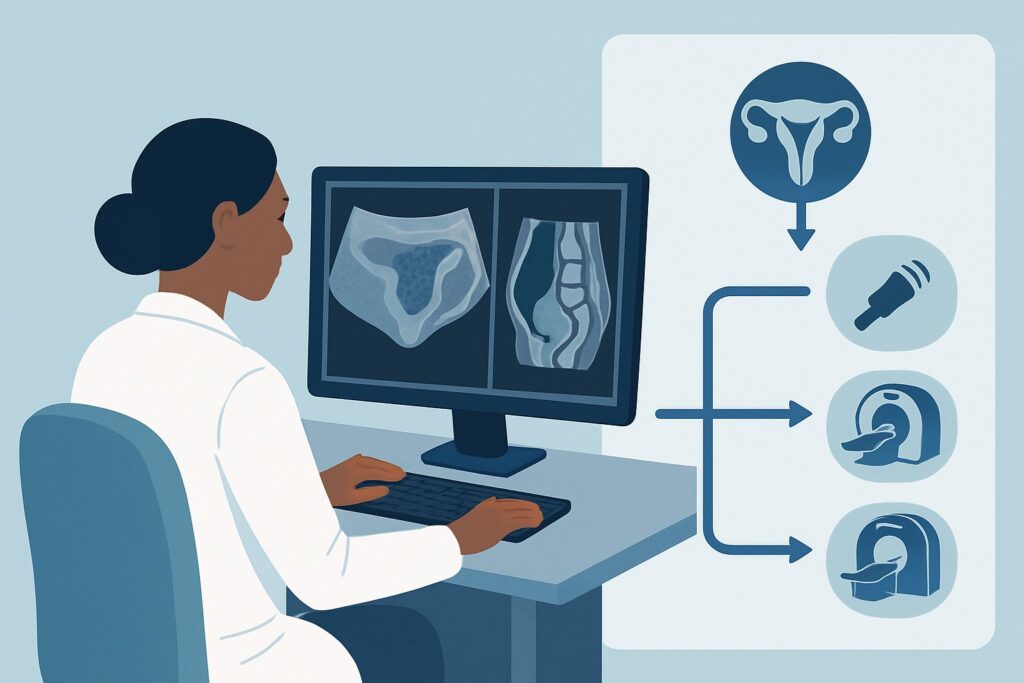
It’s Tuesday afternoon, and you’re seeing a 46-year-old patient for her scheduled 6-month follow-up. Her abnormal uterine bleeding (AUB) has been stable on medical management since her initial transvaginal ultrasound, which identified a 4 cm intramural leiomyoma. The clinical question is straightforward but critical: what is the most appropriate imaging study to order today to assess for interval change? This scenario—surveillance of a previously identified finding in AUB—requires a deliberate choice to ensure diagnostic value without unnecessary cost or radiation. This article details the American College of Radiology (ACR) Appropriateness Criteria workflow for this exact situation, where follow-up imaging is planned and surveillance is the goal. For this specific presentation, a US duplex Doppler pelvis is rated Usually Appropriate.
Who Fits This Clinical Scenario?
This guidance applies specifically to patients with abnormal uterine bleeding who have already undergone an initial imaging study (typically ultrasound) that revealed a finding for which periodic surveillance is clinically indicated. The key element is that a decision has already been made to monitor, rather than immediately intervene on, a specific finding.
Inclusion criteria for this workflow:
- A patient with a history of AUB.
- A known finding on a prior ultrasound, such as a uterine leiomyoma (fibroid), suspected adenomyosis, an endometrial polyp, or a simple ovarian cyst.
- The clinical plan is to monitor this finding for stability, growth, or change in character over a defined interval.
This workflow is distinct from other AUB scenarios. It does not apply to patients undergoing their first imaging workup for AUB, as that represents an initial diagnostic challenge. It also does not apply to patients whose initial ultrasound was inconclusive or technically limited. In those cases, the goal is not surveillance but further characterization, which often requires a different imaging modality. For example, if the initial report stated “endometrial thickening cannot be fully assessed due to uterine position,” the next step would fall under the ACR variant for inconclusive initial ultrasound, not surveillance.
What Diagnoses Are You Working Up in This Scenario?
When ordering surveillance imaging for AUB, you are primarily assessing for changes in known benign or likely benign pathologies. The goal is to detect progression that might alter clinical management while providing reassurance if findings are stable.
Uterine Leiomyoma (Fibroid)
This is one of the most common reasons for surveillance. The follow-up study aims to measure any interval growth in the fibroid(s). Rapid growth, while uncommon, can raise suspicion for a rare leiomyosarcoma, though most growth is benign. The imaging also re-evaluates the fibroid’s relationship to the endometrial cavity, as a change in position can worsen bleeding symptoms.
Endometrial Polyp
If a suspected endometrial polyp was seen on initial imaging and managed conservatively, follow-up ultrasound assesses for changes in size or vascularity. The addition of Doppler is particularly useful here to evaluate the vascular stalk, a characteristic feature. Significant growth may prompt consideration for hysteroscopic removal.
Adenomyosis
For patients with adenomyosis diagnosed on a prior ultrasound or MRI, surveillance imaging can monitor the extent of disease and response to hormonal therapy. While classic findings like myometrial heterogeneity and cysts may not change dramatically, imaging can confirm the absence of new, superimposed pathology like a polyp or fibroid.
Ovarian Cysts
Occasionally, an adnexal cyst is an incidental finding during an AUB workup. If it was characterized as simple or likely benign (e.g., a hemorrhagic cyst), follow-up ultrasound is performed to ensure resolution or stability, confirming its benign nature and distinguishing it from a neoplastic process.
Why Is US Duplex Doppler Pelvis the Recommended Study for This Presentation?
The ACR designates US duplex Doppler pelvis, which encompasses both transabdominal and transvaginal approaches, as “Usually Appropriate” for surveillance in this scenario. This recommendation is based on its high diagnostic utility, safety profile, and ability to provide a direct comparison to the initial study.
Ultrasound is the ideal modality for follow-up because it excels at visualizing the myometrium, endometrium, and ovaries without using ionizing radiation (0 mSv). Since the initial diagnosis was made with ultrasound, a repeat study allows for a precise, side-by-side comparison of measurements and morphology, which is the essence of surveillance. The transvaginal component provides high-resolution images of the endometrium and uterine architecture, while the transabdominal view offers a broader overview, which is crucial for large uteri or fibroids.
The “duplex Doppler” component is not incidental; it adds critical functional information by assessing blood flow. This is invaluable for evaluating a polyp’s vascular stalk, assessing flow within a fibroid, or characterizing an ovarian cyst. Increased or chaotic vascularity can be a subtle sign that a previously stable finding requires more aggressive workup.
Alternative Modalities Rated by the ACR:
- MRI pelvis without and with IV contrast is also rated “Usually Appropriate” but is not the typical first choice for routine surveillance. It is best reserved for cases where ultrasound provides an incomplete picture, such as mapping numerous large fibroids before a myomectomy or when there is a specific concern for malignancy that ultrasound cannot resolve. While it offers superior soft tissue contrast, it is more expensive and less accessible than ultrasound.
- CT pelvis with IV contrast is rated “Usually Not Appropriate.” CT provides poor soft-tissue differentiation of the uterine layers compared to ultrasound or MRI, making it unsuitable for evaluating subtle changes in fibroids, polyps, or the endometrium. Furthermore, it exposes the patient to unnecessary ionizing radiation (adult RRL=☢☢☢ 1-10 mSv), which is contrary to the principle of safe, repeatable surveillance imaging.
What’s Next After US Duplex Doppler Pelvis? Downstream Workflow
The results of the surveillance ultrasound will guide the next steps in management, which can range from continued observation to procedural intervention.
If the Finding is Stable or Improved:
When the ultrasound confirms that the known fibroid, polyp, or cyst is unchanged in size and character, it provides reassurance. The typical next step is to continue the current clinical management and schedule the next surveillance imaging study at an interval determined by the specific finding and the patient’s symptoms. For many stable, asymptomatic fibroids, imaging may be spaced out to every 1-2 years or discontinued altogether.
If the Finding Shows Significant Progression:
If the ultrasound reveals significant growth of a fibroid, a change in the complexity of a cyst, or increased vascularity in a polyp, the management plan must be reassessed. This result often triggers a referral to a gynecologist if the patient is not already under their care. The downstream pathway may involve more advanced imaging, such as an MRI for surgical planning, or a procedural evaluation like a hysteroscopy with biopsy or polypectomy.
If the Finding is No Longer Visualized:
In some cases, such as with a functional or hemorrhagic ovarian cyst, the follow-up study may show complete resolution. This is a definitive result that concludes the surveillance for that specific finding. No further imaging is required, and the patient can be reassured.
Pitfalls to Avoid (and When to Get Help)
Navigating surveillance imaging requires attention to detail to avoid common errors that can lead to diagnostic uncertainty or unnecessary tests.
- Missing Priors: The single most critical error is performing a follow-up study without access to the prior images and report. The radiologist must have the previous study for direct comparison; without it, a “surveillance” study becomes a new, less informative “initial” study.
- Underutilizing Doppler: Forgetting to order the study “with Doppler” can miss key information, especially when evaluating endometrial polyps or characterizing the vascularity of a fibroid.
- Inappropriate Modality Choice: Defaulting to CT for pelvic problems is a frequent pitfall. For this gynecologic indication, CT offers low diagnostic yield and unnecessary radiation exposure.
- Vague Ordering Information: Provide a clear reason for the exam on the order, such as “Follow-up of 4 cm intramural fibroid seen on ultrasound from [date].” This focuses the radiologist’s attention on the key clinical question.
If the surveillance ultrasound shows rapid interval growth of a solid mass or the development of complex features in a cyst (e.g., solid components, thick septations), escalate care immediately with a referral to a gynecologic specialist for further evaluation.
Related ACR Topics and Tools
For a comprehensive overview of imaging across all clinical presentations of abnormal uterine bleeding, please consult our parent topic guide. For other specific scenarios or to explore the underlying data, the following resources are available.
- For breadth across all scenarios in Abnormal Uterine Bleeding, see our parent guide: Abnormal Uterine Bleeding: ACR Appropriateness Decoded.
- To look up other clinical variants and their ACR-rated recommendations: ACR Appropriateness Criteria Lookup.
- To review standardized imaging techniques for recommended studies: Imaging Protocol Library.
- To discuss cumulative radiation exposure with patients when considering modalities like CT: Radiation Dose Calculator.
Frequently Asked Questions
Why not just order an MRI for surveillance to get the best possible images?
While MRI provides excellent detail, it is not the first choice for routine surveillance due to higher cost, longer scan times, and less widespread availability compared to ultrasound. Ultrasound is radiation-free and allows for a direct, effective comparison with the prior ultrasound, making it the most efficient and appropriate tool for monitoring stable or slowly changing findings.
What is the difference between a standard pelvic ultrasound and a ‘US duplex Doppler pelvis’?
A standard pelvic ultrasound uses sound waves to create grayscale images of the pelvic organs. A ‘duplex Doppler’ ultrasound adds a second component that assesses blood flow. It can show the presence, direction, and velocity of blood within vessels. This is crucial for evaluating the vascular stalk of a polyp or assessing blood flow within a fibroid or ovarian mass, which can help characterize the finding.
How often should I order surveillance imaging for a known uterine fibroid?
There is no single fixed interval for all patients. The frequency of surveillance depends on the size and location of the fibroid, the severity of the patient’s symptoms, their age, and their reproductive plans. A small, asymptomatic fibroid in a perimenopausal patient may require less frequent follow-up than a larger, symptomatic fibroid in a patient desiring future fertility. This decision should be based on clinical judgment in consultation with the patient.
My patient’s initial ultrasound was reported as ‘inconclusive’ due to a large body habitus. Does this surveillance guidance apply?
No. This guidance is for surveillance of a known, well-characterized finding. If the initial study was inconclusive or technically limited, the clinical question is one of diagnosis, not monitoring. That situation corresponds to a different ACR variant, where MRI is often the next appropriate step to achieve a definitive diagnosis.
Is sonohysterography needed for routine fibroid surveillance?
Sonohysterography, where saline is instilled into the endometrial cavity during an ultrasound, is ‘Usually Appropriate’ but not typically necessary for routine surveillance of intramural or subserosal fibroids. Its primary role is to better delineate submucosal fibroids or endometrial polyps by distending the cavity. It would be considered if the initial standard ultrasound was unclear about a finding’s relationship to the endometrium.
Reviewed by Pouyan Golshani, MD, Interventional Radiologist — May 29, 2026