What Is the Best Imaging for a Recent Ischemic Stroke After 24 Hours?
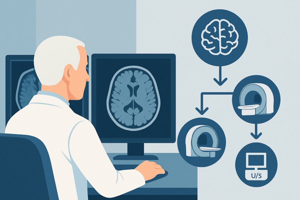
A 68-year-old patient is in your clinic for follow-up. Two days ago, he experienced sudden-onset left arm weakness and facial droop. An emergency department visit included a non-contrast head CT that ruled out hemorrhage, and he was diagnosed with an ischemic stroke. His symptoms have partially improved, but a residual deficit remains. Now, outside the hyperacute window for intervention, you must decide on the next imaging step to fully characterize the infarct and guide secondary prevention strategies. What is the most appropriate initial study in this subacute phase?
This clinical workflow guide addresses the specific American College of Radiology (ACR) scenario for an adult with a recent ischemic infarct, more than 24 hours after onset, who needs initial comprehensive imaging. For this presentation, an MRI of the head without IV contrast is rated as Usually Appropriate.
Who Fits This Clinical Scenario for a Recent Ischemic Infarct?
This guidance applies to adult patients with a confirmed or strongly suspected ischemic stroke where the initial event occurred more than 24 hours prior. The focus here is not on hyperacute intervention (like thrombolysis or thrombectomy) but on the comprehensive diagnostic workup that follows. This workup aims to define the extent of the injury, confirm the diagnosis, and investigate the underlying etiology to prevent a subsequent event.
It is crucial to distinguish this scenario from similar but distinct clinical presentations that require different imaging pathways:
- Acute Stroke (< 24 hours): If a patient presents within the window for reperfusion therapy, the imaging protocol is urgent and different, often involving CT/CTA and CT Perfusion to identify salvageable brain tissue. This subacute workflow is for patients outside that time-sensitive window.
- Transient Ischemic Attack (TIA): If the patient’s focal neurologic symptoms have completely resolved, the scenario is classified as a TIA. The imaging workup is similarly focused on identifying the stroke mechanism but follows a slightly different ACR variant.
- Hemorrhagic Stroke: If the initial imaging revealed an intraparenchymal hemorrhage rather than ischemia, the diagnostic and follow-up imaging strategy is entirely different and focuses on the cause and evolution of the bleed.
This article is specifically for the patient who has “graduated” from the hyperacute phase and now needs a definitive, detailed look at the ischemic brain injury.
What Diagnoses Are You Working Up in This Scenario?
After 24 hours, the primary imaging goal shifts from guiding acute intervention to establishing a clear diagnosis and uncovering the cause. The differential diagnosis and workup are focused on confirming the infarct and classifying its etiology, often using the TOAST (Trial of Org 10172 in Acute Stroke Treatment) criteria.
Confirming and Characterizing the Infarct: The first step is to use a high-sensitivity imaging modality to precisely delineate the size, location, and age of the ischemic territory. An MRI can visualize infarcts that were invisible on an initial non-contrast CT, especially those in the posterior fossa (cerebellum and brainstem) or small, deep lacunar infarcts.
Determining Stroke Etiology: A comprehensive workup aims to identify the underlying cause to tailor secondary prevention. Imaging helps differentiate between major categories. A pattern of multiple infarcts across different arterial territories strongly suggests a cardioembolic source, prompting a cardiac workup. Evidence of significant stenosis in the carotid bifurcation or major intracranial arteries points toward large-artery atherosclerosis. A single, small, deep infarct in the basal ganglia, thalamus, or pons is characteristic of small-vessel occlusion (lacunar stroke). Less common but critical causes, such as arterial dissection or vasculitis, may also have characteristic imaging findings. A thorough imaging evaluation is the foundation for classifying the stroke and preventing another one.
Why Is an MRI of the Head Without Contrast Usually Appropriate for This Presentation?
For a patient more than 24 hours out from an ischemic stroke, an MRI of the head without IV contrast is the superior initial study. Its high tissue contrast and specific sequences provide information that other modalities cannot, directly addressing the key clinical questions in this phase.
The ACR rates MRI head without IV contrast as Usually Appropriate because of its unparalleled sensitivity. The Diffusion-Weighted Imaging (DWI) sequence can detect cytotoxic edema from ischemia within minutes of onset and remains positive for approximately 10-14 days. This makes it exceptionally reliable for confirming a subacute infarct. The corresponding Apparent Diffusion Coefficient (ADC) map helps distinguish true restricted diffusion from other phenomena. This combination is far more sensitive than CT for visualizing the full extent of the injury.
Comparing MRI to other options highlights its value:
- CT head without IV contrast: While also rated Usually Appropriate and essential in the emergency setting to exclude hemorrhage, its sensitivity for ischemia is lower. In the first 24 hours, a CT can be normal in up to 60% of ischemic strokes. After 24 hours, signs of stroke (hypodensity) become more apparent, but small or brainstem infarcts can still be missed. It carries a radiation dose of 1-10 mSv (ACR RRL=☢☢☢).
- MRI head perfusion with IV contrast: This study is rated Usually not appropriate for this scenario. Perfusion imaging is designed to identify the “ischemic penumbra”—tissue at risk of infarction—to guide hyperacute therapies like thrombectomy. After 24 hours, the infarct is typically established, and the penumbra is no longer present, rendering perfusion imaging unnecessary. It adds the cost and potential risks of IV contrast without providing actionable information in the subacute setting.
The recommended MRI without contrast avoids both ionizing radiation (RRL=O 0 mSv) and the risks of gadolinium-based contrast agents, making it the safest and most diagnostically powerful choice for this specific clinical question.
What’s the Next Step After the MRI Result? Downstream Workflow
The findings on the non-contrast head MRI will dictate the subsequent diagnostic pathway. The goal is to move from confirming the infarct to identifying its cause.
- If the MRI confirms an infarct in a single vascular territory: The immediate next step is to evaluate the blood vessels supplying that territory. This is a search for the embolic or thrombotic source. Appropriate next studies include CTA neck with IV contrast, MRA neck without or with IV contrast, or a US duplex Doppler of the carotid arteries. These studies are rated Usually Appropriate or May be appropriate and are essential for identifying significant stenosis, plaque, or dissection that requires specific treatment (e.g., carotid endarterectomy, stenting, or specific antithrombotic therapy).
- If the MRI shows multiple infarcts in different vascular territories: This “embolic shower” pattern is highly suggestive of a central source, most commonly cardioembolic (e.g., from atrial fibrillation). While vascular imaging of the neck may still be performed, the priority shifts to a comprehensive cardiac evaluation, including an electrocardiogram, prolonged cardiac monitoring (telemetry or Holter), and an echocardiogram to look for intracardiac thrombus, valvular disease, or a patent foramen ovale.
- If the MRI is negative or findings are indeterminate: If a high-quality MRI with DWI sequences is negative despite a compelling clinical history, the diagnosis of ischemic stroke should be reconsidered. The event may have been a TIA with no resulting infarct, or a stroke mimic such as a complex migraine, post-ictal (Todd’s) paralysis, or a metabolic disturbance. In this case, further neurologic consultation is warranted before proceeding with an extensive and potentially unnecessary workup.
Pitfalls to Avoid (and When to Get Help)
Navigating the subacute stroke workup requires avoiding several common pitfalls to ensure an accurate diagnosis and effective secondary prevention plan.
- Pitfall 1: Stopping at the initial non-contrast head CT. An ED CT that is “negative for acute bleed” is not a complete stroke workup. It often misses the ischemic injury itself. Failing to proceed to MRI can lead to an incomplete diagnosis and missed opportunities for prevention.
- Pitfall 2: Forgetting the vascular workup. A brain MRI confirms the infarct but does not always reveal the cause. The workup is incomplete without imaging of the cervicocerebral vasculature (via CTA, MRA, or ultrasound) to search for the underlying pathology.
- Pitfall 3: Misinterpreting infarct age. Distinguishing a new subacute infarct from a chronic, old infarct is critical. This relies on correctly interpreting the DWI and ADC sequences. If there is uncertainty, a neuroradiologist’s interpretation is essential.
If the patient’s neurologic status worsens, if imaging reveals an unexpected finding like a tumor or evidence of vasculitis, or if the diagnosis remains unclear after initial studies, escalate immediately to a neurologist or a dedicated stroke service for specialized management.
Related ACR Topics and Tools
For a comprehensive overview of imaging for all stroke-related scenarios, please consult our parent guide. Additional tools are available to help you apply these guidelines in your practice.
- For breadth across all scenarios in Cerebrovascular Diseases-Stroke and Stroke-Related Conditions, see our parent guide: Cerebrovascular Diseases-Stroke and Stroke-Related Conditions: ACR Appropriateness Decoded.
- To look up appropriateness criteria for other clinical presentations, use the ACR Appropriateness Criteria Lookup.
- For detailed procedural techniques on the recommended study, explore the Imaging Protocol Library.
- To discuss cumulative radiation exposure with patients, the Radiation Dose Calculator can be a helpful resource.
Frequently Asked Questions
Why not just get a CT? It’s faster and was already done in the ED.
The emergency department CT is primarily performed to rapidly rule out hemorrhage before administering acute therapies. For the subacute workup after 24 hours, an MRI is far more sensitive for defining the precise size, location, and age of an ischemic infarct. This detailed information is critical for determining the underlying cause and planning effective secondary prevention.
Does this patient need IV contrast with the MRI?
For this specific scenario—evaluating a recent ischemic infarct after 24 hours—IV contrast is rated as *Usually not appropriate* by the ACR. The key MRI sequences for identifying and characterizing a subacute infarct (DWI, ADC, FLAIR) do not require contrast. Contrast would be added only if there is a specific clinical concern for an alternative diagnosis like a brain tumor, infection, or active demyelination, which is not part of a standard stroke workup.
What if the MRI shows an old stroke, but I’m clinically certain this event was new?
This is a common clinical challenge. The Diffusion-Weighted Imaging (DWI) sequence on the MRI is the key differentiator. A new (acute or subacute) infarct will demonstrate restricted diffusion, appearing bright on DWI and dark on the corresponding ADC map. A chronic, old infarct will not have this signal pattern. If the DWI sequence is negative despite a strong clinical history, the event may have been a Transient Ischemic Attack (TIA) that did not result in a permanent infarct, or a stroke mimic.
Should I order an MRA of the head and neck at the same time as the brain MRI?
Combining the brain MRI with an MRA of the head and neck is an efficient and common practice. An MRA or CTA is a crucial part of the etiologic workup to evaluate for large vessel stenosis, dissection, or other vascular abnormalities. Ordering them together can streamline the diagnostic process and provide a more complete picture in a single imaging session. The ACR rates MRA of the head and neck as *May be appropriate* as part of this initial comprehensive evaluation.
Reviewed by Pouyan Golshani, MD, Interventional Radiologist — May 30, 2026