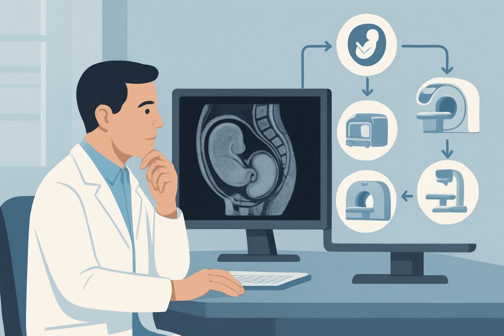
What Is the Best Imaging for Follow-up of Placenta Accreta Spectrum Disorder?
A 34-year-old G3P2 patient with a history of two prior cesarean deliveries and a known placenta previa was diagnosed with suspected placenta accreta spectrum (PAS) disorder on her 20-week anatomy scan. Now, at 32 weeks gestation, she presents for a scheduled follow-up to assess for disease progression and to inform the multidisciplinary team’s plan for delivery. As the ordering physician, you must decide on the most appropriate imaging modality to re-evaluate the depth of placental invasion and map the abnormal vasculature without exposing the fetus to unnecessary risk. This article details the clinical workflow for selecting the right imaging study in this specific follow-up scenario. According to the American College of Radiology (ACR) Appropriateness Criteria, a `US duplex Doppler pregnant uterus` is Usually Appropriate for this indication.
Who Fits This Clinical Scenario for Placenta Accreta Spectrum Follow-up?
This guidance applies specifically to pregnant patients with an already established or highly suspected diagnosis of placenta accreta spectrum disorder who are undergoing subsequent imaging for surveillance. The primary goal is not initial diagnosis, but rather monitoring the condition’s progression, typically in the late second or third trimester. This serial evaluation is crucial for refining the surgical plan for a planned cesarean delivery, often a cesarean-hysterectomy, and for assembling the necessary multidisciplinary surgical team.
This workflow is distinct from other clinical situations. This article does not apply to:
- Low-risk patients for initial screening: A patient with no known clinical risk factors (such as prior uterine surgery or placenta previa) undergoing initial evaluation would fall under the “Low risk for placenta accreta spectrum disorder. No known clinical risk factors. Initial Imaging” scenario.
- High-risk patients for initial diagnosis: A patient with significant risk factors (e.g., multiple prior cesarean sections, placenta previa) who has not yet had definitive imaging to diagnose PAS would be managed under the “High risk for placenta accreta spectrum disorder. Initial Imaging” scenario.
The key differentiator for this article’s workflow is the pre-existing diagnosis and the clinical question of interval change or progression.
What Diagnoses Are You Working Up in This Scenario?
In the follow-up setting for placenta accreta spectrum, the imaging is not meant to establish a new diagnosis but to characterize the extent and severity of the known condition. The primary goal is to provide a detailed anatomical and vascular map for the surgical team.
Progression of Myometrial Invasion: The most critical assessment is determining if the placental villi have invaded deeper into the uterine wall (placenta increta) or have breached the uterine serosa entirely to involve adjacent structures (placenta percreta). Identifying progression from a suspected accreta to a definite increta or percreta fundamentally changes the surgical risk and approach.
Bladder Wall Involvement: A key concern in anterior placentas is invasion into the posterior wall of the urinary bladder. Ultrasound aims to visualize the uterine serosa-bladder interface. Loss of this clear hypoechoic boundary or evidence of vascularity crossing into the bladder wall is a significant finding that necessitates the involvement of a urology or gynecologic oncology surgeon.
Parametrial and Adjacent Organ Invasion: In the most severe form, placenta percreta, the placental tissue can invade laterally into the broad ligament and parametrium or involve other pelvic organs. Follow-up imaging seeks to identify the extent of this lateral spread, which is a major predictor of massive hemorrhage and surgical morbidity.
Mapping of Neovascularity: Duplex Doppler is essential for evaluating the abnormal vascular patterns associated with PAS. This includes large, irregular placental lacunae with turbulent flow, hypervascularity at the serosa-bladder interface, and prominent bridging vessels that cross from the placenta through the myometrium. The extent of this neovascularity correlates with the severity of invasion and risk of bleeding.
Why US duplex Doppler pregnant uterus Is the Recommended Study for This Presentation
The ACR rates `US duplex Doppler pregnant uterus`, along with transabdominal and transvaginal ultrasound, as Usually Appropriate for the follow-up of placenta accreta spectrum. This recommendation is based on the modality’s safety, accessibility, and high diagnostic utility for the key clinical questions in this scenario.
Ultrasound is the primary imaging tool because it involves no ionizing radiation (adult and pediatric radiation relative level: O, 0 mSv) and is entirely safe for both the mother and fetus. Its real-time nature allows for a dynamic assessment of blood flow and tissue planes. The combination of transabdominal ultrasound for a global overview and transvaginal ultrasound for high-resolution imaging of the lower uterine segment and cervix provides a comprehensive evaluation.
The “duplex Doppler” component is what makes this study so powerful for PAS follow-up. Grayscale imaging identifies sonographic signs like loss of the retroplacental clear space and myometrial thinning. Color and spectral Doppler add crucial information by visualizing the chaotic, turbulent flow within placental lacunae and identifying the abnormal vessels bridging the myometrium or extending to the bladder, which are hallmarks of invasive placentation. This vascular mapping is invaluable for surgical planning.
Alternative Imaging Modalities
- MRI abdomen and pelvis without IV contrast: This study is rated as May be appropriate. MRI can be an excellent problem-solving tool when ultrasound findings are equivocal or when there is high suspicion for posterior placentation or deep pelvic invasion that is difficult to visualize with ultrasound due to fetal position or maternal body habitus. It provides excellent soft tissue contrast and a large field of view, offering a clear map of the extent of invasion. However, it is reserved as an adjunct because it is more costly, less accessible, and more time-consuming than ultrasound.
- MRI abdomen and pelvis without and with IV contrast: This study is rated as Usually not appropriate. The use of gadolinium-based contrast agents during pregnancy is generally avoided. Gadolinium crosses the placenta and enters the fetal circulation, with unknown long-term consequences. Since non-contrast MRI and duplex Doppler ultrasound can typically provide the necessary diagnostic information, the potential risk of contrast administration is not justified.
What’s Next After US duplex Doppler pregnant uterus? Downstream Workflow
The results of follow-up imaging directly inform the patient’s management plan, primarily concerning the timing and logistical complexity of delivery.
- If Findings Are Stable or Show Minimal Progression: If the degree of suspected invasion appears unchanged from prior studies, the patient can typically continue with the established plan for serial surveillance (e.g., repeat ultrasound in 2-4 weeks). The multidisciplinary team will continue planning for a scheduled cesarean-hysterectomy, usually between 34 0/7 and 35 6/7 weeks of gestation, to minimize the risk of spontaneous labor and emergent hemorrhage.
- If Findings Show Significant Progression: Evidence of deeper myometrial invasion or new involvement of the bladder or parametrium is a critical update. This finding reinforces the need for a highly experienced, multidisciplinary surgical team, potentially including gynecologic oncology, urology, and interventional radiology. It may also prompt the team to consider an earlier delivery date and ensure adequate blood products are available.
- If Findings Are Indeterminate or Equivocal: When ultrasound cannot confidently confirm or exclude deep invasion, particularly of the posterior uterine wall or bladder dome, this is the primary indication to proceed with the May be appropriate study: an MRI of the abdomen and pelvis without IV contrast. The superior soft-tissue resolution and panoramic view of MRI can clarify the anatomy and provide the definitive “road map” needed for safe surgical planning.
Pitfalls to Avoid (and When to Get Help)
Navigating the follow-up of placenta accreta spectrum requires meticulous imaging technique and interpretation. Avoiding common pitfalls can prevent mischaracterization of this high-risk condition.
- Underutilizing Transvaginal Ultrasound: Relying solely on a transabdominal approach can easily miss critical findings in the lower uterine segment, the most common location for PAS, especially in the setting of placenta previa. A transvaginal scan is essential for high-resolution assessment of this region.
- Forgetting to Optimize the Bladder: An overly distended bladder can compress the lower uterine segment, creating a false-negative appearance by obscuring the serosa-bladder interface. Conversely, a completely empty bladder can make this interface difficult to identify. A partially filled bladder is ideal for this assessment.
- Performing Grayscale Only: A grayscale ultrasound without color and spectral Doppler is an incomplete examination for PAS. The diagnosis and grading of accreta spectrum disorders are heavily dependent on identifying the characteristic abnormal vascularity.
If there is any suspicion of bladder or parametrial invasion on ultrasound that cannot be confidently excluded, escalate immediately by discussing the case with the multidisciplinary care team and strongly considering a non-contrast MRI for further evaluation.
Related ACR Topics and Tools
For a comprehensive understanding of imaging across all placenta accreta spectrum scenarios and related technical protocols, the following resources are valuable.
- For breadth across all scenarios in Placenta Accreta Spectrum Disorder, see our parent guide: Placenta Accreta Spectrum Disorder: ACR Appropriateness Decoded.
- To explore adjacent clinical scenarios and their corresponding ACR ratings, use the ACR Appropriateness Criteria Lookup.
- For detailed guidance on performing various imaging studies, consult the Imaging Protocol Library.
- To discuss cumulative radiation exposure with patients, even when ordering non-ionizing studies, the Radiation Dose Calculator can be a useful aid.
Frequently Asked Questions
How often should follow-up ultrasound be performed for a patient with known placenta accreta spectrum disorder?
There is no universal consensus, but a common approach is to perform serial ultrasounds every 2 to 4 weeks during the third trimester. This frequency allows for monitoring of disease progression and helps refine the timing for a planned delivery.
Is an MRI always necessary for surgical planning in placenta accreta spectrum?
No, an MRI is not always required. High-quality duplex Doppler ultrasound is often sufficient for diagnosis and planning. MRI is rated ‘May be appropriate’ and is best used as a problem-solving tool when ultrasound findings are equivocal, or for assessing posterior placentas or suspected deep pelvic invasion that is difficult to visualize with ultrasound.
Why is gadolinium-based contrast avoided for an MRI in this scenario?
Gadolinium-based contrast agents cross the placenta and enter the fetal circulation and amniotic fluid. While no definitive harm has been proven in humans, the long-term effects on the fetus are unknown. Therefore, its use is ‘Usually not appropriate’ during pregnancy unless the potential benefit to the mother unequivocally outweighs the potential fetal risk, which is rarely the case in PAS evaluation where non-contrast imaging suffices.
Can follow-up imaging rule out placenta accreta spectrum if a prior study was suspicious?
While follow-up imaging can provide more clarity, it is very difficult to definitively ‘rule out’ placenta accreta spectrum once there are significant sonographic signs. The primary goal of follow-up is to assess the extent and severity of invasion to plan for the safest possible delivery, rather than to reverse a prior diagnosis.
What is the most important finding to look for on a follow-up ultrasound for PAS?
While many signs are important, evidence of invasion into the bladder wall or parametrium is one of the most critical findings. This significantly increases surgical complexity and the risk of massive hemorrhage, and its identification is essential for assembling the correct multidisciplinary surgical team.
Reviewed by Pouyan Golshani, MD, Interventional Radiologist — May 29, 2026