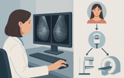
What Is the Best Imaging for Locoregional Staging of Breast Cancer During Pregnancy?

A 34-year-old patient, 22 weeks pregnant with her first child, sits in your office. The biopsy results are back, confirming invasive ductal carcinoma in her right breast. The multidisciplinary tumor board needs accurate locoregional staging to determine the optimal treatment plan—one that balances maternal oncologic safety with fetal well-being. The immediate question is which imaging study to order next to evaluate the axilla and surrounding tissues without harming the fetus. This article provides a detailed clinical workflow for this specific, high-stakes scenario, focusing on the American College of Radiology (ACR) Appropriateness Criteria. For locoregional staging in a pregnant patient with newly diagnosed breast cancer, the ACR rates axillary ultrasound as Usually appropriate.
Who Fits This Clinical Scenario?
This guidance is for a very specific patient population: a pregnant female of any gestational age who has a new, biopsy-proven diagnosis of breast cancer and requires initial locoregional staging. The primary clinical goal is to assess the extent of the disease in the breast and regional lymph node basins (primarily the axilla) to inform surgical and systemic therapy decisions.
This workflow does not apply to several related but distinct clinical situations. It is crucial to distinguish this scenario from:
- Initial workup of a palpable mass: If the patient presented with a palpable lump and cancer has not yet been diagnosed, the imaging pathway is different. That scenario focuses on diagnosis, not staging. (See ACR guidance for Pregnant female, focal pain or palpable breast mass).
- Breast cancer screening during pregnancy: This guidance is not for asymptomatic screening, even in high-risk pregnant patients. Screening protocols have their own specific recommendations. (See ACR guidance for Pregnant female, breast cancer screening).
- Evaluation for distant metastatic disease: This article covers locoregional (breast and lymph node) staging only. Assessing for distant metastases (e.g., in the liver, lungs, or bones) requires a separate workup, which must also be carefully tailored to minimize fetal risk.
Applying this staging workflow is appropriate only after a tissue diagnosis of malignancy has been established.
What Diagnoses Are You Working Up in This Scenario?
In this context, the “differential diagnosis” is less about identifying the primary disease (which is already known) and more about determining its extent. The imaging workup is designed to answer critical staging questions that directly influence treatment. The primary considerations are the presence and burden of nodal disease and the local extent of the primary tumor.
Axillary Lymph Node Metastasis
This is the most critical component of locoregional staging and the strongest prognostic factor outside of tumor characteristics. Identifying metastatic disease in the axillary lymph nodes is essential for TNM (Tumor, Node, Metastasis) staging. The presence of positive nodes typically necessitates more aggressive treatment, such as axillary lymph node dissection instead of a less invasive sentinel lymph node biopsy, and may alter recommendations for chemotherapy.
Multifocal or Multicentric Disease
Staging also involves evaluating the primary breast for additional, clinically occult tumor foci. The presence of multifocal (multiple tumors in one quadrant) or multicentric (multiple tumors in different quadrants) disease can preclude breast-conserving surgery (lumpectomy) and make a mastectomy the necessary surgical approach.
Internal Mammary or Supraclavicular Nodal Involvement
Less common than axillary involvement, metastasis to other nodal basins like the internal mammary or supraclavicular chains signifies a higher disease burden and a worse prognosis. While not always the primary target of initial imaging, these areas may be assessed if axillary disease is extensive or if the primary tumor is located medially in the breast.
Why Is Axillary Ultrasound the Recommended Study for This Presentation?
For locoregional staging of newly diagnosed breast cancer in a pregnant patient, the ACR designates axillary ultrasound (US) as a Usually appropriate imaging study. This recommendation is rooted in the modality’s excellent safety profile during pregnancy and its high diagnostic utility for the primary clinical question: assessing axillary lymph node status.
Ultrasound uses no ionizing radiation (0 mSv) and does not require intravenous contrast, making it the safest initial imaging choice for both the mother and the fetus. It provides high-resolution imaging of the axillary lymph nodes, allowing radiologists to assess for morphologic features suspicious for malignancy, such as cortical thickening, loss of the fatty hilum, and rounded shape. Crucially, if suspicious nodes are identified, ultrasound provides real-time guidance for a fine-needle aspiration (FNA) or core needle biopsy to obtain a definitive cytological or histological diagnosis. This ability to diagnose and stage the axilla non-surgically is a major advantage.
Alongside axillary US, a comprehensive breast US is also rated Usually appropriate to fully characterize the known cancer and search for additional ipsilateral lesions. Diagnostic mammography and digital breast tomosynthesis are also rated Usually appropriate and can be safely performed with appropriate abdominal shielding. The radiation dose to the fetus from a shielded mammogram is negligible (adult RRL ☢☢ 0.1-1mSv). Mammography is particularly valuable for assessing the extent of ductal carcinoma in situ (DCIS) and identifying suspicious microcalcifications that may not be visible on ultrasound.
In contrast, other powerful imaging tools are rated lower for this specific scenario due to safety concerns:
- MRI breast without and with IV contrast is rated Usually not appropriate. While breast MRI is highly sensitive for detecting additional cancer foci and assessing nodal disease, its use in pregnancy is limited by the need for a gadolinium-based contrast agent. Gadolinium can cross the placenta and enter the fetal circulation, and while no definitive harm has been proven in humans, its use is generally avoided during pregnancy unless absolutely essential.
- Sestamibi molecular breast imaging (MBI) is also rated Usually not appropriate. This nuclear medicine study involves injecting a radioactive tracer and carries a significant radiation dose (adult RRL ☢☢☢ 1-10 mSv), posing an unacceptable risk to the fetus.
What’s Next After Axillary Ultrasound? Downstream Workflow
The results of the axillary ultrasound and any subsequent biopsies will directly guide the next steps in the patient’s multidisciplinary care plan. The pathway diverges based on whether nodal metastases are confirmed.
If the axillary US is positive (suspicious nodes confirmed by biopsy):
A positive axillary node biopsy upstages the patient. This finding typically means the patient will require an axillary lymph node dissection (ALND) at the time of breast surgery to clear all cancerous nodes. It also solidifies the recommendation for systemic chemotherapy, which can often be administered safely during the second and third trimesters.
If the axillary US is negative (no morphologically suspicious nodes):
In this case, the patient is considered clinically node-negative. The standard of care is to proceed with a sentinel lymph node biopsy (SLNB) at the time of surgery (mastectomy or lumpectomy). During an SLNB, a tracer (technetium-99m sulfur colloid and/or blue dye) is injected to identify the first one to three “sentinel” nodes that drain the tumor. These nodes are removed and examined by a pathologist. If they are negative, it is highly likely the rest of the axilla is also negative, and a full ALND can be avoided, sparing the patient significant morbidity like lymphedema.
If the findings are indeterminate:
Occasionally, a lymph node may appear equivocal on ultrasound—not clearly benign but not definitively malignant. In these cases, a US-guided FNA or core biopsy is the best next step to resolve the ambiguity and ensure accurate staging before surgery.
Pitfalls to Avoid (and When to Get Help)
Navigating a cancer diagnosis during pregnancy is complex, and several pitfalls can compromise care. First, avoid the temptation to delay staging and treatment due to the pregnancy. Modern oncologic protocols can be safely adapted for pregnant patients, and delays can worsen maternal prognosis. Second, do not rely solely on a clinical axillary examination; imaging is more sensitive for detecting occult nodal metastases. Third, be aware that pregnancy-related hormonal changes can cause reactive lymphadenopathy, which can mimic malignancy on ultrasound. Therefore, any morphologically suspicious node warrants biopsy for confirmation. Finally, if mammography is performed, always ensure meticulous lead shielding of the patient’s abdomen to minimize fetal radiation exposure. If there is any uncertainty about the best imaging pathway or interpretation of results, consultation with a breast imaging radiologist is critical.
Related ACR Topics and Tools
This article addresses one specific clinical scenario. For a comprehensive overview of all related presentations, from screening to palpable masses during pregnancy, refer to the parent topic guide. The following GigHz tools can also support clinical decision-making for this and other imaging scenarios.
- For breadth across all scenarios in Breast Imaging During Pregnancy, see our parent guide: Breast Imaging During Pregnancy: ACR Appropriateness Decoded.
- ACR Appropriateness Criteria Lookup — for adjacent scenarios
- Imaging Protocol Library — for technique on the recommended study
- Radiation Dose Calculator — for cumulative dose conversations
Frequently Asked Questions
Why is MRI not the first choice for staging breast cancer in pregnancy?
Breast MRI is rated ‘Usually not appropriate’ primarily because optimal evaluation requires a gadolinium-based contrast agent. These agents can cross the placenta and enter the fetal circulation. While the risk of harm is theoretical, their use is avoided during pregnancy as a precaution, especially when excellent non-contrast alternatives like ultrasound and shielded mammography exist.
Is mammography safe during pregnancy for staging?
Yes, diagnostic mammography (including tomosynthesis) is considered safe and is rated ‘Usually appropriate’ by the ACR for this scenario. When performed with proper lead shielding over the abdomen and pelvis, the radiation dose to the fetus is negligible and well below the threshold known to cause harm.
If the axillary ultrasound is negative, do we still need to sample the lymph nodes?
Yes. A negative axillary ultrasound means the patient is clinically node-negative, but it does not rule out microscopic metastases. The standard of care for clinically node-negative patients is a sentinel lymph node biopsy (SLNB) performed at the time of breast surgery. This procedure is more accurate than imaging for detecting micrometastatic disease.
Does this guidance apply to staging for distant metastases?
No, this workflow is specifically for locoregional staging (the breast and nearby lymph nodes). Staging for distant metastases is a separate clinical question. The workup typically involves a chest radiograph (with shielding), liver ultrasound, and targeted imaging for specific symptoms, while avoiding high-radiation studies like CT and PET/CT whenever possible.
How do pregnancy-related changes affect the accuracy of axillary ultrasound?
Pregnancy and lactation can cause benign reactive lymphadenopathy, where lymph nodes become enlarged due to hormonal and physiological changes. This can make it more challenging to distinguish benign nodes from metastatic ones. Radiologists rely on specific morphological criteria (like cortical thickness and loss of the fatty hilum) rather than size alone. Any node with suspicious features, regardless of the background of reactive changes, should be considered for biopsy.
Reviewed by Pouyan Golshani, MD, Interventional Radiologist — May 30, 2026