What Is the Best Initial Imaging for a New, Palpable, Unilateral Axillary Lump?

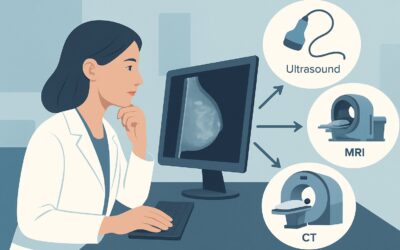
A 45-year-old female patient presents to your clinic on a Tuesday afternoon. During a self-exam last week, she discovered a new, firm, non-tender lump in her left axilla. She reports no changes to her breasts and feels otherwise well. You perform a physical exam and confirm a palpable, mobile, 1.5 cm nodule in the axilla. The immediate clinical question is clear: what is the most appropriate and effective imaging study to order first? This scenario requires a precise diagnostic approach to differentiate benign causes from potentially malignant ones. According to the American College of Radiology (ACR) Appropriateness Criteria, for the initial imaging of a new, palpable, unilateral axillary lump in a female, US axilla is rated Usually Appropriate.
Who Fits This Clinical Scenario for an Axillary Lump?
This clinical workflow is specifically designed for a female patient presenting with a new, palpable, unilateral axillary lump as their primary complaint. The key inclusion criteria are a distinct, touchable abnormality in the armpit on one side that has not been previously evaluated. The patient may or may not have associated symptoms like tenderness, but the lump itself is the reason for the visit.
It is critical to distinguish this presentation from several related but distinct clinical scenarios that follow different diagnostic pathways:
- Bilateral Axillary Lumps: This guidance does not apply if the patient has palpable lumps in both axillae. Bilateral findings are more suggestive of a systemic process, such as a viral illness, autoimmune condition, or a reaction to a recent vaccination, and the workup may be different.
- Known Breast Cancer Diagnosis: This workflow is not for patients who already have a diagnosed breast cancer. In that context, axillary imaging is performed for staging purposes, not for the initial workup of a new lump. The ACR has separate, specific criteria for axillary staging based on tumor size and clinical node status.
- Lump Within the Breast Tissue: If the palpable lump is clearly within the breast parenchyma and not isolated to the axilla, the workup should follow the guidelines for a palpable breast mass, which typically begins with diagnostic mammography and/or ultrasound of the breast.
This article focuses exclusively on the initial imaging decision for an isolated, one-sided axillary lump.
What Diagnoses Are You Working Up in This Scenario?
When a female patient presents with a unilateral axillary lump, the imaging workup is designed to evaluate a specific differential diagnosis, ranging from common benign conditions to serious malignancies.
The most common cause of a palpable axillary lump is reactive lymphadenopathy. Lymph nodes in the axilla drain the arm, chest wall, and breast, and they can enlarge in response to local inflammation, infection (e.g., from a skin lesion on the arm), or recent vaccination. While common, this is a diagnosis of exclusion after more significant pathology has been ruled out. Ultrasound is key to identifying the reassuring features of a reactive node, such as a preserved fatty hilum and a uniformly thin cortex.
The most consequential diagnosis to consider is metastatic disease, most commonly from an occult (hidden) breast cancer. The axillary lymph nodes are the first site of spread for most breast cancers. Less commonly, metastases can originate from melanoma or other malignancies. Imaging aims to identify suspicious nodal features like a rounded shape, loss of the fatty hilum, focal or diffuse cortical thickening, and abnormal vascularity.
Another important consideration is accessory axillary breast tissue. This is ectopic glandular tissue that persists in the axilla after embryonic development. It is a common anatomical variant and can become palpable or tender, particularly in response to hormonal fluctuations. On imaging, it appears as tissue identical to normal breast parenchyma. While usually benign, it can, in rare cases, be the site of a primary axillary breast cancer.
Finally, the differential includes a variety of benign skin and subcutaneous lesions. These can include lipomas (fatty tumors), sebaceous cysts (epidermal inclusion cysts), or inflammatory conditions like hidradenitis suppurativa. Ultrasound is highly effective at characterizing these superficial lesions and confirming they are not of lymph node origin.
Why Is Axillary Ultrasound the Recommended First Study for a Palpable Lump?
The ACR designates axillary ultrasound (US) as Usually Appropriate for this scenario because it directly and effectively addresses the key clinical questions with no radiation exposure and high diagnostic yield.
The primary strength of ultrasound is its exceptional soft-tissue resolution, which allows for detailed morphologic assessment of the palpable finding. It can reliably:
- Confirm the origin: Is the lump an enlarged lymph node, accessory breast tissue, a simple cyst, or a solid subcutaneous mass like a lipoma?
- Characterize lymph nodes: US is the best modality for evaluating the internal architecture of a lymph node. It can distinguish the reassuring features of a benign reactive node (oval shape, preserved central fatty hilum) from suspicious features of malignancy (rounded shape, effaced hilum, thickened cortex).
- Guide intervention: If a suspicious solid mass or abnormal lymph node is identified, ultrasound provides real-time guidance for a fine-needle aspiration (FNA) or core needle biopsy during the same visit or as a scheduled follow-up, accelerating the diagnostic timeline.
In contrast, other imaging modalities are rated lower for this specific initial workup:
- Mammography or Digital Breast Tomosynthesis (DBT): Rated as May be appropriate, these studies are not the ideal first step for evaluating an isolated axillary lump. While an axillary view can be included, mammography is less sensitive than ultrasound for assessing lymph node morphology. It is often used as a complementary study to search for an occult primary breast cancer if the ultrasound reveals a suspicious axillary node.
- MRI of the Breast: Rated as Usually not appropriate for this initial workup. While highly sensitive, breast MRI has lower specificity and can lead to a cascade of further testing for incidental findings. Its use is typically reserved for problem-solving after initial imaging is complete or for staging in a patient with biopsy-proven cancer.
From a safety perspective, ultrasound is the clear choice. It involves no ionizing radiation (adult relative radiation level: O, 0 mSv), unlike mammography (RRL: ☢☢, 0.1-1 mSv) or CT (RRL: ☢☢☢☢, 10-30 mSv). When ordering, it is helpful to specify “Targeted US of the palpable [right/left] axillary lump” to ensure the sonographer and radiologist focus directly on the area of clinical concern.
What’s Next After US axilla? Downstream Workflow
The results of the axillary ultrasound will dictate the next steps in the patient’s care. The workflow branches based on whether the findings are clearly benign, suspicious, or indeterminate.
- If the US is definitively benign: If the ultrasound identifies a simple cyst, a classic-appearing lipoma, or a lymph node with clearly benign features (e.g., a preserved fatty hilum and thin cortex in the setting of a recent vaccination), no further imaging is typically required. The patient can be reassured, and clinical follow-up may be recommended to ensure the lump resolves or remains stable.
- If the US is suspicious for malignancy: If the ultrasound reveals a lymph node with suspicious features (e.g., rounded, hypoechoic, thickened cortex, absent hilum) or a solid mass, the next step is an image-guided biopsy. An ultrasound-guided core needle biopsy is the standard procedure to obtain a tissue diagnosis. A diagnostic mammogram should also be performed on the ipsilateral breast to search for a potential primary cancer.
- If the US identifies accessory breast tissue: If the palpable lump is confirmed to be accessory axillary breast tissue without suspicious features, it is generally considered a benign finding. No further workup is needed unless the patient is experiencing significant symptoms. A diagnostic mammogram may still be considered, particularly in women of screening age, to provide a baseline evaluation of this tissue.
- If the US is negative or indeterminate: If the ultrasound fails to identify a sonographic correlate for the palpable lump, or if the findings are equivocal, a diagnostic mammogram of the ipsilateral breast and axilla is a reasonable next step. This is particularly important in women over 30. If both studies are negative and clinical suspicion remains, short-term clinical follow-up in 3-6 months is often recommended.
Pitfalls to Avoid (and When to Get Help)
Navigating the workup of an axillary lump requires careful attention to detail to avoid common diagnostic errors.
- Pitfall 1: Dismissing a lump after a negative mammogram. A mammogram can miss pathology in the axilla, especially dense lymph nodes or lesions obscured by surrounding tissue. Ultrasound is more sensitive for nodal morphology; do not skip it if there is a true palpable finding.
- Pitfall 2: Not correlating with clinical history. Always consider recent events like infections, skin trauma, or vaccinations (especially COVID-19 or flu shots in the ipsilateral arm), as these are common causes of transient reactive adenopathy.
- Pitfall 3: Assuming a lump in accessory breast tissue is benign. While usually benign, primary breast cancer can arise in accessory axillary tissue. Any suspicious mass within this tissue warrants a biopsy, just as it would in the breast.
If a biopsy confirms malignancy in an axillary lymph node but the mammogram and ultrasound of the breast are negative, this is considered occult breast cancer (axillary presentation). At this point, escalation to a breast surgeon and further imaging with breast MRI is warranted to search for the primary tumor.
Related ACR Topics and Tools
For a comprehensive overview of all clinical scenarios related to axillary imaging, further reading and specialized tools can provide additional context and support for clinical decision-making.
For breadth across all scenarios in Imaging of the Axilla, see our parent guide: Imaging of the Axilla: ACR Appropriateness Decoded.
The following GigHz resources can also help streamline your workflow:
- ACR Appropriateness Criteria Lookup — for quickly checking adjacent or alternative clinical scenarios.
- Imaging Protocol Library — for detailed technical guidance on performing the recommended studies.
- Radiation Dose Calculator — for discussing cumulative radiation exposure with patients when considering studies like mammography or CT.
Frequently Asked Questions
Should I order a mammogram at the same time as the axillary ultrasound?
For an isolated palpable axillary lump, the ACR recommends starting with a targeted axillary ultrasound. A diagnostic mammogram is rated ‘May be appropriate’ and is often performed as the next step if the ultrasound shows a suspicious lymph node, to look for a primary breast cancer. Some centers may perform both on the same day for efficiency, but ultrasound is the key initial test for characterizing the lump itself.
What if the patient recently had a COVID-19 vaccine in that arm?
Unilateral axillary lymphadenopathy is a well-documented and common reaction to mRNA COVID-19 vaccines, as well as other vaccinations. It is crucial to obtain this history. If a patient has a new lump and a recent ipsilateral vaccination, and the ultrasound shows reactive-appearing nodes, short-term clinical or imaging follow-up (e.g., in 6-12 weeks) to ensure resolution is a reasonable approach to avoid unnecessary biopsies.
Is an MRI a good first test for an axillary lump?
No, for the initial evaluation of a palpable axillary lump, breast MRI is rated ‘Usually not appropriate’ by the ACR. While very sensitive, it lacks specificity and can lead to false positives and unnecessary anxiety and biopsies. Its role is typically reserved for cases where a biopsy has already confirmed cancer, to help with staging or searching for an occult primary tumor.
What if the ultrasound is negative but I can still feel the lump?
This situation requires careful clinical judgment. A palpable finding that is ‘ultrasound-occult’ can occur. The next steps may include a diagnostic mammogram (if not already done) and close clinical follow-up. If the lump persists or grows, a repeat ultrasound or consultation with a breast specialist or surgeon for consideration of excisional biopsy may be necessary.
Does this guidance apply to men with an axillary lump?
This specific ACR variant is for female patients. While the initial imaging study for a male patient with an axillary lump would also likely be ultrasound, the differential diagnosis and pre-test probability of malignancy (e.g., male breast cancer) are different, and the clinical context must be carefully considered. This guidance should not be directly extrapolated without accounting for these differences.
Reviewed by Pouyan Golshani, MD, Interventional Radiologist — May 29, 2026