What Is the Best Initial Imaging for a Perianal Fistula in a Child with Crohn Disease?
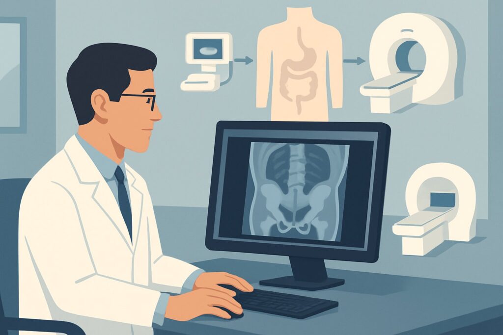
It’s late in the afternoon at the pediatric gastroenterology clinic. You are seeing a 12-year-old with a known diagnosis of Crohn disease, well-controlled for the past year on biologics. Today, their parent reports new, persistent perianal pain and intermittent, non-purulent drainage from a small skin opening near the anus. On examination, you confirm an external fistula opening. Before you can coordinate care with pediatric surgery and adjust medical therapy, you need to understand the anatomy of the tract and rule out an abscess. The immediate question is which imaging study will provide the most accurate and safest assessment. According to the American College of Radiology (ACR) Appropriateness Criteria, for a child with known Crohn disease and a perianal fistula, MRI pelvis with IV contrast is rated Usually Appropriate.
Who Fits This Clinical Scenario?
This guidance is specifically for a pediatric patient with an already established diagnosis of Crohn disease who now presents with clinical signs or symptoms of a perianal fistula. This includes findings like a visible external opening, perianal or gluteal pain, a palpable cord-like tract, swelling, or drainage of pus, stool, or mucus from a perianal site. The key elements are the known underlying inflammatory bowel disease (IBD) and the new perianal complication, for which this is the initial imaging workup.
This workflow should be distinguished from several related but distinct clinical situations:
- Child with suspected Crohn disease: If the patient does not have a confirmed diagnosis of Crohn disease and presents with perianal symptoms as part of their initial workup, the imaging strategy may differ. That scenario focuses on establishing the primary diagnosis of IBD.
- Child with known Crohn disease and suspected acute intra-abdominal exacerbation: If the primary concern is a flare within the abdomen (e.g., increased abdominal pain, diarrhea, fever) without specific perianal signs, the focus of imaging shifts from the pelvis to the entire abdomen, often using Magnetic Resonance Enterography (MRE) or Computed Tomography Enterography (CTE).
- Child with known Crohn disease for disease surveillance: Routine imaging to monitor therapy response in an asymptomatic or stable patient follows a different protocol, aimed at assessing overall disease burden rather than a specific, acute complication like a fistula.
Applying this guidance is most effective when the primary clinical question is to define the anatomy of a suspected perianal fistula and to identify any associated abscess in a child with confirmed Crohn disease.
What Diagnoses Are You Working Up in This Scenario?
When ordering imaging for a perianal fistula in a child with Crohn disease, the goal is to answer specific questions that directly influence surgical and medical management. The differential is less about “what is the cause” (which is almost certainly Crohn’s) and more about “what is the extent and complexity of the disease.”
Perianal Fistula Classification and Complexity: This is the primary reason for imaging. The study must accurately delineate the fistula’s path in relation to the anal sphincter complex. Radiologists typically use the Parks classification to describe tracts as intersphincteric, transsphincteric, suprasphincteric, or extrasphincteric. Identifying a “complex” fistula—one with multiple tracts, a high tract (suprasphincteric/extrasphincteric), or association with an abscess—is critical, as it often necessitates more aggressive medical therapy (e.g., anti-TNF agents) and specialized surgical approaches like seton placement to avoid incontinence.
Perianal or Ischiorectal Abscess: The most urgent finding to identify or exclude is an associated abscess. A fistula tract can become blocked, leading to a collection of pus. An undrained abscess can cause severe pain, systemic illness, and extensive tissue damage. Its presence mandates prompt surgical drainage, often before or concurrent with an escalation in medical therapy. MRI is highly sensitive for detecting these fluid collections, which appear as well-defined, rim-enhancing areas.
Rectal and Anal Canal Inflammation: The imaging study also provides a detailed view of the adjacent rectum and anal canal, allowing for assessment of active proctitis or stricturing. The presence of significant rectal inflammation can impact the choice of surgical intervention and medical management, as healing may be impaired in areas of severe active disease.
Why Is MRI of the Pelvis with IV Contrast the Recommended Study for This Presentation?
The ACR designates MRI pelvis with IV contrast as Usually Appropriate because it offers the best combination of diagnostic accuracy and safety for evaluating perianal Crohn disease in children. Its superior soft-tissue contrast resolution is unmatched for visualizing the fine details of fistula tracts and their relationship to the pelvic floor musculature.
The rationale for its top rating is multi-faceted:
- Superior Tract Delineation: T2-weighted sequences excel at showing fluid-filled fistula tracts as bright signals against the surrounding tissue. This allows for precise mapping of the primary tract, any secondary branches, and their course relative to the internal and external anal sphincters—information that is essential for surgical planning to preserve fecal continence.
- High Sensitivity for Abscesses: After the administration of intravenous gadolinium-based contrast, the inflamed walls of an abscess cavity will brightly enhance on T1-weighted, fat-suppressed images, making even small collections conspicuous. Non-contrast MRI is significantly less sensitive for this critical finding.
- Zero Ionizing Radiation: This is a paramount consideration in pediatric patients. Children with Crohn disease will likely require multiple imaging studies over their lifetime. Choosing a radiation-free modality like MRI (0 mSv) whenever possible is crucial for minimizing cumulative radiation exposure and the associated long-term risks.
Alternative studies are rated lower for specific, important reasons in this scenario:
- CT pelvis with IV contrast is rated May be appropriate. While it can identify large abscesses, its soft-tissue resolution is far inferior to MRI for delineating fistula tracts. More importantly, it involves a significant radiation dose (pediatric RRL ☢☢☢☢ 3-10 mSv), making it a suboptimal choice for a young patient with a chronic condition unless MRI is unavailable or contraindicated.
- Transperineal US pelvis is also rated May be appropriate. It is a radiation-free and accessible option that can identify superficial tracts and collections. However, it is highly operator-dependent, can be uncomfortable for the patient, and has a limited field of view, often failing to visualize deep or complex tracts that extend into the ischiorectal fossa or above the levator ani muscles.
When ordering, it is crucial to specify “pelvic MRI for perianal fistula” to ensure the radiology department uses a dedicated protocol with high-resolution, small field-of-view images through the anal canal. The inclusion of IV contrast is not optional; it is essential for assessing inflammatory activity and detecting abscesses.
What’s Next After MRI of the Pelvis with IV Contrast? Downstream Workflow
The results of the pelvic MRI create a clear branch point in the patient’s management plan, guiding collaboration between the pediatric gastroenterologist and pediatric surgeon.
If the MRI is positive for a complex fistula or an abscess:
This finding typically triggers an urgent consultation with pediatric surgery. An abscess requires prompt incision and drainage, often performed as an examination under anesthesia (EUA). During the EUA, the surgeon can also directly visualize the fistula anatomy and place a draining seton if needed. Medically, this finding often signals a need to optimize or escalate immunosuppressive therapy, as complex fistulas are a marker of more aggressive disease.
If the MRI is positive for a simple, low fistula without an abscess:
Management may be less urgent. The surgical and gastroenterology teams can discuss options, which may include medical therapy alone, fistulotomy for a very simple tract, or seton placement. The decision depends on the specific anatomy, the severity of symptoms, and the patient’s overall disease activity.
If the MRI is negative:
A high-quality negative MRI is very reassuring and makes a clinically significant fistula or abscess highly unlikely. If symptoms persist despite a negative MRI, the next step is often a diagnostic EUA, which remains the ultimate gold standard for direct visualization. The clinical picture should be re-evaluated to consider other causes of perianal pain, such as anal fissures or skin conditions, though these are less common in this context.
If the MRI is indeterminate:
This is uncommon with modern MRI techniques. However, if the findings are equivocal, a direct EUA is the most logical next step to resolve the diagnostic uncertainty and proceed with treatment.
Pitfalls to Avoid (and When to Get Help)
Navigating the workup of perianal Crohn disease requires avoiding several common pitfalls to ensure timely and accurate diagnosis.
- Ordering a non-contrast MRI: Forgetting to request IV contrast is a frequent error. A non-contrast study is inadequate for assessing inflammatory activity and can easily miss an abscess, delaying critical treatment.
- Defaulting to CT: Reaching for CT out of habit or perceived speed exposes the child to unnecessary ionizing radiation. Reserve CT for situations where MRI is absolutely contraindicated or immediately unavailable in an unstable patient.
- Vague Ordering Information: Simply ordering “MRI Pelvis” without providing the specific clinical context (“known Crohn’s, rule out perianal fistula/abscess”) may result in a generic protocol that lacks the high-resolution sequences needed to answer the clinical question.
- Delaying Imaging: If an abscess is suspected based on signs like fever, severe pain, or a tender, fluctuant mass, imaging should be obtained urgently.
If the MRI confirms an abscess or a complex, high fistula, or if the patient shows any signs of systemic toxicity (fever, tachycardia), immediate escalation to a pediatric surgeon and inpatient admission are warranted.
Related ACR Topics and Tools
This article focuses on a single, specific clinical scenario. For a comprehensive overview of all pediatric Crohn disease imaging variants, from initial diagnosis to surveillance, please consult our parent topic hub article. For additional decision support, the following resources are available:
- Parent Topic Hub: For breadth across all scenarios in Crohn Disease-Child, see our parent guide: Crohn Disease-Child: ACR Appropriateness Decoded.
- ACR Criteria Lookup: To explore other clinical variants or search different topics, use the ACR Appropriateness Criteria Lookup.
- Imaging Protocols: For detailed technical specifications on MRI and other modalities, see the Imaging Protocol Library.
- Dose Conversations: To discuss radiation exposure from alternative studies like CT, use the Radiation Dose Calculator.
Frequently Asked Questions
Why not just proceed directly to an examination under anesthesia (EUA) instead of getting an MRI first?
While EUA is the gold standard for diagnosis and treatment, a pre-operative MRI provides a crucial roadmap for the surgeon. It delineates the full extent of deep tracts and identifies any unsuspected abscesses, allowing for more precise and comprehensive surgical planning. This can reduce the risk of missing a complex component of the fistula and may decrease the need for repeat procedures.
Is an MRI without contrast ever sufficient for evaluating a perianal fistula?
No, an MRI without IV contrast is considered an incomplete study for this indication. While non-contrast T2-weighted images are excellent for showing the fluid-filled fistula tract, they are unreliable for detecting an associated abscess or assessing the degree of active inflammation. IV contrast is essential for highlighting abscess walls and inflamed tissue, which are critical findings for management.
What if the child is claustrophobic or too young to tolerate an MRI without moving?
This is a common challenge in pediatric imaging. Most imaging centers have well-established protocols for performing MRIs with sedation or general anesthesia for young children or those who cannot remain still. The benefits of obtaining a high-quality, motion-free MRI almost always outweigh the risks of sedation in this clinical context. This should be coordinated with the radiology department and, if needed, a pediatric anesthesiologist.
How does imaging for a perianal fistula differ from imaging for an intra-abdominal Crohn’s flare?
The imaging target and protocol are completely different. For a perianal fistula, the study is a dedicated pelvic MRI with a small field-of-view focused on the anal canal and surrounding soft tissues. For an intra-abdominal flare, the standard is typically MR Enterography (MRE), which involves drinking oral contrast and imaging the entire small and large bowel to assess for inflammation, strictures, and penetrating complications within the abdomen.
Does this same imaging guidance apply to adults with Crohn disease and a perianal fistula?
Yes, the principles are identical. MRI of the pelvis with and without IV contrast is also the ‘Usually Appropriate’ first-line imaging modality for evaluating perianal fistulas in adults with Crohn disease. The emphasis on avoiding radiation is even more pronounced in children, but the superior diagnostic accuracy of MRI makes it the preferred test for all age groups.
Reviewed by Pouyan Golshani, MD, Interventional Radiologist — May 29, 2026