What Is the Best Initial Imaging for a Suspected Cardiac Mass in an Adult?

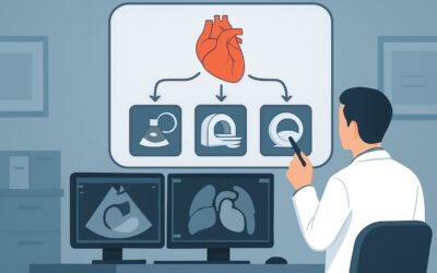
It’s late in your clinic day when you see a 58-year-old patient with new-onset exertional dyspnea and intermittent palpitations. A recent outpatient chest radiograph, ordered for a persistent cough, was reported as showing an “abnormal cardiac contour,” raising suspicion for an intracardiac mass. The patient has no prior cardiac history. You now face the critical decision of which initial imaging study to order to confirm or exclude a cardiac mass and begin to characterize it. This article provides a step-by-step clinical workflow for this exact scenario, guided by the American College of Radiology (ACR) Appropriateness Criteria. For this presentation, the ACR rates `US echocardiography transthoracic resting` as Usually Appropriate.
Who Fits This Clinical Scenario?
This guidance is for clinicians evaluating an adult patient with a new suspicion of a cardiac mass where no mass has been previously confirmed by imaging. The key inclusion criteria are:
- Patient: Adult
- Presentation: Clinical signs or symptoms (e.g., new heart failure, arrhythmia, embolic event) or an incidental finding on other imaging (like a chest X-ray or CT) that raises suspicion for a cardiac mass.
- History: No previously known or diagnosed cardiac mass. This is the initial diagnostic step.
This workflow is distinct from similar, but clinically different, scenarios. This article does not apply if:
- A cardiac mass has already been identified on an echocardiogram. In that case, the question is about further characterization, which falls under the ACR variant “Adult. Known cardiac mass in echocardiography. Unknown etiology. Next imaging study.”
- The patient has an established diagnosis and is undergoing surveillance. That follow-up imaging workflow is covered in the variant “Adult. Known cardiac mass. Established etiology. Follow-up imaging.”
Correctly identifying your patient’s scenario is crucial for selecting the most appropriate and resource-effective imaging pathway from the start.
What Diagnoses Are You Working Up in This Scenario?
When you suspect a cardiac mass, the differential diagnosis is broad, ranging from benign tumors to aggressive malignancies and non-neoplastic mimics. The goal of initial imaging is to detect the lesion and provide clues to its etiology.
Benign Primary Tumors: The most common primary cardiac tumor in adults is the atrial myxoma, typically a pedunculated, mobile mass arising from the interatrial septum in the left atrium. While histologically benign, myxomas can cause significant morbidity through obstruction of blood flow or systemic embolization. Other less common benign tumors include papillary fibroelastomas (often on valve leaflets), lipomas, and fibromas.
Malignant Tumors: Malignant tumors of the heart are rare but carry a poor prognosis. Secondary, or metastatic, tumors are substantially more common than primary cardiac malignancies. Cancers that frequently metastasize to the heart include lung cancer, breast cancer, melanoma, and lymphoma. Primary cardiac malignancies are most often sarcomas (e.g., angiosarcoma), which are typically large, infiltrative masses with associated pericardial effusions.
Non-Neoplastic Mass Mimics: It is critical to remember that not all intracardiac masses are tumors. Thrombus is a very common finding, particularly in the left atrial appendage in patients with atrial fibrillation or in ventricular apices in the setting of wall motion abnormalities from prior myocardial infarction. Other mimics include large vegetations from endocarditis, prominent normal anatomic variants (like the crista terminalis or Chiari network), and pericardial cysts.
Why Is US Echocardiography Transthoracic Resting the Recommended Initial Study?
For the initial evaluation of a suspected cardiac mass in an adult, the ACR designates `US echocardiography transthoracic resting` (TTE) as a Usually Appropriate first step. This recommendation is based on its excellent balance of diagnostic capability, safety, and accessibility.
A TTE is a non-invasive, real-time imaging modality that uses no ionizing radiation (0 mSv). It is widely available, relatively inexpensive, and can be performed at the bedside if necessary. TTE is highly effective for the initial detection of a mass, providing crucial information about its size, location, mobility, attachment point, and any associated hemodynamic consequences, such as valvular obstruction or regurgitation. It can also assess overall cardiac function and identify associated findings like pericardial effusions.
While TTE is the primary starting point, other advanced imaging modalities are also highly rated but are typically reserved for problem-solving or subsequent characterization:
- MRI heart function and morphology without and with IV contrast: Also rated Usually Appropriate, cardiac MRI (CMR) offers superior soft tissue contrast and is the gold standard for tissue characterization. It can help differentiate thrombus from tumor and benign from malignant lesions based on signal characteristics and gadolinium enhancement patterns. However, it is less available, more expensive, and has a longer acquisition time than TTE, making it a better second-line or problem-solving tool in most initial workups.
- CT heart function and morphology with IV contrast: This is another Usually Appropriate option. Cardiac CT provides excellent spatial resolution and is particularly useful for assessing calcification within a mass and evaluating for extracardiac extension or coronary artery involvement. Its major drawback is the significant radiation dose (☢☢☢☢ 10-30 mSv), making TTE or MRI preferable initial choices when feasible.
- Radiography chest: A chest X-ray is rated Usually not appropriate for this indication. While it may be the study that first raises suspicion, it lacks the spatial and contrast resolution to confirm, localize, or characterize an intracardiac mass.
The logical first step is the TTE. Its ability to confirm the presence of a mass and assess its immediate functional impact provides the necessary information to guide the subsequent, more advanced, diagnostic pathway.
What’s Next After US Echocardiography Transthoracic Resting? Downstream Workflow
The results of the initial transthoracic echocardiogram will dictate the next steps in the patient’s management and imaging journey. The workflow branches based on whether the study is positive, negative, or indeterminate.
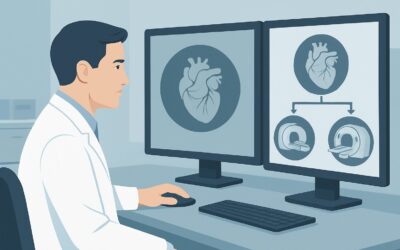
If the TTE is positive and identifies a mass: The primary goal shifts from detection to characterization. The findings on TTE (e.g., location, mobility, appearance) will narrow the differential. At this point, the patient’s clinical scenario transitions to “Adult. Known cardiac mass in echocardiography. Unknown etiology. Next imaging study.” The most common next step is a `MRI heart function and morphology without and with IV contrast`. Cardiac MRI excels at tissue characterization, helping to distinguish myxoma, thrombus, lipoma, or a suspected malignancy, which is critical for surgical or therapeutic planning.
If the TTE is negative but clinical suspicion remains high: A negative TTE is often reassuring, but it is not perfect. Acoustic windows can be poor, and small masses or those in posterior structures (like the left atrial appendage) can be missed. If clinical suspicion persists (e.g., a patient with a known primary cancer and a suspected embolic event), the next step is often `US echocardiography transesophageal` (TEE), which is rated May be appropriate (Disagreement). TEE provides superior resolution of posterior cardiac structures and is more sensitive for small masses, vegetations, and thrombi.
If the TTE is indeterminate or technically limited: In cases where acoustic windows are suboptimal or the findings are equivocal, proceeding directly to cardiac MRI is the most logical step. As a Usually Appropriate study for initial evaluation, it serves as the definitive problem-solving tool, bypassing the need for a TEE in many cases where tissue characterization, rather than just improved visualization, is the primary goal.
Pitfalls to Avoid (and When to Get Help)
Navigating the workup of a suspected cardiac mass requires careful interpretation and awareness of common challenges. Here are several pitfalls to avoid:
- Mistaking normal anatomy for pathology: Prominent normal structures like the crista terminalis, Eustachian valve, Chiari network, or moderator band can be mistaken for masses on TTE. An experienced sonographer and interpreting physician are key to avoiding this error.
- Stopping with a limited study: If a TTE is technically limited due to poor acoustic windows (common in patients with obesity or lung disease) and clinical suspicion is moderate to high, do not accept the study as negative. Proceed to TEE or cardiac MRI.
- Overlooking thrombus: Thrombus is the great mimicker and a common intracardiac “mass.” Always consider it in the differential, especially with regional wall motion abnormalities or atrial fibrillation. The management (anticoagulation) is vastly different from that of a tumor (surgery).
- Ignoring the clinical context: A new mass in a patient with a history of melanoma is highly suspicious for metastasis. A mass on a valve in a patient with fever and positive blood cultures is likely a vegetation. The imaging findings must be integrated with the full clinical picture.
If you identify a large, mobile mass with high embolic potential or one causing significant hemodynamic compromise, this constitutes a clinical emergency. Escalate immediately to a cardiology and cardiothoracic surgery service for urgent management.
Related ACR Topics and Tools
For a comprehensive overview of all clinical variants related to the evaluation of cardiac masses, please consult the parent topic hub article. For further exploration of imaging guidelines, protocols, and safety, the following resources are available:
- For breadth across all scenarios in Evaluation of Cardiac Masses, see our parent guide: Evaluation of Cardiac Masses: ACR Appropriateness Decoded.
- To explore adjacent clinical scenarios and their corresponding ACR ratings, use the ACR Appropriateness Criteria Lookup tool.
- For detailed procedural techniques on recommended studies, see the Imaging Protocol Library.
- To discuss radiation exposure with patients, especially when considering CT, use the Radiation Dose Calculator.
Frequently Asked Questions
Why is TTE the first choice over Cardiac MRI, since both are rated ‘Usually Appropriate’?
Transthoracic echocardiography (TTE) is recommended as the initial study primarily due to its widespread availability, lower cost, portability, and speed. It provides excellent real-time information on mass location, mobility, and hemodynamic impact without radiation or contrast. Cardiac MRI, while superior for tissue characterization, is more resource-intensive and is best used as a second step to characterize a mass found on TTE or when TTE is non-diagnostic.
What should I do if my patient’s TTE is inconclusive because of poor acoustic windows?
If a TTE is technically limited and cannot confidently confirm or exclude a mass, the next step is typically a Cardiac MRI. MRI is not limited by acoustic windows and provides excellent anatomical detail and tissue characterization. A Transesophageal Echocardiogram (TEE) is another option, particularly if the suspicion is for a small mass on a valve or in the left atrial appendage.
Is a chest X-ray ever useful in this scenario?
According to the ACR, a chest X-ray is ‘Usually not appropriate’ for the primary evaluation or characterization of a cardiac mass. However, it often serves as the initial test that incidentally raises suspicion due to an abnormal cardiac silhouette or calcification, thereby triggering the more specific cardiac imaging workflow starting with TTE.
When should I consider ordering a Cardiac CT scan first?
While Cardiac CT is also rated ‘Usually Appropriate,’ it’s generally not the first choice due to its significant radiation dose. A CT might be considered first-line if MRI is contraindicated (e.g., incompatible implanted device), if there is a strong suspicion of calcification within the mass (e.g., fibroma), or if assessment of the coronary arteries or extracardiac structures is a primary concern from the outset.
Does the term ‘suspected cardiac mass’ include a suspected thrombus?
Yes. From an initial imaging perspective, the workup is the same. A thrombus is a non-neoplastic intracardiac mass, and TTE is an excellent first-line tool to detect it. The subsequent characterization with TTE, contrast echo, or Cardiac MRI helps differentiate thrombus from a tumor, which is critical as their management pathways are completely different.
Reviewed by Pouyan Golshani, MD, Interventional Radiologist — May 30, 2026