What Is the Best Initial Imaging for a Suspected Rectovesicular or Rectovaginal Fistula?
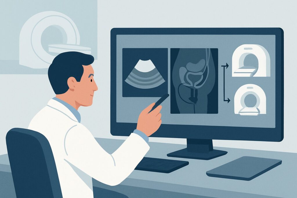
A 58-year-old woman with a history of pelvic radiation for cervical cancer presents to your clinic with several weeks of foul-smelling vaginal discharge and recurrent urinary tract infections. She reports passing gas from her vagina. You are concerned about a post-radiation rectovaginal fistula, an abnormal communication between the rectum and the vagina. The immediate clinical question is how to confirm this suspicion and, more importantly, to delineate the anatomy for surgical planning. This requires an imaging study with excellent soft-tissue resolution that can visualize the fistula tract, its relationship to adjacent organs, and any associated inflammation or abscess. According to the American College of Radiology (ACR) Appropriateness Criteria, MRI pelvis without and with IV contrast is rated Usually Appropriate for this initial workup.
Who Fits This Clinical Scenario for a Suspected Rectal Fistula?
This guidance applies to patients where there is a clinical suspicion of a fistula connecting the rectum to an adjacent pelvic organ, specifically the urinary bladder (rectovesicular) or the vagina (rectovaginal). The clinical presentation is key. Patients typically report symptoms that are highly specific for an abnormal connection between the gastrointestinal tract and the genitourinary system.
Inclusion criteria for this workflow:
- Pneumaturia: Passing air during urination.
- Fecaluria: Passing fecal matter in the urine.
- Feculent vaginal discharge: Stool exiting the vagina.
- Recurrent, polymicrobial urinary tract infections, especially in a patient with known risk factors like diverticulitis, Crohn’s disease, pelvic malignancy, or prior pelvic radiation.
Exclusion criteria (patients who fit a different ACR variant):
- Suspected simple perianal fistula: Patients with external skin openings, pain, and drainage near the anus without signs of deeper organ involvement. This presentation falls under the Suspected perianal disease. Abscess or fistula scenario.
- Post-surgical anastomotic leak: Patients with recent colorectal surgery presenting with fever, pain, or sepsis. This is covered by the Suspected complication postproctectomy or coloproctectomy variant.
- Symptoms of proctitis: Patients with rectal bleeding, tenesmus, and discharge without specific signs of a fistula to the bladder or vagina. This aligns with the Suspected proctitis or pouchitis scenario.
Disambiguating these presentations ensures the correct imaging pathway is chosen from the outset.
What Diagnoses Are You Working Up in This Scenario?
When ordering imaging for a suspected rectovesicular or rectovaginal fistula, you are primarily confirming its presence and evaluating for underlying causes and complications. The differential diagnosis guides the radiologist’s search pattern.
Rectovesicular or Rectovaginal Fistula
This is the primary diagnosis to confirm or exclude. The most common cause of rectovesicular fistulas in adults is complicated diverticulitis, where an inflamed colonic diverticulum erodes into the bladder. For rectovaginal fistulas, obstetric trauma is a leading cause worldwide, while in developed nations, Crohn’s disease, pelvic malignancy (e.g., cervical, rectal, or vaginal cancer), and complications from pelvic radiation therapy are more frequent etiologies. The imaging study must clearly define the fistula’s origin, course, and termination.
Complex Pelvic Abscess
An abscess can be both a cause and a consequence of a fistula. A large, uncontained pelvic abscess can erode into adjacent structures, creating a fistulous tract. Conversely, a fistula can lead to leakage of enteric contents, resulting in a secondary abscess. Imaging must identify any fluid collections that require percutaneous or surgical drainage, as this is a critical component of treatment.
Anorectal or Pelvic Malignancy
A primary or recurrent tumor is a crucial diagnosis to consider, as it completely changes the management pathway. Advanced colorectal, cervical, bladder, or prostate cancer can directly invade adjacent organs, leading to fistula formation. Imaging helps stage the tumor, assess its extent, and detect metastatic disease, which is vital for oncologic and surgical planning.
Inflammatory Bowel Disease (IBD)
Crohn’s disease is well-known for its penetrating, transmural inflammation that can lead to complex fistulizing disease. While more commonly associated with perianal fistulas, it can also cause rectovesicular and rectovaginal fistulas. Imaging can reveal other signs of Crohn’s, such as bowel wall thickening, strictures, and mesenteric inflammation (“creeping fat”), supporting the diagnosis.
Why Is MRI of the Pelvis the Recommended Initial Study for a Suspected Rectal Fistula?
The ACR designates MRI pelvis without and with IV contrast as Usually Appropriate because of its unparalleled ability to visualize pelvic soft-tissue anatomy and pathology without using ionizing radiation. This makes it the premier modality for characterizing the complex relationships between the rectum, bladder, vagina, and surrounding muscles and fat planes.
The superior soft-tissue contrast of MRI is essential for directly visualizing the thin, often subtle, fistulous tract. T2-weighted sequences are particularly effective at highlighting the fluid-filled or inflamed tract against the background of normal pelvic structures. The addition of intravenous gadolinium-based contrast is critical. Post-contrast T1-weighted fat-suppressed images will show enhancement of the fistula tract walls and any associated inflammatory changes or abscess collections, significantly increasing the study’s diagnostic confidence and accuracy.
While MRI is the top-rated study, other modalities are considered:
- CT pelvis with IV contrast is also rated Usually Appropriate. It is a widely available and rapid imaging technique that can reliably identify fistulas, especially when there is air in the bladder or vagina. However, its soft-tissue resolution is inferior to MRI, which can make it difficult to see small or non-inflamed tracts. It also involves significant ionizing radiation (adult RRL=☢☢☢ 1-10 mSv), a key consideration in younger patients or those with IBD who may require multiple follow-up scans. CT is an excellent alternative when MRI is contraindicated (e.g., incompatible implants) or not readily available.
- Fluoroscopy contrast enema, cystography, or vaginography are rated May be appropriate. These studies involve instilling contrast directly into the rectum, bladder, or vagina to see if it crosses into an adjacent organ. While they can functionally confirm a fistula’s presence, they provide no information about the surrounding soft tissues, such as an underlying tumor or abscess. They are now typically reserved for cases where cross-sectional imaging is equivocal or as a problem-solving tool.
- MRI pelvis without IV contrast is rated May be appropriate. Omitting contrast significantly reduces the study’s sensitivity for detecting active inflammation and abscesses, which are crucial for guiding management. It should generally be avoided unless the patient has a severe contraindication to gadolinium.
Ultimately, contrast-enhanced MRI provides the most comprehensive single examination, offering detailed anatomical mapping that is essential for effective surgical or medical treatment planning.
What’s Next After a Pelvic MRI? Downstream Workflow for Rectal Fistulas
The results of the pelvic MRI will directly guide the subsequent clinical and surgical workflow. The radiologist’s report should detail the fistula’s location, size, and complexity, as well as any underlying pathology.
- If the MRI is positive for a fistula: The next step is typically a multidisciplinary consultation. For a rectovesicular fistula, this involves both colorectal surgery and urology. For a rectovaginal fistula, colorectal surgery and gynecology are involved. The detailed anatomical map provided by the MRI allows surgeons to plan the optimal approach, which may range from a primary repair to a more complex reconstruction, often with a diverting ostomy to allow the area to heal. If an underlying malignancy is found, an oncology consult is paramount.
- If the MRI is negative for a fistula but identifies another cause: The workflow shifts to address the identified pathology. For example, if a pelvic abscess is found without a clear fistula, the patient may be referred for interventional radiology-guided drainage. If signs of active Crohn’s disease are present, the patient should be managed by a gastroenterologist to optimize medical therapy.
- If the MRI is negative and clinical suspicion remains high: A negative MRI is highly reliable, but no test is perfect. If symptoms persist and are highly specific (e.g., witnessed fecaluria), the next step may be an examination under anesthesia (EUA) by the relevant surgical specialist. During an EUA, the surgeon can directly visualize the rectal, bladder, and vaginal mucosa and may perform intraoperative tests like instilling methylene blue dye to identify a small or intermittent fistula tract. A functional study like a fluoroscopic cystogram could also be considered.
Common Pitfalls to Avoid (and When to Get Help)
Navigating the workup for a suspected rectal fistula requires careful attention to detail to avoid common diagnostic and management errors.
- Ordering a non-contrast study: Forgetting to order the MRI “without and with IV contrast” is a frequent pitfall. The contrast-enhanced sequences are vital for identifying inflammation and abscesses that define the fistula’s activity and guide treatment.
- Accepting a non-diagnostic report: If the imaging report is equivocal, don’t hesitate to speak directly with the radiologist. Providing specific clinical details (e.g., “patient has pneumaturia after pelvic radiation”) can help them review the images with a more focused search pattern.
- Misinterpreting the underlying cause: A fistula is often a symptom of a more significant underlying disease (e.g., diverticulitis, cancer, Crohn’s). Ensure the entire report is reviewed for ancillary findings that point to the root cause, as this dictates the ultimate treatment strategy.
When to escalate: If a patient with a suspected fistula presents with signs of sepsis, such as fever, tachycardia, hypotension, or an acute abdomen, this constitutes a surgical emergency. Expedite imaging if the patient is stable enough, but obtain an immediate surgical consultation, as they may require emergent exploration and diversion.
Related ACR Topics and Tools
This article focuses on a single clinical scenario. For a comprehensive overview of all anorectal disease variants, from abscesses to proctitis, please consult our parent topic guide. You can also use the tools below to explore adjacent ACR criteria, review imaging protocols, and discuss radiation dose with patients.
- For breadth across all scenarios in Anorectal Disease, see our parent guide: Anorectal Disease: ACR Appropriateness Decoded.
- ACR Appropriateness Criteria Lookup — For exploring related or alternative clinical scenarios.
- Imaging Protocol Library — For technical details on how specific studies are performed.
- Radiation Dose Calculator — For estimating cumulative radiation exposure and facilitating patient conversations.
Frequently Asked Questions
Why is MRI preferred over CT when both are rated ‘Usually Appropriate’ for suspected rectal fistulas?
While both are appropriate, MRI is generally preferred due to its superior soft-tissue contrast, which allows for better visualization of the fistula tract, associated inflammation, and any underlying pathology like a small abscess or tumor. Additionally, MRI does not use ionizing radiation, which is a significant advantage, especially in younger patients or those with conditions like Crohn’s disease who may need repeated imaging over time.
Is a non-contrast pelvic MRI sufficient if my patient has a severe gadolinium allergy?
A non-contrast MRI is rated ‘May be appropriate’ and can still be useful, as T2-weighted sequences can often identify the fluid-filled fistula tract. However, it is significantly less sensitive for detecting active inflammation and abscesses. In a patient with a severe contrast allergy, a non-contrast MRI is a reasonable choice, but you should discuss the limitations with the radiologist. A contrast-enhanced CT could also be an alternative, assuming no contraindication to iodinated contrast.
What if the patient’s symptoms are more consistent with a simple perianal fistula near the skin?
If the symptoms are limited to an external opening, pain, and drainage near the anus without signs of deeper organ involvement (like air in the urine or stool from the vagina), the clinical scenario is different. You should refer to the ACR guidelines for ‘Suspected perianal disease. Abscess or fistula. Initial imaging.’ In that scenario, MRI is also the preferred study, but the protocol may be tailored differently to focus on the anal sphincter complex.
Can oral or rectal contrast help improve the diagnostic accuracy of MRI or CT for fistulas?
The use of oral or rectal contrast is not routinely recommended in the ACR guidelines for this specific scenario. Intravenous (IV) contrast is the most critical component for highlighting inflammation. While rectal contrast could theoretically opacify the fistula, it can also create artifacts that obscure the surrounding soft tissues, and it is often not necessary for diagnosis with modern high-resolution MRI or CT techniques.
Does the cause of the suspected fistula (e.g., Crohn’s vs. diverticulitis vs. radiation) change the choice of initial imaging?
No, the initial choice of imaging—contrast-enhanced pelvic MRI—remains the same regardless of the suspected underlying cause. The strength of MRI is its ability to not only confirm the fistula but also to provide crucial clues about the etiology. It can demonstrate the characteristic findings of diverticulitis, the transmural inflammation of Crohn’s disease, or the fibrotic changes associated with radiation, all within a single examination.
Reviewed by Pouyan Golshani, MD, Interventional Radiologist — May 29, 2026