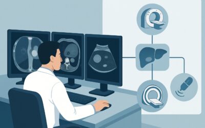
Which Imaging Study Is Best for an Indeterminate Liver Lesion in a Healthy Patient?

A 48-year-old male undergoes a noncontrast computed tomography (CT) of the abdomen and pelvis for suspected kidney stones. The kidneys are clear, but the radiologist notes an incidental, indeterminate 2.5 cm lesion in the right hepatic lobe. The patient has no history of liver disease, no known malignancy, and normal liver function tests. You are now faced with a common clinical question: what is the most appropriate next step to characterize this finding and avoid unnecessary procedures? This article provides a step-by-step workflow for this specific scenario, grounded in the American College of Radiology (ACR) Appropriateness Criteria. For this presentation, the ACR designates MRI abdomen without and with IV contrast as a Usually appropriate examination.
## Who Fits This Clinical Scenario?
This guidance applies to a very specific patient population. Correctly identifying if your patient fits this scenario is the critical first step to ensure you order the right test and avoid diagnostic delays.
Inclusion Criteria for This Workflow:
- An indeterminate liver lesion greater than 1 cm was discovered on initial imaging.
- The initial imaging was a noncontrast CT, a single-phase contrast-enhanced CT, or a noncontrast MRI. These studies are insufficient for full characterization.
- The patient has a normal liver parenchyma, with no signs of cirrhosis, steatosis, or other chronic liver disease.
- There is no clinical suspicion or known history of an extrahepatic malignancy.
Exclusion Criteria (These Patients Require a Different Workflow):
- Known Chronic Liver Disease: If the patient has cirrhosis or other chronic liver conditions, the risk of hepatocellular carcinoma (HCC) is much higher. The workup follows a different pathway, often using the Liver Imaging Reporting and Data System (LI-RADS).
- Known Extrahepatic Malignancy: If the patient has a known cancer (e.g., colon, breast, lung), any new liver lesion is considered a potential metastasis until proven otherwise, altering the diagnostic algorithm.
- Lesion Found on Ultrasound: If the initial imaging was an ultrasound, the subsequent recommended steps may differ.
- Lesion Less Than 1 cm: Small incidental lesions (<1 cm) in this patient population are overwhelmingly benign and often do not require immediate characterization, with surveillance being a more common approach.
## What Diagnoses Are You Working Up in This Scenario?
In a patient with a normal liver and no known cancer, an incidentally discovered liver lesion is most likely benign. The goal of the imaging workup is to confidently diagnose a benign entity to halt further unnecessary tests, or to identify the rare lesion that requires further management.
The primary differential diagnoses include:
Hepatic Hemangioma: This is the most common benign liver tumor. It is a collection of abnormal blood vessels and is typically asymptomatic. On multiphasic imaging, it has a very characteristic enhancement pattern (peripheral, discontinuous, nodular enhancement that fills in over time) that allows for a definitive, non-invasive diagnosis.
Focal Nodular Hyperplasia (FNH): The second most common benign liver lesion, FNH is a regenerative mass of hepatocytes. It is also typically asymptomatic and has no malignant potential. It is characterized by intense, uniform enhancement in the arterial phase and often contains a non-enhancing central scar, features that are well-visualized on multiphasic MRI.
Hepatic Adenoma: This is a less common benign tumor, but it is clinically significant due to its potential for hemorrhage and, rarely, malignant transformation. It is most frequently seen in women with a history of oral contraceptive use. Distinguishing it from FNH is crucial, and MRI with specific contrast agents is highly effective at this differentiation.
Hepatocellular Carcinoma (HCC): While HCC is the most common primary liver cancer, it is very rare in patients without underlying chronic liver disease or cirrhosis. However, it remains a remote possibility in the differential and must be excluded.
## Why Is MRI Abdomen Without and With IV Contrast the Recommended Study?
The ACR rates MRI abdomen without and with IV contrast as Usually appropriate because it offers the highest diagnostic accuracy for this clinical problem without using ionizing radiation.
The rationale is based on several key advantages:
- Superior Soft Tissue Contrast: MRI provides exceptional detail of the liver parenchyma and the internal characteristics of a lesion, far exceeding that of CT. This allows for the confident identification of features like the T2-hyperintense signal of a hemangioma or the central scar in an FNH.
- Dynamic Contrast Enhancement: A multiphasic liver MRI involves acquiring images before and at multiple time points after the injection of a gadolinium-based contrast agent (arterial, portal venous, and delayed phases). The specific way a lesion enhances and washes out contrast over time is often pathognomonic. For example, the classic “lightbulb bright” T2 signal and peripheral nodular enhancement of a hemangioma are best seen on MRI.
- Hepatobiliary Agents: Specialized MRI contrast agents (e.g., gadoxetate disodium) are taken up by normal hepatocytes. FNH contains functional hepatocytes and will therefore retain this contrast on delayed images, whereas adenomas and malignant lesions will not. This property significantly increases the accuracy in differentiating FNH from adenoma.
- No Ionizing Radiation: Since the patient is otherwise healthy and the lesion is likely benign, avoiding the radiation dose of CT is a significant benefit. The ACR lists the radiation level for multiphasic CT as ☢☢☢☢ (10-30 mSv), while MRI is 0 mSv.
Why are other studies rated lower for this specific scenario?
- CT abdomen with IV contrast multiphase: While also rated Usually appropriate, it is generally considered the second-line option. It is a viable alternative if MRI is contraindicated (e.g., incompatible pacemaker, severe claustrophobia) or unavailable. However, its lower soft tissue contrast can sometimes leave a diagnosis indeterminate, and it imparts a significant radiation dose.
- Image-guided biopsy liver: This is rated Usually not appropriate as a primary diagnostic step. Biopsy is an invasive procedure with risks of bleeding, infection, and (rarely) tumor seeding. Given the high accuracy of non-invasive characterization with multiphasic MRI, biopsy is reserved for the rare cases where imaging remains inconclusive.
When ordering the study, it is crucial to specify a “multiphasic liver protocol MRI” to ensure the radiology department performs the correct dynamic sequences for lesion characterization.
## What’s Next After MRI Abdomen Without and With IV Contrast? Downstream Workflow
The results of the multiphasic MRI will guide the next steps in a clear, evidence-based manner. The goal is to provide a definitive answer and a clear management plan.
- Result: Definitive Benign Lesion (e.g., Hemangioma, FNH): If the MRI report confidently identifies a classic hemangioma or FNH, the workup is complete. No further imaging, follow-up, or treatment is required. The most important next step is to communicate this benign result clearly to the patient to provide reassurance and prevent future anxiety or redundant imaging.
- Result: Probable Hepatic Adenoma: The management of a hepatic adenoma depends on its size and patient-specific factors. For women, discontinuation of oral contraceptives is typically recommended. Small adenomas (<5 cm) are often managed with surveillance imaging. Larger adenomas may be considered for surgical resection due to the increased risk of hemorrhage or malignant transformation. A consultation with a hepatologist or liver surgeon is appropriate.
- Result: Indeterminate After MRI: In a small number of cases, a lesion may have atypical features and remain indeterminate even after a high-quality MRI. The first step should be a direct conversation with the interpreting radiologist. Further options may include a follow-up MRI in 3-6 months to assess for stability, consideration of contrast-enhanced ultrasound (May be appropriate), or discussion at a multidisciplinary tumor board. Biopsy remains a final option if a definitive diagnosis is required and cannot be reached non-invasively.
- Result: Suspicious for Malignancy: Although rare in this scenario, if the MRI features are suspicious for malignancy (e.g., HCC or metastasis from an unknown primary), an urgent referral to a specialist (hepatology or surgical oncology) is warranted. A biopsy would likely be the next step to obtain a tissue diagnosis and guide treatment.
## Pitfalls to Avoid (and When to Get Help)
Navigating the workup of an incidental liver lesion requires avoiding several common missteps that can lead to diagnostic errors or unnecessary procedures.
- Ordering the Wrong Study: Do not order a standard “CT abdomen with contrast.” A single-phase (portal venous) study is inadequate for characterizing liver lesions and is the reason this workup was initiated in the first place. Specify a “multiphasic liver protocol” for either CT or MRI.
- Ignoring the Clinical Context: Applying this workflow to a patient with cirrhosis or a known cancer is a critical error. Those scenarios have entirely different probabilities and require distinct diagnostic algorithms (e.g., LI-RADS).
- Premature Biopsy: Resist the urge to biopsy an indeterminate lesion before obtaining a high-quality, multiphasic cross-sectional imaging study. Most lesions can be diagnosed non-invasively, avoiding procedural risks.
- Inadequate Communication: Failing to reassure a patient after a definitive benign diagnosis can lead to persistent anxiety and “victim of medical imaging technology” (VOMIT) syndrome.
If the MRI results are indeterminate or concerning for malignancy, escalate care by consulting with the radiologist and referring the patient to a hepatologist or liver surgeon.
## Related ACR Topics and Tools
For further reading on related scenarios and to explore imaging decision support tools, please see the resources below.
- For breadth across all scenarios in Liver Lesion-Initial Characterization, see our parent guide: Liver Lesion-Initial Characterization: ACR Appropriateness Decoded.
- To explore other clinical presentations, use the ACR Appropriateness Criteria Lookup.
- For technical details on imaging techniques, browse the Imaging Protocol Library.
- To discuss radiation exposure with patients, consult the Radiation Dose Calculator.
Frequently Asked Questions
Why not just get a biopsy first to get a definitive answer?
Biopsy is rated ‘Usually not appropriate’ as a first step in this scenario because it is an invasive procedure with risks like bleeding and infection. Modern multiphasic MRI is highly accurate (often over 95% for common benign lesions like hemangiomas and FNH) and can provide a definitive diagnosis non-invasively, making biopsy unnecessary for the vast majority of these patients.
My patient has a pacemaker and cannot get an MRI. What is the next best test?
If MRI is contraindicated, the ACR rates ‘CT abdomen with IV contrast multiphase’ as ‘Usually appropriate.’ It is the best alternative. It is critical to order a multiphasic liver protocol CT, not a standard single-phase scan, to properly assess the lesion’s enhancement pattern. The main drawback is the radiation exposure (10-30 mSv).
What if the lesion is exactly 1.0 cm? Does this workflow still apply?
Yes, this workflow applies to lesions greater than or equal to 1.0 cm. Lesions smaller than 1.0 cm in a patient with a normal liver and no cancer history are almost always benign (e.g., small cysts or hemangiomas) and are typically managed with surveillance or may not require any follow-up, depending on institutional guidelines and radiologist recommendation.
The initial CT report mentioned the lesion was ‘hypodense.’ What does that mean?
‘Hypodense’ simply means the lesion appears darker than the surrounding liver tissue on a CT scan. This is a non-specific finding. Cysts, hemangiomas, metastases, and other lesions can all appear hypodense on a noncontrast or single-phase CT. This is precisely why it is considered ‘indeterminate’ and requires further characterization with a dedicated multiphasic study like MRI or CT.
Does the patient need to have their creatinine checked before a contrast-enhanced MRI?
Yes, institutional policies generally require a recent creatinine level to assess kidney function before administering a gadolinium-based contrast agent for an MRI. This is to screen for patients at risk of nephrogenic systemic fibrosis (NSF), a rare but serious complication. The specific GFR cutoff for administering contrast may vary by institution and the type of contrast agent used.
Reviewed by Pouyan Golshani, MD, Interventional Radiologist — May 29, 2026