What Is the Best Initial Imaging for a Symptomatic Primary Shoulder Arthroplasty?

A 68-year-old patient with a total shoulder arthroplasty placed three years ago presents to your clinic with six weeks of new, worsening deep shoulder pain and a grinding sensation with movement. They deny any recent trauma or fevers. On exam, their range of motion is limited by pain, but there are no overlying skin changes. You need to begin the diagnostic workup, and the first decision is which imaging study to order. This is a common clinical crossroads where choosing the right initial test is critical for an efficient and accurate evaluation. For this specific scenario—the initial imaging of a symptomatic primary shoulder arthroplasty—the American College of Radiology (ACR) finds that Radiography shoulder is Usually appropriate.
Who Fits This Clinical Scenario?
This guidance applies to a specific patient population: individuals with a primary (not revision) shoulder arthroplasty who have become symptomatic. Symptoms may include new or worsening pain, decreased range of motion, instability, weakness, or audible/palpable clicking or grinding. This article addresses the crucial initial imaging step in their workup.
It is essential to distinguish this presentation from several related but distinct clinical situations that follow different diagnostic pathways:
- Asymptomatic Routine Follow-up: This guidance does not apply to patients undergoing routine, scheduled surveillance imaging without any new symptoms. That scenario has its own set of recommendations.
- High Suspicion of Infection: While infection is on the differential, this initial pathway assumes infection is not the clear leading diagnosis based on systemic signs (fever, chills) or local signs (erythema, warmth, draining sinus). Patients with high clinical suspicion for infection may require a different or accelerated workup.
- Workup After Initial Radiographs: This article covers the first imaging test. If initial radiographs are negative or equivocal, subsequent imaging choices (e.g., for suspected loosening, infection, or rotator cuff tear) are covered in separate, downstream ACR Appropriateness Criteria variants.
What Diagnoses Are You Working Up in This Scenario?
When a patient with a shoulder replacement develops new symptoms, the differential diagnosis centers on the implant, the surrounding bone, and the adjacent soft tissues. The initial imaging choice is designed to efficiently evaluate the most common and consequential mechanical causes of failure.
Aseptic Loosening: This is one of the most common long-term failure modes for any joint arthroplasty. It involves the failure of fixation between the implant and the bone without an infectious cause. Radiographs are the primary tool for detecting signs of loosening, such as progressive radiolucent lines at the bone-cement or bone-implant interface, component migration, subsidence, or a change in component alignment compared to prior studies.
Periprosthetic Fracture: A fracture can occur around the components, either from significant trauma or from chronic stress and bone weakening. Radiographs are highly sensitive for identifying most periprosthetic fractures, which are critical to diagnose as they often require urgent surgical management.
Instability or Dislocation: The prosthetic joint can subluxate or dislocate. This is often clinically apparent but requires imaging to confirm the direction and degree of displacement and to assess for any associated component failure or fracture. An appropriate radiographic series is the definitive diagnostic test.
Component Failure: Though less common, the implant itself can fail. This can include fracture of the metal components or, more commonly, wear of the polyethylene (plastic) liner of the glenoid. While polyethylene is radiolucent, its wear can be inferred on radiographs by observing asymmetric joint space or eccentric positioning of the humeral head on the glenoid component.
Periprosthetic Joint Infection (PJI): While advanced imaging or joint aspiration is often needed to confirm PJI, initial radiographs are still a necessary first step. They can show aggressive bone loss (osteolysis), periosteal reaction, or rapid component loosening that can raise suspicion for an infectious process over aseptic loosening.
Why Is Shoulder Radiography the Recommended Initial Study?
For the initial evaluation of a symptomatic primary shoulder arthroplasty, the ACR designates Radiography shoulder as Usually appropriate. This recommendation is based on the modality’s high diagnostic utility for the primary differential diagnoses, its wide availability, low cost, and minimal radiation exposure.
A standard arthroplasty series, typically including a true anteroposterior (AP) view in the scapular plane (Grashey view) and an axillary lateral view, provides a comprehensive assessment of the key areas of concern. These views are optimized to evaluate component alignment, the bone-implant interfaces, joint congruity, and the integrity of the surrounding bone. Radiographs are highly effective for detecting dislocation, periprosthetic fracture, and significant component migration or loosening. Comparing the new images to previous postoperative radiographs is crucial for detecting subtle but progressive changes, such as widening radiolucent lines, that indicate implant failure.
Conversely, more advanced imaging modalities are rated as Usually not appropriate for this initial step for several key reasons:
- CT shoulder (without or with contrast): While CT provides excellent cross-sectional detail of bone, it imparts a significantly higher radiation dose (☢☢☢ 1-10 mSv vs. ☢ <0.1 mSv for radiography) and is not necessary for the initial screen. Metal artifact from the prosthesis can also degrade image quality, though specialized metal artifact reduction sequence (MARS) protocols can mitigate this. CT is reserved as a second-line, problem-solving tool if radiographs are inconclusive but suspicion for loosening or subtle fracture remains high.
- MRI shoulder (without or with contrast): MRI is the gold standard for soft tissue evaluation, but its utility in assessing a metal implant is severely limited by magnetic susceptibility artifact. Even with MARS techniques, the critical bone-implant interface is often obscured. Therefore, it is not the right initial test when the primary questions relate to hardware integrity and fixation. It is considered in a different clinical pathway when a rotator cuff tear is the specific concern after initial radiographs are negative.
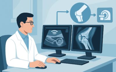
- US shoulder: Ultrasound is unable to penetrate the metallic and polyethylene components of the arthroplasty, meaning it cannot visualize the bone-implant interface at all. While it can assess for adjacent fluid collections or evaluate parts of the rotator cuff, its inability to assess for loosening, fracture, or dislocation makes it Usually not appropriate as the primary imaging tool.
What’s Next After Shoulder Radiography? Downstream Workflow
The results of the initial shoulder radiographs will dictate the subsequent clinical pathway. The goal of this first step is to triage the patient toward the correct diagnosis and management plan.
If radiographs are clearly positive: When radiographs reveal an obvious cause for the patient’s symptoms, such as a periprosthetic fracture, dislocation, or frank signs of component loosening (e.g., significant migration or widely progressive radiolucent lines), the next step is typically a consultation with an orthopedic surgeon. The surgeon may order a preoperative CT scan for better characterization of bone stock and planning, but the initial radiograph has already established the primary diagnosis.
If radiographs are negative or equivocal: This is a very common outcome. A patient can have significant pathology, such as early-stage loosening or infection, with normal-appearing radiographs. If the patient remains symptomatic despite unremarkable x-rays, the workup must continue. The next step is guided by the leading clinical suspicion:
- If infection is suspected: The next step involves obtaining inflammatory markers (Erythrocyte Sedimentation Rate and C-Reactive Protein). If these are elevated, a diagnostic joint aspiration for culture and cell count is often the next procedure. This aligns with the ACR variant for a symptomatic patient where infection is not excluded.
- If aseptic loosening is suspected: If infection is considered unlikely (normal inflammatory markers), the next imaging study to consider is often a CT scan with metal artifact reduction sequences to look for subtle signs of loosening or osteolysis not visible on plain films.
- If a soft tissue cause is suspected: If the clinical picture suggests a rotator cuff tear or other soft tissue pathology, an MRI with MARS protocols or a dedicated ultrasound may be considered, following the specific ACR variant for that indication.
Pitfalls to Avoid (and When to Get Help)
Navigating the workup of a painful shoulder arthroplasty requires careful attention to detail to avoid common missteps.
- Pitfall 1: An Incomplete Radiographic Series. Ordering a single AP view is insufficient. An axillary lateral view is essential for assessing glenohumeral alignment and detecting posterior subluxation or eccentric polyethylene wear.
- Pitfall 2: Forgetting to Compare with Priors. The most valuable information often comes from change over time. Always compare current radiographs with the initial postoperative films and any other prior studies to detect progressive radiolucency or component shift.
- Pitfall 3: Over-reliance on a “Normal” Report. Radiographs have limited sensitivity for early-stage loosening and infection. If a patient has persistent, unexplained pain despite normal radiographs, the workup is not over. Clinical symptoms should drive the decision to proceed with lab work or advanced imaging.
If radiographs are negative but clinical suspicion for an urgent issue like infection or fracture remains high based on the patient’s symptoms or exam, an immediate orthopedic consultation or escalation to advanced imaging is warranted.
Related ACR Topics and Tools
For a comprehensive overview of all clinical scenarios related to imaging after shoulder arthroplasty, from asymptomatic follow-up to specific complications, please refer to our parent guide. For other tools to help refine your imaging decisions, see the resources below.
- For breadth across all scenarios in Imaging After Shoulder Arthroplasty, see our parent guide: Imaging After Shoulder Arthroplasty: ACR Appropriateness Decoded.
- To explore other clinical presentations, use the Imaging Appropriateness Selector.
- For details on imaging techniques, browse the Imaging Protocol Library.
- To discuss radiation exposure with patients, consult the Radiation Dose Calculator.
Frequently Asked Questions
What specific radiographic views are essential for evaluating a shoulder arthroplasty?
A standard shoulder arthroplasty series should include, at minimum, a true anteroposterior (AP) view in the scapular plane (a Grashey view) and an axillary lateral view. The Grashey view provides a true profile of the glenohumeral joint space, while the axillary view is critical for assessing anterior-posterior alignment and detecting subluxation or eccentric wear.
Why isn’t CT the first test if it shows more detail than an x-ray?
While CT provides superior detail of bone, it is not the appropriate initial test due to significantly higher radiation dose and cost. Standard radiographs are highly effective and sufficient for diagnosing the most common mechanical complications like dislocation, fracture, and significant loosening. CT is reserved as a second-line, problem-solving tool for cases where radiographs are negative or equivocal but clinical suspicion remains high.
My patient’s x-ray is normal, but they still have significant pain. What should I do next?
This is a common and important clinical scenario. A normal radiograph does not rule out pathology. The next step is driven by your clinical suspicion. If you suspect an infection, order inflammatory labs (ESR, CRP) and consider a joint aspiration. If you suspect mechanical loosening, a CT with metal artifact reduction may be the next best step. If a soft tissue problem like a rotator cuff tear is suspected, an MRI may be warranted. This moves the workup into a different ACR clinical variant.
How does this guidance change for a reverse total shoulder arthroplasty (rTSA)?
The initial imaging modality remains the same: radiography is the first and most appropriate step. However, the specific complications and radiographic findings to look for differ with an rTSA. For example, radiologists and surgeons will specifically look for signs of scapular notching, acromial stress fracture, and different patterns of loosening unique to the reverse prosthesis design.
Can ultrasound be used at all in a patient with a shoulder replacement?
While ultrasound is rated ‘Usually not appropriate’ for the initial global assessment of a painful arthroplasty, it can have a niche role for specific questions. For instance, it can be used to evaluate for a periprosthetic fluid collection or abscess, or to assess the integrity of the deltoid muscle. However, it cannot visualize the bone-implant interface, which is the most critical area, making it unsuitable as a primary screening tool.
Reviewed by Pouyan Golshani, MD, Interventional Radiologist — May 26, 2026