What Is the Best Initial Imaging for Axillary Adenopathy in Patients with Silicone Implants?
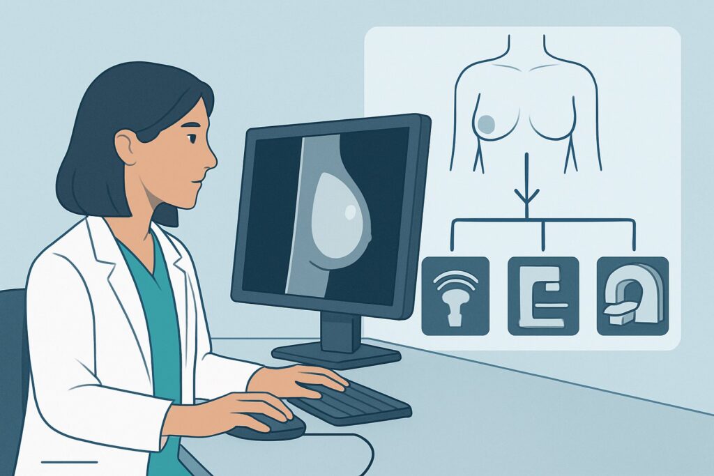
A 48-year-old woman with silicone breast implants placed 12 years ago presents for evaluation of a new, palpable, non-tender lymph node in her left axilla, discovered during a self-exam. She has no palpable breast masses, skin changes, or other symptoms. As the ordering clinician, you must decide on the most appropriate initial imaging study to evaluate this unexplained adenopathy, balancing the need to assess for malignancy against the specific considerations introduced by the presence of breast implants. This article provides a focused, evidence-based workflow for this exact clinical scenario. According to the American College of Radiology (ACR) Appropriateness Criteria, a breast ultrasound is the Usually Appropriate first step to evaluate the axillary finding.
Who Fits This Clinical Scenario for Axillary Adenopathy with Implants?
This guidance is specifically for patients who meet all the following criteria:
- Age: 40 years or older.
- Patient Identity: Female or transfeminine.
- Clinical Presentation: Unexplained axillary adenopathy (e.g., a palpable lymph node or nodes found on physical exam or incidentally on other imaging).
- Implant History: Current or prior history of silicone breast implants.
- Imaging Stage: This is the initial imaging workup for this specific problem.
It is crucial to distinguish this presentation from similar but distinct clinical situations that follow different diagnostic pathways. This workflow does not apply if:
- A palpable breast mass is also present: The workup would then be dictated by the breast mass, typically starting with diagnostic mammography and targeted ultrasound.
- The patient has saline implants: While the workup may be similar, the differential diagnosis for adenopathy related to implant material is different.
- The primary concern is implant rupture: Symptoms like a change in breast shape, pain, or hardening would point toward a different ACR variant focused on implant integrity, where MRI is often considered.
- The patient is under age 40: While the imaging choice might be the same, the pre-test probability of malignancy is different, which can influence downstream management.
What Diagnoses Are You Working Up in This Scenario?
When evaluating isolated axillary adenopathy in a patient over 40 with silicone implants, the differential diagnosis is broad, but several key possibilities drive the imaging strategy. The goal is to differentiate benign, implant-related causes from potentially malignant ones.
The most critical diagnosis to exclude is metastatic breast cancer. Axillary lymph nodes are the most common site of regional metastasis from an occult (non-palpable) primary breast tumor. In patients over 40, this is a primary concern, and the imaging workup is designed to identify suspicious nodal features and guide biopsy while also evaluating the breast itself.
A common and unique consideration in this population is silicone lymphadenopathy. This is a benign, foreign-body granulomatous reaction to microscopic amounts of silicone (“gel bleed”) or free silicone from an extracapsular implant rupture that has migrated to the regional lymph nodes. The nodes can become enlarged and firm, mimicking malignancy on physical exam. Imaging is key to identifying specific features of silicone within the node.
A less common but highly consequential diagnosis is Breast Implant-Associated Anaplastic Large Cell Lymphoma (BIA-ALCL). While it typically presents with a delayed seroma, it can, in some cases, present as a mass or lymphadenopathy. This T-cell lymphoma is a known, though rare, complication associated with textured-surface breast implants.
Finally, the adenopathy could be due to other causes unrelated to the breast or implants, such as systemic lymphoma, melanoma metastases, or benign reactive/inflammatory changes from infection or autoimmune conditions. The initial imaging focuses on characterizing the nodes to determine if they are suspicious enough to warrant a biopsy.
Why Is Breast Ultrasound the Recommended First Study for This Presentation?
The ACR rates breast ultrasound (US) as Usually Appropriate for this scenario because it directly and effectively addresses the primary clinical questions without radiation or intravenous contrast.
The primary strength of ultrasound is its high-resolution characterization of lymph node morphology. A radiologist can assess features that help distinguish benign from malignant nodes, such as:
- Cortical thickness: Malignant infiltration often causes focal or diffuse cortical thickening.
- Fatty hilum: The preservation of the central, bright-appearing fatty hilum is a reassuring sign of a benign node. Its effacement or loss is suspicious.
- Shape: Benign nodes are typically oval or reniform, while malignant nodes tend to become more rounded.
- Vascularity: Doppler evaluation can assess blood flow patterns within the node.
Crucially for this specific scenario, ultrasound is highly effective at identifying intramammary and axillary free silicone. Silicone within a lymph node creates a characteristic, intensely echogenic, and “dirty” shadowing appearance often described as a “snowstorm” sign. Identifying this feature strongly suggests silicone lymphadenopathy as the etiology, potentially avoiding an unnecessary biopsy of a benign entity.
Furthermore, if a node appears morphologically suspicious, ultrasound is the ideal modality to guide a percutaneous core needle biopsy in real-time, providing a safe and accurate method for obtaining a tissue diagnosis.
Why Other Studies Are Rated Differently
- Diagnostic Mammography and Digital Breast Tomosynthesis (DBT): These are also rated Usually Appropriate. In practice, a diagnostic mammogram is often performed in conjunction with the targeted axillary ultrasound. While ultrasound is superior for evaluating the palpable node itself, mammography is essential for examining the entire breast parenchyma to search for an occult primary cancer that may have metastasized to the axilla. The two tests are complementary. Ultrasound is often the first and most direct way to evaluate the palpable axillary finding, with mammography providing the broader breast context. Both involve a low dose of ionizing radiation (ACR RRL ☢☢, 0.1-1 mSv).
- MRI Breast (with or without contrast): The ACR rates breast MRI as Usually not appropriate as the initial imaging test for this problem. While MRI is the most sensitive test for detecting both breast cancer and silicone implant rupture, it is not the right first step for isolated adenopathy. It is more costly, less widely available, and has a higher rate of false positives that can lead to unnecessary downstream interventions. It is typically reserved for problem-solving after initial imaging is complete or for staging once a malignancy has been diagnosed.
What’s Next After the Ultrasound? Downstream Clinical Workflow
The results of the breast and axillary ultrasound will guide the subsequent management steps. The clinical pathway typically branches based on the sonographic findings in the abnormal lymph node(s).
- If the node appears morphologically benign: If the lymph node has a normal oval shape, thin cortex, and a preserved fatty hilum, and no other suspicious findings are seen in the breast, a short-term imaging follow-up (e.g., in 6 months) may be recommended to ensure stability.
- If the node shows the “snowstorm” sign of silicone: This finding is highly specific for silicone lymphadenopathy. If the node is not otherwise morphologically suspicious (e.g., markedly enlarged or rounded), this is considered a benign finding. The patient can be reassured, and further workup may not be necessary unless there is a clinical change.
- If the node is morphologically suspicious: If the node is rounded, has a thickened cortex, and/or has a lost fatty hilum, it is suspicious for malignancy regardless of the presence of silicone. The next step is an ultrasound-guided core needle biopsy of the most abnormal-appearing node to obtain a tissue diagnosis.
- If the ultrasound is negative/inconclusive but clinical suspicion remains high: If a palpable abnormality persists despite a negative or equivocal ultrasound, further evaluation is warranted. This may involve a diagnostic mammogram if not already performed, or potentially a referral to a breast surgeon for clinical assessment and possible excisional biopsy.
Pitfalls to Avoid (and When to Get Help)
Navigating this scenario requires careful attention to the specific details of the patient’s history and imaging findings. Here are a few common pitfalls to avoid:
- Stopping the workup after identifying silicone: Do not assume that the presence of silicone in a lymph node excludes malignancy. Metastatic cancer and silicone lymphadenopathy can coexist in the same node. The decision to biopsy should be based on nodal morphology, not just the presence of silicone.
- Forgetting the mammogram: While ultrasound is excellent for the targeted axillary evaluation, it is not a screening tool for the entire breast. A concurrent or subsequent diagnostic mammogram is crucial to evaluate for a primary breast cancer.
- Dismissing BIA-ALCL: Although rare, keep BIA-ALCL on the differential, especially in patients with a history of textured implants who present with a mass or adenopathy. Ensure the pathologist is aware of the implant history if a biopsy is performed.
If imaging findings are equivocal or if there is a discrepancy between the clinical exam and imaging results, escalation to a breast imaging specialist or a breast surgeon is the appropriate next step.
Related ACR Topics and Tools
For a comprehensive overview of all clinical variants related to breast implant imaging, or to explore the tools used to develop these recommendations, the following resources are available:
- For breadth across all scenarios in Breast Implant Evaluation, see our parent guide: Breast Implant Evaluation: ACR Appropriateness Decoded.
- ACR Appropriateness Criteria Lookup — for adjacent scenarios
- Imaging Protocol Library — for technique on the recommended study
- Radiation Dose Calculator — for cumulative dose conversations
Frequently Asked Questions
Why isn’t MRI the first choice if it’s the best test for silicone implant rupture?
While breast MRI is the most sensitive imaging test for evaluating silicone implant integrity, this specific clinical scenario is focused on evaluating unexplained axillary adenopathy, not suspected rupture. Ultrasound is a more direct, cost-effective, and highly effective initial test for characterizing lymph nodes and can readily identify silicone within them. MRI is rated ‘Usually not appropriate’ as a first step here because it’s less specific and can lead to more false positives, reserving it for problem-solving or post-diagnosis staging.
If the ultrasound shows silicone in the lymph node, is a biopsy still needed?
Not always. If the lymph node shows the classic ‘snowstorm’ sign of silicone but is otherwise morphologically benign (normal size, shape, thin cortex, preserved fatty hilum), it can often be confidently diagnosed as benign silicone lymphadenopathy, and biopsy can be avoided. However, if the node is also morphologically suspicious (e.g., rounded, with a thickened cortex), a biopsy is necessary because malignancy can coexist with silicone deposits.
Should I order a diagnostic mammogram at the same time as the axillary ultrasound?
Yes, in a patient aged 40 or older, ordering a diagnostic mammogram (including tomosynthesis) along with the targeted axillary ultrasound is considered ‘Usually Appropriate’ and is standard practice. The ultrasound directly evaluates the palpable node, while the mammogram evaluates the entire breast for an occult primary cancer that could be the source of a malignant axillary node.
Does this guidance change if the patient has a history of textured implants?
The initial imaging choice (ultrasound and mammogram) remains the same. However, a history of textured implants raises the clinical suspicion for the rare but important diagnosis of Breast Implant-Associated Anaplastic Large Cell Lymphoma (BIA-ALCL). It is critical to communicate this history to the radiologist and, if a biopsy is performed, to the pathologist, as special tissue analysis may be required.
What if the patient is under 40 years old?
This specific ACR variant applies to patients aged 40 and older. While the initial imaging modality (ultrasound) would likely be the same for a younger patient with a palpable axillary node, the pre-test probability of breast cancer is significantly lower. The decision-making for follow-up versus biopsy might be different, and the role of mammography might be adjusted based on the ultrasound findings and the patient’s overall risk profile.
Reviewed by Pouyan Golshani, MD, Interventional Radiologist — May 29, 2026