What Is the Best Initial Imaging for Chronic or Progressive Myelopathy? An ACR-Guided Workflow
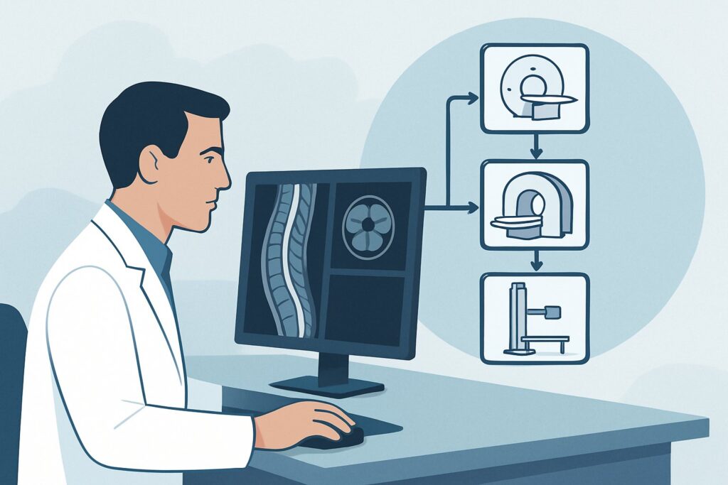
A 64-year-old patient presents to your clinic with a nine-month history of slowly worsening gait imbalance, stiffness in his legs, and a new “pins and needles” sensation in his hands. His neurologic exam is notable for brisk reflexes in the lower extremities and a positive Babinski sign. You suspect a process affecting the spinal cord—a myelopathy—but the cause is unclear. Deciding on the right initial imaging study is critical to avoid diagnostic delays and unnecessary tests. This article provides a focused workflow for this exact situation: the initial imaging workup for chronic or progressive myelopathy. According to the American College of Radiology (ACR) Appropriateness Criteria, an MRI of the spine area of interest without and with IV contrast is Usually Appropriate and the recommended first step.
Who Fits This Clinical Scenario?
This guidance applies to patients presenting with a subacute, chronic, or progressive myelopathy. The key feature is the timeline: symptoms have developed and worsened over a period of weeks, months, or even years. The clinical picture is one of upper motor neuron dysfunction, which can include spasticity, hyperreflexia, weakness, gait disturbance, and sensory deficits that may correspond to a specific spinal cord level. The term “progressive” implies a steady worsening, while “chronic” refers to the long-standing nature of the symptoms.
It is crucial to distinguish this from an acute presentation. This workflow does NOT apply to patients with:
- Acute Onset Myelopathy: Patients whose symptoms develop over hours to a few days. This is a neurologic emergency that suggests a different differential diagnosis, such as spinal cord infarction, acute transverse myelitis, hemorrhage, or sudden cord compression from trauma or an acute disc herniation. The workup for acute myelopathy is a distinct clinical scenario with its own imaging considerations.
- Radiculopathy Without Myelopathy: Patients with symptoms of a pinched nerve root (e.g., shooting pain down one arm or leg) but without signs of spinal cord involvement like hyperreflexia or spasticity.
This article is for the patient whose insidious symptom progression points toward a structural, inflammatory, or neoplastic cause that requires detailed anatomical imaging to uncover.
What Diagnoses Are You Working Up in This Scenario?
When ordering imaging for chronic myelopathy, you are investigating a broad differential. The goal of the initial study is to differentiate between compressive and non-compressive causes, as this distinction fundamentally alters the subsequent management pathway.
Compressive Myelopathy: This is the most common category and involves physical impingement on the spinal cord. The leading cause is degenerative disease, such as cervical or thoracic spondylosis, where bone spurs (osteophytes), thickened ligaments, and bulging or herniated discs narrow the spinal canal. Other compressive causes include spinal tumors, either metastatic disease from another primary cancer or primary tumors arising from the spinal cord or its coverings.
Inflammatory and Demyelinating Conditions: Multiple Sclerosis (MS) is a primary consideration, especially in younger patients. MS plaques can form in the spinal cord, disrupting nerve signals and causing progressive disability. Other inflammatory conditions like sarcoidosis or certain forms of transverse myelitis can also present with a subacute or progressive course.
Spinal Vascular Malformations: Though less common, conditions like dural arteriovenous fistulas (dAVFs) can cause progressive myelopathy. These abnormal connections between arteries and veins lead to venous congestion and swelling in the spinal cord, producing symptoms that worsen over time.
Other Intrinsic Cord Abnormalities: This category includes primary spinal cord tumors like ependymomas and astrocytomas, or a syrinx (a fluid-filled cavity within the cord), which can expand slowly and cause progressive neurologic decline.
Why Is MRI Without and With IV Contrast the Recommended Study for This Presentation?
The ACR rates MRI of the spine area of interest without and with IV contrast as Usually Appropriate because it provides the most comprehensive evaluation for the wide range of potential causes in chronic myelopathy. Its superior soft-tissue contrast is unmatched for visualizing the spinal cord parenchyma, surrounding cerebrospinal fluid spaces, intervertebral discs, and ligaments.
The rationale for the specific components of this study is clear:
- MRI without contrast: The non-contrast sequences (particularly T1-weighted and T2-weighted images) are excellent for assessing the structural anatomy. They can clearly define the extent of spinal canal stenosis from degenerative changes, identify cord compression, and reveal abnormal T2 signal within the cord, which indicates edema, inflammation, or gliosis.
- MRI with IV contrast: The administration of gadolinium-based contrast is essential for detecting any disruption of the blood-brain (or blood-spinal cord) barrier. This is critical for identifying and characterizing enhancing lesions such as tumors, active demyelinating plaques in MS, or inflammatory processes like sarcoidosis. It also helps delineate vascular malformations.
An MRI of the spine area of interest without IV contrast is also rated Usually Appropriate, but it may be insufficient if an inflammatory, infectious, or neoplastic process is suspected. Omitting contrast in these cases can lead to a missed or delayed diagnosis.
Why Are Other Studies Rated Lower?
Alternative imaging modalities are generally less suitable for the initial evaluation of chronic myelopathy:
- CT Myelography: Rated as May be appropriate, this study involves injecting intrathecal contrast via lumbar puncture, followed by a CT scan. It is an invasive procedure that carries a risk of headache and other complications. While it provides excellent detail of the bony anatomy and can show cord compression, it offers poor visualization of the cord parenchyma itself. It is typically reserved for patients who have contraindications to MRI (e.g., non-compatible implanted devices). It also involves significant radiation (RRL: Varies).
- Radiography (X-ray): Rated as Usually not appropriate, plain X-rays cannot visualize the spinal cord or other soft tissues. They can show alignment and severe degenerative bony changes but will miss the vast majority of causes of myelopathy, leading to a false sense of security if negative.
Given that MRI provides superior diagnostic information with no ionizing radiation (RRL: 0 mSv), it is the clear choice for the initial workup. Once you’ve decided on the study, proper protocol is key. For a detailed look at the technique, contrast considerations, and reading principles for a common spinal segment, see our guide: MRI Lumbar Spine Without Contrast.
What’s Next After MRI? Downstream Workflow
The results of the MRI will guide your next steps and determine the necessary consultations. The workflow typically branches based on whether a compressive or non-compressive cause is identified.
If the MRI shows significant cord compression:
The next step is a prompt referral to a neurosurgeon or orthopedic spine surgeon. The imaging findings, correlated with the patient’s clinical exam, will determine the urgency and type of surgical intervention required to decompress the spinal cord and prevent further neurologic decline.
If the MRI shows a non-compressive lesion (e.g., inflammation, tumor):
A referral to a neurologist is warranted.
- If findings suggest demyelination (MS), the neurologist may order an MRI of the brain to look for characteristic lesions, as well as a lumbar puncture for cerebrospinal fluid analysis (e.g., for oligoclonal bands).
- If a spinal cord tumor is identified, the neurologist and neurosurgeon will collaborate on further management, which may include biopsy, surgical resection, and/or radiation therapy.
- If a vascular malformation is suspected, a spinal angiogram may be necessary for definitive diagnosis and treatment planning.
If the MRI is negative:
A normal MRI in a patient with clear signs of myelopathy is a diagnostic challenge. The focus shifts to non-structural causes. The next steps may include blood work to rule out metabolic or nutritional deficiencies (e.g., Vitamin B12, copper), genetic testing, and neurophysiologic studies like electromyography (EMG) and nerve conduction studies (NCS) to further characterize the neurologic dysfunction. A neurology consultation is essential in this scenario.
Pitfalls to Avoid (and When to Get Help)
Navigating the workup for chronic myelopathy requires careful clinical-radiologic correlation. Here are a few common pitfalls to avoid:
- Imaging the Wrong Segment: The most critical error is ordering an MRI of the wrong part of the spine. A thorough neurologic exam to identify the sensory and motor level is paramount to ensure the imaging covers the area of suspected pathology.
- Omitting Contrast Inappropriately: Forgoing IV contrast in a patient where tumor or inflammation is a reasonable possibility can miss the diagnosis. Unless there is a strong contraindication, a study with and without contrast is preferred.
- Satisfaction of Search: It is common for older patients to have degenerative changes on their MRI. Be cautious not to attribute the patient’s myelopathy to mild spondylosis when another, more subtle pathology (like an MS plaque) might be the true cause.
If the clinical picture is confusing or the MRI findings are ambiguous, do not hesitate to escalate. A direct conversation with the reading neuroradiologist can provide valuable insights, and a consultation with a neurologist can help refine the differential diagnosis and plan further investigation.
Related ACR Topics and Tools
For a comprehensive overview of imaging for all myelopathy presentations and to explore related clinical scenarios, please refer to the resources below. These tools can help ensure you are ordering the most appropriate study for every patient.
- For breadth across all scenarios in Myelopathy, see our parent guide: Myelopathy: ACR Appropriateness Decoded.
- To look up other clinical variants and their ACR-rated studies, visit the ACR Appropriateness Criteria Lookup.
- For detailed procedural techniques, explore our Imaging Protocol Library.
- To discuss cumulative radiation exposure with patients for whom CT is considered, use the Radiation Dose Calculator.
Frequently Asked Questions
Why is it important to order the MRI both without and with IV contrast?
Ordering the study both ‘without and with’ contrast provides a complete picture. The non-contrast images are best for evaluating anatomy, degenerative changes, and cord swelling (edema). The post-contrast images are essential for detecting processes that disrupt the blood-spinal cord barrier, such as tumors, active inflammatory plaques from multiple sclerosis, or infections. Omitting contrast can lead to missing these critical diagnoses.
What should I order if my patient has a non-MRI-conditional pacemaker or other contraindication to MRI?
In cases where a patient cannot undergo an MRI, CT myelography is rated as ‘May be appropriate’ by the ACR. This is an invasive test requiring a lumbar puncture to inject contrast into the spinal canal before the CT scan. While it is inferior to MRI for visualizing the spinal cord itself, it is a valuable second-line option for identifying significant cord compression.
The patient’s symptoms are in their legs. Why would I need to image their cervical or thoracic spine?
Myelopathy is an upper motor neuron problem. The location of the symptoms (e.g., legs) indicates the spinal tracts that are affected, but the lesion causing it can be anywhere at or above that level. Signs like hyperreflexia in the legs point to a lesion in the thoracic or cervical cord. A thorough neurologic exam to determine the ‘sensory level’ is the best way to guide which part of the spine to image.
How does this imaging workup differ from that for acute myelopathy?
The workup for acute myelopathy is a medical emergency. While MRI is also the primary imaging modality, the differential diagnosis is different and includes acute causes like spinal cord infarction, traumatic injury, hemorrhage, or a rapidly expanding abscess. The urgency is much higher, and the interpretation focuses on identifying time-sensitive conditions that may require immediate intervention.
Can I just start with a plain X-ray to screen for problems?
No, plain radiography (X-ray) is rated ‘Usually not appropriate’ for this scenario. X-rays can only visualize bones and are incapable of showing the spinal cord, discs, or nerves. A normal X-ray provides no reassurance that a serious cause of myelopathy is absent, and it should not be used as a screening tool in this clinical context.
Reviewed by Pouyan Golshani, MD, Interventional Radiologist — May 29, 2026