Which Initial Imaging Is Best for New Ophthalmoplegia or Diplopia? An ACR Workflow
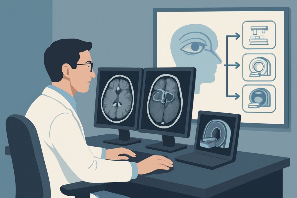
A 68-year-old male with a history of hypertension and type 2 diabetes presents to your clinic with three days of new, binocular double vision. On examination, he has an isolated left sixth nerve palsy, causing horizontal diplopia that worsens on leftward gaze. You suspect a microvascular ischemic cause given his risk factors, but a compressive lesion, such as an aneurysm or tumor, must be excluded. The immediate clinical question is which imaging study provides the most definitive and efficient initial evaluation. This article provides a deep dive into the American College of Radiology (ACR) Appropriateness Criteria for this specific scenario, explaining the rationale for the recommended studies. For this presentation, the ACR rates MRI head without and with IV contrast as Usually Appropriate.
Who Fits This Clinical Scenario?
This guidance applies to adult and pediatric patients presenting with new-onset ophthalmoplegia (weakness of the eye muscles) or diplopia (double vision) where the initial history and physical examination do not suggest an obvious traumatic or infectious cause. The key inclusion criterion is the new onset of symptoms related to extraocular muscle dysfunction. This could manifest as an isolated cranial nerve palsy (affecting the oculomotor, trochlear, or abducens nerves), multiple nerve palsies, or a more complex gaze abnormality.
It is crucial to distinguish this presentation from similar, but distinct, clinical scenarios that follow different diagnostic pathways:
- Traumatic Visual Defect: If the patient has a history of recent head or facial trauma, the workup shifts to the “Traumatic visual defect. Suspect orbital injury” variant, where CT is often the primary modality to assess for fractures.
- Proptosis or Globe Displacement: If the primary finding is nontraumatic orbital asymmetry, exophthalmos (bulging eye), or enophthalmos (sunken eye), the evaluation should follow the guidance for that specific scenario, which focuses more on orbital mass or thyroid eye disease.
- Signs of Infection: Patients with fever, eyelid erythema, swelling, and pain are more likely to have orbital cellulitis. This presentation routes to the “Suspected orbital cellulitis, uveitis, or scleritis” variant, where imaging goals are to define the extent of infection.
What Diagnoses Are You Working Up in This Scenario?
When a patient presents with new ophthalmoplegia or diplopia, the differential diagnosis is broad, spanning from benign, self-limiting conditions to life-threatening emergencies. Imaging is critical for differentiating these possibilities. The primary goal is to evaluate the entire pathway of ocular motor control, from the brainstem nuclei to the cranial nerves, through the cavernous sinus and superior orbital fissure, to the extraocular muscles themselves.
Cranial Nerve Palsy (Compressive vs. Ischemic): This is a leading consideration. While microvascular ischemia is a common cause of isolated nerve palsies in older adults with vascular risk factors, a compressive lesion must be ruled out. This includes intracranial aneurysms (particularly of the posterior communicating artery for a third nerve palsy), meningiomas, schwannomas, or metastatic tumors along the nerve’s path.
Cavernous Sinus Pathology: The cavernous sinus houses multiple cranial nerves (III, IV, V1, V2, VI) in a compact space. A single pathology here, such as a tumor, carotid-cavernous fistula, thrombosis, or an inflammatory process like Tolosa-Hunt syndrome or sarcoidosis, can cause complex ophthalmoplegia involving multiple nerves.
Thyroid Eye Disease (Graves’ Orbitopathy): As one of the most common causes of diplopia in adults, this autoimmune condition leads to inflammation and enlargement of the extraocular muscles. The characteristic pattern of muscle involvement (inferior and medial recti most commonly) is often readily identifiable on cross-sectional imaging.
Myasthenia Gravis: While primarily a clinical and serologic diagnosis, imaging is essential in the initial workup of ophthalmoplegia to exclude structural mimics. Myasthenia can present with fluctuating ptosis and diplopia that can mimic nearly any pattern of ocular motor weakness.
Brainstem Lesions: A less common but critical consideration is a lesion within the brainstem, such as a stroke, demyelinating plaque (e.g., from multiple sclerosis), or tumor, affecting the cranial nerve nuclei or internuclear pathways. This highlights the need for imaging that includes the posterior fossa.
Why Is MRI Head Without and With IV Contrast the Recommended Study?
The ACR designates MRI head without and with IV contrast as Usually Appropriate for the initial imaging of ophthalmoplegia or diplopia. This recommendation is based on MRI’s superior soft-tissue contrast, which is essential for visualizing the small and complex structures responsible for eye movement.
MRI provides exquisite detail of the cranial nerves, brainstem, cavernous sinuses, and orbital soft tissues. It is highly sensitive for detecting the subtle inflammation, demyelination, or small tumors that are often responsible for these symptoms. The addition of intravenous contrast is critical; it enhances the visibility of inflammatory processes (like neuritis or sarcoidosis), neoplasms, and vascular abnormalities. Without contrast, many of these key diagnoses could be missed, making a non-contrast study significantly less valuable for the initial workup.
Several other MRI protocols, including MRI orbits without and with IV contrast, are also rated Usually Appropriate. The choice between a head and orbits protocol depends on the clinical suspicion. If a brainstem or central cause is suspected, an MRI of the head is superior. If pathology is thought to be confined to the orbit (e.g., suspected thyroid eye disease), a dedicated orbital MRI with thin slices can be optimal.
Let’s consider the alternatives and why they are rated differently for this specific scenario:
- CT orbits with IV contrast: This study is also rated Usually Appropriate. It is an excellent alternative when MRI is contraindicated (e.g., incompatible pacemaker) or not immediately available. CT is faster than MRI and superior for evaluating bony anatomy. However, it provides less detail of the cranial nerves and brainstem and involves ionizing radiation (☢☢☢ 1-10 mSv).
- CT head without IV contrast: Rated as May be appropriate, this study has significant limitations. While it can identify acute hemorrhage or large masses, it is insensitive to the most common causes of nontraumatic diplopia, such as small infarcts, cranial nerve enhancement, or cavernous sinus inflammation. It is generally insufficient as a standalone initial test.
The key advantage of MRI is its ability to comprehensively evaluate the relevant anatomy without exposing the patient to ionizing radiation (0 mSv). When ordering, specifying “without and with IV contrast” and providing a clear clinical history of “new-onset diplopia, rule out compressive lesion” helps the radiology team tailor the protocol for optimal diagnostic yield.
What’s Next After MRI? Downstream Workflow
The results of the initial MRI will guide the subsequent clinical pathway. The workflow branches based on whether the findings are positive, negative, or indeterminate.
If the study is positive for a structural lesion:
A finding like a tumor, aneurysm, or evidence of a carotid-cavernous fistula requires urgent specialist consultation. A compressive tumor will typically be referred to neuro-oncology and neurosurgery. An aneurysm requires immediate neurosurgical or neurointerventional radiology evaluation. Inflammatory findings, such as nerve enhancement suggestive of neuritis or findings consistent with Tolosa-Hunt syndrome, warrant a referral to neurology or neuro-ophthalmology for further workup and potential immunosuppressive therapy.
If the study is negative:
A normal MRI is a crucial and reassuring finding, as it effectively rules out most compressive and acute structural causes. The diagnostic focus then shifts to non-structural etiologies. This is the point to strongly consider:
- Microvascular ischemia: In patients with appropriate risk factors (diabetes, hypertension, age), a negative MRI supports a presumptive diagnosis of an ischemic cranial nerve mononeuropathy. The typical management is observation for spontaneous improvement over 6-12 weeks.
- Neuromuscular junction disease: Myasthenia gravis is a primary consideration. The next steps would include serologic testing for acetylcholine receptor antibodies and muscle-specific kinase (MuSK) antibodies, and potentially an electromyogram (EMG).
- Systemic inflammatory or autoimmune conditions: Further laboratory workup may be indicated to investigate conditions like sarcoidosis or vasculitis.
If the study is indeterminate:
Occasionally, MRI findings may be nonspecific. In these cases, or if a vascular abnormality is suspected despite a non-diagnostic MRI, a vascular imaging study like MRA head and neck or CTA head and neck (both rated May be appropriate) may be the logical next step.
Pitfalls to Avoid (and When to Get Help)
Navigating the workup for diplopia requires careful consideration to avoid common diagnostic traps.
- Omitting IV Contrast: Ordering a non-contrast MRI or CT as the initial study is a frequent pitfall. Many of the key pathologies, including inflammation and small tumors, are only visible after contrast administration.
- Choosing the Wrong Modality: Defaulting to CT without a specific contraindication to MRI can lead to a missed diagnosis and unnecessary radiation exposure, particularly for younger patients. MRI’s superior soft-tissue resolution is purpose-built for this differential.
- Tunnel Vision on the Orbits: Focusing exclusively on the orbits can cause you to miss a central cause in the brainstem or a compressive lesion along the cranial nerve’s path through the skull base. An MRI of the head often provides a more comprehensive initial survey.
- Underestimating Red Flags: Diplopia accompanied by a “thunderclap” headache, pupillary involvement in a third nerve palsy, or signs of multiple cranial nerve deficits is a neurologic emergency. If you suspect a ruptured aneurysm, escalate immediately for the fastest available neurovascular imaging (often CTA) and neurosurgical consultation.
Related ACR Topics and Tools
This article covers a single, focused clinical scenario. For a comprehensive overview of all related presentations and to explore the evidence behind other imaging decisions, please consult the resources below. For breadth across all scenarios in Orbits, Vision, and Visual Loss, see our parent guide: Orbits, Vision, and Visual Loss: ACR Appropriateness Decoded.
- ACR Appropriateness Criteria Lookup — for adjacent scenarios
- Imaging Protocol Library — for technique on the recommended study
- Radiation Dose Calculator — for cumulative dose conversations
Frequently Asked Questions
Why is MRI preferred over CT for the initial imaging of nontraumatic diplopia?
MRI is preferred because of its superior ability to visualize soft tissues. It provides detailed images of the cranial nerves, brainstem, extraocular muscles, and cavernous sinus without using ionizing radiation. This makes it more sensitive than CT for detecting the most common causes of diplopia, such as inflammation, small tumors, or demyelination, which may be invisible on CT.
Is intravenous contrast always necessary for an MRI in this scenario?
Yes, for the initial diagnostic workup, IV contrast is highly recommended. The ACR rates both ‘MRI head without and with IV contrast’ and ‘MRI orbits without and with IV contrast’ as ‘Usually Appropriate.’ A non-contrast MRI is only rated ‘May be appropriate’ because it can miss key pathologies like cranial nerve enhancement from inflammation or small, enhancing tumors.
What imaging should I order if my patient has a pacemaker or another contraindication to MRI?
If MRI is contraindicated, ‘CT orbits with IV contrast’ is the best alternative and is also rated ‘Usually Appropriate’ by the ACR. While it involves radiation and has lower soft-tissue contrast than MRI, it is a robust study for identifying larger masses, bony abnormalities, and significant inflammatory changes like those seen in thyroid eye disease.
My patient’s diplopia is isolated and painless, and I suspect a microvascular cause. Do they still need imaging?
Yes, initial imaging is still recommended. While an isolated, pupil-sparing third, fourth, or sixth nerve palsy in an older patient with vascular risk factors is often due to microvascular ischemia, it is critical to rule out a compressive lesion like an aneurysm or tumor. A negative MRI provides the reassurance needed to manage the patient conservatively with observation.
Should I order an ‘MRI Orbits’ or an ‘MRI Head’ for diplopia?
The choice depends on the clinical context. If you suspect the problem is isolated to the eye muscles or orbital contents (e.g., thyroid eye disease), a dedicated ‘MRI Orbits’ provides high-resolution images of that area. However, if the cause could be central (in the brainstem) or along the cranial nerve’s path (in the cavernous sinus), an ‘MRI Head’ provides a more comprehensive evaluation of the entire neuro-ocular axis and is often the better initial choice for an undifferentiated presentation.
Reviewed by Pouyan Golshani, MD, Interventional Radiologist — June 27, 2026