What Is the Best Initial Imaging for Clinically Suspected Fibroids?
A 38-year-old G2P2 presents to your clinic with a six-month history of progressively heavier menstrual bleeding and a sensation of pelvic pressure. On examination, you palpate a firm, enlarged, and irregular uterus. Your leading clinical suspicion is uterine leiomyomas, commonly known as fibroids. You need to confirm the diagnosis, assess the number, size, and location of the fibroids, and rule out other potential causes for her symptoms. This raises the critical question: what is the most appropriate initial imaging study to order? This article provides a detailed workflow for this specific scenario, anchored in the American College of Radiology (ACR) Appropriateness Criteria, which rate a US duplex Doppler pelvis as Usually Appropriate for the initial evaluation of clinically suspected fibroids.
Who Fits This Clinical Scenario?
This guidance applies specifically to patients presenting for the initial diagnostic imaging of clinically suspected uterine fibroids. The typical patient is a premenopausal or perimenopausal individual with signs and symptoms such as heavy menstrual bleeding (menorrhagia), pelvic pain or pressure, bulk-related symptoms like increased urinary frequency, or a palpably enlarged uterus on physical exam. This workflow is designed for the first imaging study in a patient without a previously confirmed diagnosis of fibroids.
It is crucial to distinguish this situation from related but distinct clinical questions. This article does not apply to:
- Patients with known fibroids requiring treatment planning. Once fibroids are diagnosed, the choice of imaging to prepare for a procedure like uterine artery embolization (UAE), myomectomy, or radiofrequency ablation follows a different set of recommendations. This is covered in the ACR guidelines for treatment planning.
- Patients undergoing surveillance or post-treatment imaging. Following a procedure or during a period of watchful waiting, the imaging strategy is different from the initial diagnostic workup.
- Patients with acute, severe pelvic pain or suspected gynecologic emergencies. Presentations concerning for ovarian torsion, ectopic pregnancy, or tubo-ovarian abscess require an emergent workup that may prioritize different imaging features or modalities.
What Diagnoses Are You Working Up in This Scenario?
When ordering initial imaging for suspected fibroids, the goal is not only to confirm their presence but also to evaluate for other conditions that can present with similar symptoms. The differential diagnosis is key to interpreting the results and planning next steps.
Uterine Leiomyomas (Fibroids)
This is the most common diagnosis and the primary target of the investigation. Fibroids are benign monoclonal tumors of uterine smooth muscle. Imaging aims to confirm their presence and document their number, size, and location (submucosal, intramural, subserosal, or pedunculated), as this information is critical for correlating with symptoms and guiding future management.
Adenomyosis
A frequent cause of heavy, painful periods, adenomyosis occurs when endometrial glands and stroma are present within the myometrium. It often coexists with fibroids and can be challenging to distinguish clinically. Ultrasound can often identify characteristic features like an enlarged, globular uterus with myometrial heterogeneity or cysts, but the findings can sometimes overlap with fibroids.
Endometrial Polyps or Hyperplasia
Abnormal uterine bleeding can also be caused by pathology within the endometrial cavity itself. Endometrial polyps or generalized thickening (hyperplasia) are important considerations. A high-quality transvaginal ultrasound is essential for evaluating the endometrial stripe and identifying focal intracavitary lesions.
Adnexal Mass
While less likely to cause heavy menstrual bleeding, an adnexal mass (e.g., an ovarian cyst or solid neoplasm) can cause pelvic pain and pressure. A pedunculated subserosal fibroid can also mimic an adnexal mass. Imaging must carefully evaluate the ovaries and adnexa to exclude a separate pathologic process.
Uterine Sarcoma
Though rare, this malignant differential is a critical consideration, particularly in postmenopausal patients with a rapidly enlarging pelvic mass, bleeding, and pain. While imaging cannot definitively diagnose or exclude sarcoma, certain features on ultrasound or MRI may raise suspicion and prompt an urgent referral to a gynecologic oncologist.
Why Is Pelvic Ultrasound the Recommended Study for This Presentation?
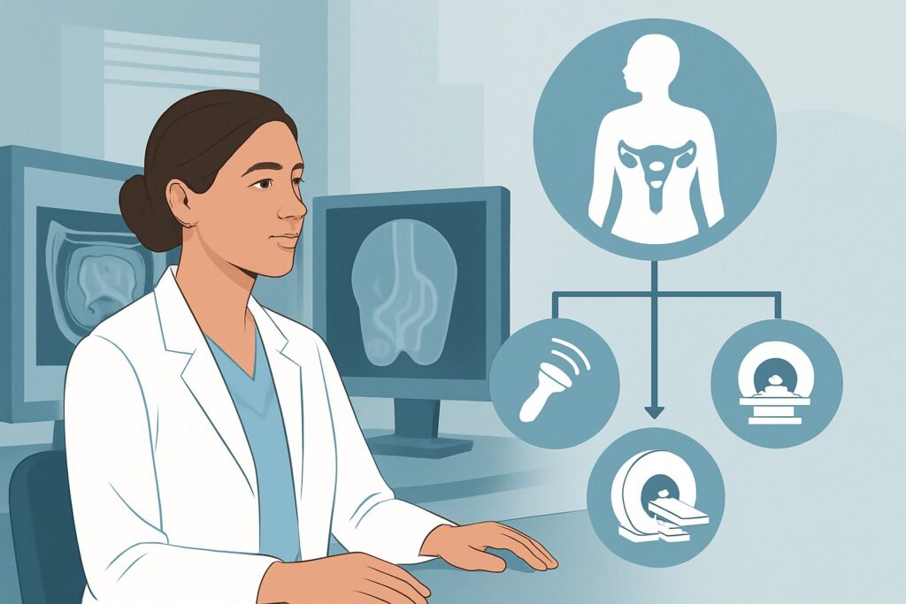
The ACR rates three variations of pelvic ultrasound as Usually Appropriate for the initial workup of suspected fibroids: US pelvis transabdominal, US pelvis transvaginal, and US duplex Doppler pelvis. In practice, a complete examination often combines these components. This recommendation is based on the modality’s high diagnostic accuracy, wide availability, cost-effectiveness, and lack of ionizing radiation.
A comprehensive pelvic ultrasound provides the necessary information for initial diagnosis and management. The transabdominal approach offers a wide field of view, which is essential for assessing a large, fibroid-distorted uterus and identifying very large or superiorly located fibroids that may be beyond the reach of a transvaginal probe. The transvaginal approach provides superior spatial resolution, offering detailed views of the myometrial texture, endometrial lining, and ovaries. This is critical for detecting smaller fibroids, characterizing their relationship to the endometrium, and evaluating for co-existing conditions like adenomyosis or endometrial polyps.
The inclusion of Duplex Doppler is not incidental. Color and spectral Doppler imaging assesses blood flow within the uterus and adnexa. This is invaluable for differentiating a pedunculated fibroid from a solid adnexal mass by identifying the uterine vascular pedicle. It also helps characterize the vascularity of myometrial masses, which can be relevant for future treatment planning.
Why are other modalities rated lower for initial imaging?
- MRI pelvis (without or with IV contrast) is rated as May be appropriate. While MRI offers superior soft tissue contrast and is the gold standard for fibroid mapping (size, number, location), it is not the recommended first step. Its higher cost and lower availability make it better suited as a problem-solving tool when ultrasound is inconclusive or for detailed pre-procedural planning for interventions like myomectomy or uterine artery embolization.
- CT pelvis (with or without IV contrast) is rated as Usually not appropriate. CT has poor soft-tissue contrast for evaluating myometrial and endometrial pathology, making it significantly inferior to both ultrasound and MRI for characterizing fibroids. More importantly, it exposes the patient to significant ionizing radiation (☢☢☢ 1-10 mSv), which is unwarranted for a benign condition in a typically reproductive-age population when a superior, radiation-free alternative exists.
What’s Next After Pelvic Ultrasound? Downstream Workflow
The results of the initial pelvic ultrasound will guide the subsequent clinical pathway. The workflow branches based on whether the findings are definitive, negative, or indeterminate.
If the study is positive for fibroids:
When ultrasound confirms the presence of one or more leiomyomas that correlate with the patient’s symptoms, the next step is clinical decision-making. This is a conversation between the clinician and the patient, weighing symptom severity, fibroid characteristics (size, location), and the patient’s fertility goals. Management options range from watchful waiting for minimal symptoms, to medical therapies (e.g., hormonal agents, tranexamic acid), to referral to a gynecologist to discuss procedural interventions.
If the study is negative or symptoms persist:
If the ultrasound is unremarkable but the patient’s symptoms of heavy bleeding or pelvic pressure continue, further evaluation is warranted. This may involve a clinical reassessment for non-uterine causes or proceeding to a more advanced imaging study. In this “problem-solving” context, an MRI of the pelvis (rated May be appropriate) is often the logical next step to better characterize subtle adenomyosis or small fibroids missed on ultrasound.
If the study is indeterminate or complex:
Sometimes, ultrasound cannot definitively distinguish between a large, complex fibroid, focal adenomyosis, or a potential sarcoma. It may also be difficult to precisely map submucosal fibroids. In these cases, MRI is the ideal follow-up study to clarify the anatomy, which is essential before considering surgical or interventional procedures. If an adnexal mass is found, the workflow shifts to characterizing that finding, potentially involving tumor markers and a gynecologic consultation.
Pitfalls to Avoid (and When to Get Help)
Navigating the initial workup for suspected fibroids requires avoiding several common pitfalls to ensure an efficient and accurate diagnosis.
- Pitfall: Ordering a limited ultrasound. Requesting only a transabdominal ultrasound can miss significant endometrial pathology or small fibroids. A complete study including a transvaginal component is standard of care unless contraindicated.
- Pitfall: Defaulting to CT for pelvic pain. In a non-emergent setting where fibroids are on the differential, ordering a CT scan exposes the patient to unnecessary radiation and provides suboptimal diagnostic information for this specific question.
- Pitfall: Attributing all bleeding to fibroids. Do not assume that the presence of fibroids is the sole cause of abnormal uterine bleeding. The endometrial lining must be carefully evaluated to rule out concurrent polyps or hyperplasia, especially in perimenopausal or postmenopausal women.
If imaging reveals features suspicious for malignancy (e.g., rapid growth on serial exams, unusual features in a postmenopausal patient) or identifies a complex adnexal mass, prompt escalation with a referral to a gynecologic specialist or gynecologic oncologist is critical.
Related ACR Topics and Tools
This article covers one specific scenario within the broader topic of fibroid imaging. For a comprehensive overview of all clinical variants, including treatment planning and surveillance, please consult our parent guide. For additional resources on imaging selection, protocols, and radiation safety, the following tools are available.
- For breadth across all scenarios in Fibroids, see our parent guide: Fibroids: ACR Appropriateness Decoded.
- To look up other clinical scenarios, use the ACR Appropriateness Criteria Lookup.
- For detailed imaging techniques, explore the Imaging Protocol Library.
- To discuss radiation exposure with patients, see the Radiation Dose Calculator.
Frequently Asked Questions
Why not just start with an MRI for suspected fibroids since it provides more detail?
While MRI is more detailed, pelvic ultrasound is the recommended initial study because it is highly accurate for initial diagnosis, widely available, significantly less expensive, and does not use ionizing radiation. It provides sufficient information to confirm the diagnosis and guide initial management in the vast majority of cases. MRI is reserved as a second-line, problem-solving tool for when ultrasound is inconclusive or for detailed pre-procedural planning.
Is a transvaginal ultrasound always necessary if a transabdominal one is done?
In most cases, yes. The transvaginal portion of the exam provides high-resolution images of the endometrium, myometrium, and ovaries that are not achievable with the transabdominal approach alone. It is crucial for detecting small fibroids, assessing for submucosal fibroids that affect the endometrial cavity, and evaluating for co-existing conditions like adenomyosis or endometrial polyps. It may be deferred in patients who are not sexually active or who decline it, but this limitation should be noted.
What if the patient is postmenopausal and presents with a new pelvic mass?
While fibroids can be present in postmenopausal women, any new or growing pelvic mass in this population requires a higher index of suspicion for malignancy, such as a uterine sarcoma or ovarian cancer. Pelvic ultrasound is still the appropriate first imaging step, but the findings should be interpreted with caution. Any features concerning for malignancy warrant an urgent referral to a gynecologic oncologist for further management, which may include MRI and surgical evaluation.
Does the presence of an IUD interfere with a pelvic ultrasound for fibroids?
An intrauterine device (IUD) does not prevent a thorough evaluation for fibroids. The IUD itself will be visible within the endometrial cavity and can create some acoustic shadowing, but experienced sonographers can typically work around this to assess the myometrium and adnexa effectively. The IUD’s position can also be confirmed during the exam.
Reviewed by Pouyan Golshani, MD, Interventional Radiologist — May 29, 2026