What’s the Best Initial Imaging for Suspected Tubal Occlusion in Female Infertility?
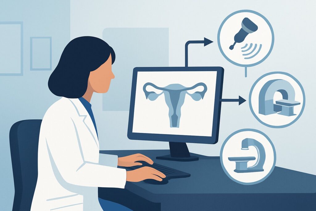
A 32-year-old woman presents to your clinic with her partner. They have been trying to conceive for 18 months without success. Her menstrual cycles are regular, and preliminary hormonal testing suggests normal ovulatory function. Her partner’s semen analysis is unremarkable. The clinical focus now shifts to evaluating structural causes, specifically the patency of her fallopian tubes and the integrity of her uterus. You need to choose the most appropriate initial imaging study to guide the next steps in her fertility workup. This article details the American College of Radiology (ACR) guided workflow for this exact scenario. For the initial evaluation, the ACR rates a transvaginal pelvic ultrasound as Usually Appropriate.
Who Fits This Clinical Scenario for Suspected Tubal Occlusion?
This guidance applies to patients presenting with female infertility where the initial workup points toward a potential structural or tubal factor. Typically, this includes individuals who have been unable to conceive after 12 months of regular, unprotected intercourse (or 6 months if the female partner is over 35 years old). The scenario assumes that more easily identifiable causes, such as anovulation or significant male factor infertility, have either been ruled out or are being investigated concurrently, making uterine and tubal assessment the logical next step.
It is crucial to distinguish this presentation from related but distinct clinical situations that follow different imaging pathways. This workflow is NOT for patients whose primary presentation is:
- Dominated by ovulatory dysfunction: If the patient has irregular cycles or clinical features of Polycystic Ovary Syndrome (PCOS), the workup prioritizes hormonal assessment and ovarian morphology, which falls under the ACR variant for evaluating ovulatory function.
- Highly suggestive of endometriosis: A patient with a dominant history of severe, cyclic pelvic pain, deep dyspareunia, and dyschezia may be evaluated under the specific ACR scenario for suspected endometriosis, which has a different imaging decision tree.
- Characterized by recurrent pregnancy loss: This article addresses the failure to conceive. The workup for patients who can conceive but experience multiple miscarriages is different and is covered in its own ACR variant.
What Diagnoses Are You Working Up in This Scenario?
When ordering initial imaging for suspected tubal factor infertility, you are investigating several potential anatomic barriers to conception. The differential diagnosis guides the choice of imaging modality and what the radiologist will be looking for.
Tubal Occlusion: This is the primary diagnosis of concern. The fallopian tubes can become blocked as a result of prior pelvic inflammatory disease (PID), adhesions from previous abdominal or pelvic surgery, or endometriosis. Occlusion can be proximal (near the uterus) or distal (near the ovary), preventing the sperm and egg from meeting or a fertilized embryo from reaching the uterus.
Hydrosalpinx: This is a specific and consequential form of distal tubal occlusion where the blocked tube fills with fluid. A hydrosalpinx is not only a sign of a non-functional tube but the fluid itself is considered embryotoxic, which can lower the success rates of subsequent treatments like in vitro fertilization (IVF).
Intrauterine Pathology: The initial imaging study also serves as a screen for uterine cavity abnormalities that can prevent embryo implantation. This includes submucosal fibroids (leiomyomas) that distort the endometrial cavity, endometrial polyps, and intrauterine adhesions (Asherman syndrome), which can result from prior uterine surgery or infection.
Congenital Uterine Anomalies: Less common but important to identify are Müllerian duct anomalies, such as a septate, bicornuate, or unicornuate uterus. These structural differences can affect fertility and increase the risk of pregnancy complications. An initial ultrasound can often detect or raise suspicion for these anomalies.
Why Is Transvaginal Ultrasound the Recommended Initial Study for This Presentation?
The ACR Appropriateness Criteria designate US pelvis transvaginal as Usually Appropriate for the initial imaging of a patient with infertility and suspected tubal occlusion. This recommendation is based on its excellent safety profile, accessibility, and diagnostic utility for evaluating the primary differential diagnoses in this setting.
A transvaginal ultrasound provides high-resolution images of the uterus, endometrium, and ovaries without using ionizing radiation (0 mSv). It is the best initial modality for identifying uterine fibroids, endometrial polyps, and ovarian pathologies like endometriomas. Crucially, while a standard 2D ultrasound cannot directly confirm tubal patency, it can detect a hydrosalpinx—a dilated, fluid-filled fallopian tube—which is a definitive sign of distal tubal blockage. Finding a hydrosalpinx provides a key piece of diagnostic information that directly impacts treatment planning.
Two other procedures are also rated Usually Appropriate but serve different, often subsequent, roles:
- Fluoroscopy hysterosalpingography (HSG): This is the classic test for tubal patency. It involves injecting iodinated contrast through the cervix under fluoroscopy (X-ray) to visualize the uterine cavity and fallopian tubes, demonstrating spill into the peritoneum if the tubes are open. While it directly answers the patency question, it involves ionizing radiation (☢☢ 0.1-1mSv) and provides less detail of the uterine wall and ovaries compared to ultrasound.
- US sonohysterography with tubal contrast agent (also known as HyCoSy): This ultrasound-based technique involves instilling saline and then a microbubble contrast agent into the uterine cavity to assess both the cavity and tubal patency. It avoids radiation but is more specialized and may not be as widely available as HSG.
An alternative rated lower is MRI pelvis without and with IV contrast, which is May be appropriate. MRI offers superb soft tissue detail and is an excellent problem-solving tool for complex cases, such as characterizing a complex adnexal mass or mapping deep infiltrating endometriosis. However, its higher cost and lower accessibility make it unsuitable as a first-line screening tool for this common clinical question.
What’s Next After Transvaginal Ultrasound? Downstream Workflow
The results of the initial transvaginal ultrasound will guide your subsequent diagnostic and management steps. The workflow branches based on the findings.
If the ultrasound is positive for a hydrosalpinx: This finding strongly suggests distal tubal occlusion. The patient should be counseled that the affected tube is non-functional and may negatively impact IVF outcomes. The next step is often a referral to a reproductive endocrinologist for discussion of options, which may include laparoscopic salpingectomy (removal of the affected tube) prior to proceeding with IVF.
If the ultrasound shows uterine pathology (e.g., suspected submucosal fibroid or large polyp): The next step is often a saline-infusion sonohysterogram (SIS) or hysteroscopy to better characterize the lesion and determine if it is impinging on the endometrial cavity. Surgical removal may be recommended before further fertility treatment.
If the ultrasound is completely normal: This is a very common outcome. The uterus and ovaries appear structurally normal, and there is no evidence of a hydrosalpinx. However, a normal ultrasound does not rule out tubal occlusion. In this case, the next step is a direct test of tubal patency. The choice is typically between a hysterosalpingogram (HSG) or a hysterosalpingo-contrast-sonography (HyCoSy), both of which are rated Usually Appropriate for this purpose.
If the findings are indeterminate or suggest a complex anomaly: For example, if the ultrasound suggests a complex congenital uterine anomaly or findings are suspicious for deep endometriosis, an MRI of the pelvis may be the appropriate next step to fully characterize the anatomy before surgical planning.
Pitfalls to Avoid (and When to Get Help)
Navigating the infertility workup requires careful interpretation and sequencing of tests. Here are a few common pitfalls to avoid in this specific scenario:
- Stopping the workup after a normal ultrasound: Remember that a normal transvaginal ultrasound does not confirm tubal patency. It is an excellent first step for uterine and ovarian evaluation, but a functional tubal assessment (HSG or HyCoSy) is still required if the ultrasound is negative.
- Misinterpreting a paratubal cyst for a hydrosalpinx: These can sometimes appear similar on ultrasound. A true hydrosalpinx often has a characteristic convoluted, tubular shape, sometimes with incomplete septa or the “beads-on-a-string” sign. If in doubt, correlation with a follow-up patency test is key.
- Ordering MRI as a first-line test: While a powerful tool, ordering an MRI for every initial infertility workup is not cost-effective and can lead to unnecessary delays. Reserve it for problem-solving after initial ultrasound findings are unclear or complex.
If the combination of non-invasive imaging studies yields conflicting or confusing results, or if surgical intervention is being considered, escalation to a reproductive endocrinology and infertility (REI) specialist is the appropriate next step.
Related ACR Topics and Tools
The ACR Appropriateness Criteria are a powerful resource for ensuring evidence-based imaging decisions. For a comprehensive overview of all clinical variants related to female infertility, please consult our parent topic hub article. For further exploration of specific scenarios or tools, the following GigHz resources are available:
- For breadth across all scenarios in Female Infertility, see our parent guide: Female Infertility: ACR Appropriateness Decoded.
- ACR Appropriateness Criteria Lookup — for adjacent scenarios
- Imaging Protocol Library — for technique on the recommended study
- Radiation Dose Calculator — for cumulative dose conversations
Frequently Asked Questions
Does a normal transvaginal ultrasound mean the fallopian tubes are open?
No. A standard transvaginal ultrasound is excellent for evaluating the uterus and ovaries, but it cannot confirm tubal patency. It can only detect indirect signs of blockage, such as a hydrosalpinx (a fluid-filled tube). If the ultrasound is normal, a dedicated patency test like a hysterosalpingogram (HSG) or a HyCoSy is still necessary to complete the tubal evaluation.
Why not just order a hysterosalpingogram (HSG) first for everyone?
While HSG directly assesses tubal patency, it uses ionizing radiation and provides less detailed information about the uterine walls (e.g., for fibroids) and ovaries compared to a transvaginal ultrasound. The ACR recommends ultrasound first because it is radiation-free and can diagnose many significant uterine and ovarian issues that would require treatment regardless of tubal status, making it a more comprehensive initial screening test.
What is the difference between a sonohysterogram and a HyCoSy?
A standard sonohysterogram (or saline-infusion sonogram) involves instilling saline into the uterine cavity to better visualize the endometrial lining for polyps or submucosal fibroids; it does not assess the tubes. A HyCoSy (Hysterosalpingo-contrast-sonography) is a more advanced procedure that uses a special microbubble contrast agent after the saline to visualize flow through the fallopian tubes via ultrasound, thereby assessing patency.
If a patient has a history of pelvic inflammatory disease (PID), should I skip the ultrasound and go straight to HSG?
Not necessarily. A history of PID significantly increases the risk of tubal occlusion and hydrosalpinx. A transvaginal ultrasound is still the recommended first step because it is the best non-invasive way to screen for a hydrosalpinx. Identifying a hydrosalpinx on ultrasound may alter management (e.g., proceeding to laparoscopy) and could potentially make a subsequent HSG unnecessary for that tube.
When is an MRI the right choice in this workup?
MRI is considered a second-line, problem-solving tool in this scenario. It is rated ‘May be appropriate’ and should be reserved for cases where ultrasound findings are inconclusive or when there is suspicion of complex pathology. Examples include characterizing a complex uterine anomaly, mapping the extent of deep infiltrating endometriosis, or evaluating an indeterminate adnexal mass found on ultrasound.
Reviewed by Pouyan Golshani, MD, Interventional Radiologist — May 29, 2026