What Is the Best Initial Imaging for Newly Diagnosed DCIS? An ACR-Guided Workflow

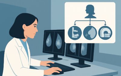
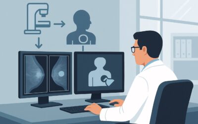
It’s late afternoon, and the pathology report from a stereotactic biopsy confirms the diagnosis: ductal carcinoma in situ (DCIS). Your patient, a 58-year-old woman, is scheduled for a surgical consultation next week. Before that appointment, you need to order the right imaging to accurately define the extent of her disease, information critical for planning breast-conserving surgery versus mastectomy. The core question is which study will best delineate the boundaries of the DCIS, identify any additional suspicious areas, and screen the contralateral breast. For this specific clinical scenario—initial imaging for newly diagnosed DCIS—the American College of Radiology (ACR) rates Digital Breast Tomosynthesis (DBT) diagnostic as Usually Appropriate, providing a clear starting point for the preoperative workup.
Who Fits This Clinical Scenario?
This workflow applies specifically to adult patients with a new, biopsy-proven diagnosis of pure Ductal Carcinoma in Situ (DCIS). The key inclusion criteria are:
- A definitive histopathologic diagnosis of DCIS from a recent breast biopsy (e.g., stereotactic, ultrasound-guided, or MRI-guided).
- The patient has not yet undergone definitive surgical treatment (lumpectomy or mastectomy).
- The primary goal of imaging is to assess the extent of disease for initial surgical planning.
It is crucial to distinguish this situation from similar but distinct clinical presentations that require different imaging pathways. This guidance does not apply if:
- Microinvasion is known: If the biopsy report explicitly states “DCIS with microinvasion,” the workup shifts to a different ACR variant that includes dedicated axillary evaluation.
- The patient is undergoing surveillance: This workflow is for initial diagnosis, not for routine surveillance after treatment for DCIS, whether that treatment was breast conservation therapy or mastectomy.
- Recurrence is suspected: Patients with a history of DCIS who present with new symptoms (e.g., a palpable lump, skin changes) fall under scenarios for suspected local recurrence, which have their own imaging recommendations.
Applying this workflow to the correct patient population ensures the most effective and appropriate use of imaging resources for preoperative planning.
What Diagnoses Are You Working Up in This Scenario?
While the diagnosis of DCIS is already established by biopsy, the purpose of initial preoperative imaging is not to re-diagnose but to accurately characterize the disease. The imaging study is designed to answer several critical questions that directly influence surgical management. The “differential” in this context refers to the potential findings that imaging must confirm or exclude.
Extent of the Index Lesion: The most fundamental goal is to determine the true size and distribution of the known DCIS. Often, the extent of microcalcifications or other mammographic findings underestimates the actual pathologic extent of the disease. Accurate measurement is essential for achieving negative surgical margins during breast-conserving surgery.
Multifocal or Multicentric Disease: Imaging must assess for additional, separate foci of DCIS within the same breast. Multifocal disease (multiple foci in the same quadrant) may still be amenable to lumpectomy, whereas multicentric disease (foci in different quadrants) often necessitates a mastectomy. Identifying these additional sites preoperatively prevents positive margins and the need for re-excision.
Occult Invasive Carcinoma: A core-needle biopsy samples only a small portion of the lesion. A significant minority of cases diagnosed as pure DCIS on biopsy are upgraded to invasive ductal carcinoma upon final surgical pathology. Imaging seeks to identify findings suggestive of invasion, such as an associated mass, architectural distortion, or suspicious enhancement, which would alter surgical plans, particularly regarding axillary lymph node assessment.
Contralateral Breast Cancer: A small but significant percentage of patients with DCIS will have a synchronous cancer in the opposite breast. Preoperative imaging includes a thorough evaluation of the contralateral breast to detect any occult malignancy that would require simultaneous treatment.
Why Is Diagnostic Digital Breast Tomosynthesis the Recommended Study?
For the initial workup of newly diagnosed DCIS, the ACR designates diagnostic Digital Breast Tomosynthesis (DBT) as Usually Appropriate. This recommendation is based on its superior ability to characterize the extent of disease compared to standard 2D mammography, which is also rated Usually Appropriate but is often considered a less optimal alternative if DBT is available.
DBT, or 3D mammography, acquires multiple low-dose images of the breast from different angles, which are then reconstructed into thin slices. This technique minimizes the effect of overlapping breast tissue, a common limitation of 2D mammography, especially in dense breasts. For DCIS, this provides a clearer depiction of the distribution and extent of microcalcifications. More importantly, it significantly improves the detection of subtle associated findings that may suggest an underlying invasive component, such as architectural distortion or a developing asymmetry.
Let’s consider the alternatives and why they are rated lower for this specific scenario:
- MRI breast without and with IV contrast is rated May be appropriate. While breast MRI has the highest sensitivity for detecting both invasive cancer and DCIS, its specificity is lower. This can lead to false-positive findings, resulting in additional biopsies and patient anxiety. Its use is typically reserved for specific situations, such as when the extent of disease on mammography is underestimated, particularly in dense breasts, or to resolve equivocal mammographic findings before surgery.
- US breast is also rated May be appropriate. Ultrasound is not a primary tool for evaluating the extent of DCIS, as most DCIS presents as microcalcifications, which are poorly visualized with sonography. However, it is an essential adjunct. If mammography or DBT shows an associated mass or distortion, a targeted ultrasound is necessary to characterize the finding, assess for an invasive component, and guide a potential biopsy. It is also the primary modality for evaluating the axillary lymph nodes if invasion is suspected.
From a safety perspective, diagnostic DBT involves a low level of ionizing radiation (ACR Relative Radiation Level ☢☢, corresponding to an effective dose of 0.1-1 mSv), which is considered a small and acceptable risk in the context of a cancer diagnosis. The decision to use DBT is driven by its diagnostic utility in providing the surgeon with the most accurate map of the disease before the first incision.
What’s Next After Diagnostic Digital Breast Tomosynthesis? Downstream Workflow
The results of the diagnostic DBT will guide the subsequent steps in the patient’s management, primarily influencing the surgical plan and the potential need for further imaging or procedures.
If the findings are concordant and localized: When DBT confirms the extent of DCIS is limited to the area of the initial biopsy and no other suspicious lesions are found in either breast, the patient can proceed directly to surgical consultation. The surgeon will use the imaging to plan for breast-conserving surgery (lumpectomy) with the goal of achieving clear margins.
If the extent is larger than expected or multifocal: If DBT reveals the DCIS is significantly more extensive than initially thought, or if there are additional suspicious foci in the same breast, the surgical plan may change. This information is critical for the surgeon to decide if a wider excision is feasible or if a mastectomy is the more appropriate oncologic procedure to ensure complete removal of the disease.
If an invasive component is suspected: Should the DBT identify a suspicious mass or architectural distortion associated with the DCIS, the next step is a targeted breast ultrasound. If a sonographic correlate is found, an ultrasound-guided core needle biopsy is performed to rule out or confirm an invasive carcinoma. A confirmed invasive component would necessitate a sentinel lymph node biopsy at the time of surgery, a procedure not typically required for pure DCIS.
If contralateral findings are present: If a new, suspicious lesion is identified in the contralateral breast, that lesion must be worked up with further imaging (e.g., spot compression views, ultrasound) and likely biopsied before definitive surgical planning for the known DCIS. This prevents the need for a second, unexpected surgery shortly after the first.
Pitfalls to Avoid (and When to Get Help)
Navigating the initial workup for DCIS requires careful attention to detail to avoid common missteps that can delay care or lead to suboptimal surgical outcomes.
- Underestimating Disease Extent: Relying solely on the appearance of microcalcifications can be misleading. Pay close attention to any associated non-calcified findings like masses or architectural distortion, as these may indicate a larger area of involvement or an invasive component.
- Forgetting the Contralateral Breast: The diagnostic workup is a bilateral evaluation. A focused review on the side of the known cancer should not come at the expense of a meticulous examination of the contralateral breast.
- Not Correlating with Pathology: Ensure the imaging findings are concordant with the pathology report. If a large, suspicious mass is seen on imaging but the biopsy returns only low-grade DCIS, a discussion with the radiologist about potential sampling error is warranted.
- Omitting Targeted Ultrasound: If DBT or mammography shows a non-calcified suspicious finding, do not proceed to surgery without a targeted ultrasound. Ultrasound is critical for characterizing these lesions and guiding a biopsy if needed.
If there is a significant discrepancy between the imaging extent and the clinical picture, or if findings are complex and multicentric, a multidisciplinary discussion involving the surgeon, radiologist, and pathologist is the best way to formulate a clear and comprehensive management plan.
Related ACR Topics and Tools
The ACR Appropriateness Criteria are a comprehensive resource for evidence-based imaging decisions. For this scenario and related ones, the following GigHz tools and resources can streamline your workflow and provide deeper context:
- For breadth across all scenarios in Imaging of Ductal Carcinoma in Situ (DCIS), see our parent guide: Imaging of Ductal Carcinoma in Situ (DCIS): ACR Appropriateness Decoded.
- To look up adjacent scenarios or other clinical questions, use the ACR Appropriateness Criteria Lookup.
- For detailed technical parameters of the recommended study, consult the Imaging Protocol Library.
- To discuss radiation exposure with your patients, the Radiation Dose Calculator can help quantify cumulative dose.
Frequently Asked Questions
Why isn’t breast MRI the first-line imaging for all newly diagnosed DCIS?
While breast MRI is highly sensitive, it has lower specificity, leading to a higher rate of false positives. This can result in unnecessary biopsies and patient anxiety. The ACR rates it as ‘May be appropriate,’ reserving its use for specific cases, such as when mammographic findings are equivocal, in very dense breasts, or to assess the extent of lobular carcinoma, but not as a routine first step for DCIS.
If the biopsy shows DCIS, why do I need more imaging instead of going straight to surgery?
A needle biopsy only samples a small part of the lesion. Preoperative imaging is crucial to determine the full extent of the DCIS, check for other suspicious spots in the same or opposite breast, and look for signs of a hidden invasive cancer. This information is essential for the surgeon to plan the correct operation—lumpectomy versus mastectomy—and achieve clean margins, reducing the risk of needing a second surgery.
What is the difference between ‘diagnostic’ and ‘screening’ tomosynthesis?
‘Screening’ tomosynthesis is performed on asymptomatic patients to detect early signs of cancer. ‘Diagnostic’ tomosynthesis is a problem-solving examination performed to evaluate a known abnormality, like a new DCIS diagnosis. It often includes additional views, such as spot compression or magnification, tailored by the radiologist to fully characterize the lesion’s size, morphology, and extent.
Does a diagnosis of DCIS require a full-body scan like a PET/CT?
No. Pure DCIS is a non-invasive, stage 0 breast cancer, meaning the abnormal cells are confined to the milk ducts and have not spread. Therefore, systemic staging with PET/CT or other distant imaging is not indicated and is rated as ‘Usually not appropriate’ by the ACR for this scenario.
If the diagnostic tomosynthesis shows a suspicious finding, does the patient need another mammogram?
Not necessarily another full mammogram. If the tomosynthesis identifies a new or associated finding like a mass, the next step is typically a targeted breast ultrasound to better characterize it. If that finding needs to be biopsied, it will be done under ultrasound guidance. Additional mammographic views, like magnification views of calcifications, are often part of the initial diagnostic study itself.
Reviewed by Pouyan Golshani, MD, Interventional Radiologist — May 30, 2026