What Is the Best Initial Imaging for Suspected Bronchiectasis in Adults?
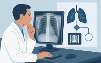
A 68-year-old male with a history of severe pneumonia five years ago presents to your clinic with a persistent, productive cough and recurrent episodes of purulent sputum over the past year. He reports no fevers or weight loss but is fatigued by the constant coughing. Physical exam reveals scattered crackles at the lung bases. You suspect bronchiectasis, a condition of permanent, abnormal bronchial dilation. The immediate clinical question is which imaging study to order first to confirm the diagnosis and guide management. According to the American College of Radiology (ACR), the initial study, `Radiography chest`, is rated Usually appropriate for this presentation, serving as a critical first step in the diagnostic workflow.
Who Fits This Clinical Scenario?
This guidance applies to adult patients for whom there is a new clinical suspicion of bronchiectasis, warranting initial diagnostic imaging. The typical presentation includes a chronic productive cough, copious sputum production (often mucopurulent), and a history of recurrent respiratory tract infections. Patients may also report dyspnea or hemoptysis. This scenario is specifically for the initial workup, where bronchiectasis is on the differential but has not been previously confirmed by imaging.
This workflow is distinct from several related clinical situations. It does not apply to:
- Patients with known bronchiectasis: If bronchiectasis is already diagnosed, and you are assessing for acute complications (like an exacerbation or severe hemoptysis) or monitoring treatment response, the imaging choice follows a different ACR variant. See the guidance for “Bronchiectasis. Assessment of complications or treatment response.”
- Patients with suspected central airway obstruction: If the primary concern is stridor, wheezing localized to a single area, or other signs pointing to a focal blockage, the workup for “Clinically suspected tracheal or bronchial stenosis” is more appropriate.
- Patients with dynamic airway collapse: If the patient’s symptoms are primarily exertional dyspnea and a “barking” cough suggestive of excessive airway collapsibility, the scenario for “Clinically suspected tracheomalacia or bronchomalacia” should be consulted.
What Diagnoses Are You Working Up in This Scenario?
When ordering initial imaging for suspected bronchiectasis, you are primarily investigating for irreversible bronchial dilation, but the findings can also point toward underlying etiologies or alternative diagnoses.
Bronchiectasis: This is the primary diagnosis of concern. It represents the final common pathway of various insults to the bronchial walls, leading to impaired mucociliary clearance and a vicious cycle of infection and inflammation. Imaging aims to identify characteristic signs like bronchial wall thickening (“tram tracking”), lack of bronchial tapering, and the presence of ring shadows or cystic changes. The distribution (focal vs. diffuse, upper vs. lower lobe) can suggest the underlying cause.
Underlying Etiologies: While imaging may not pinpoint the exact cause, it can provide crucial clues. For instance, upper-lobe predominant bronchiectasis might suggest cystic fibrosis or post-tuberculous changes. Central bronchiectasis is classic for allergic bronchopulmonary aspergillosis (ABPA). Diffuse disease could be related to an immunodeficiency or ciliary dyskinesia.
Chronic Obstructive Pulmonary Disease (COPD) / Chronic Bronchitis: These conditions share significant clinical overlap with bronchiectasis, particularly chronic cough and sputum production. A chest radiograph can show signs of hyperinflation or bronchial wall thickening common in COPD, but it is less sensitive for the definitive bronchial dilation of bronchiectasis.
Nontuberculous Mycobacterial (NTM) Infection: NTM is a common cause of bronchiectasis, particularly in older women (Lady Windermere syndrome). Imaging may reveal a classic combination of bronchiectasis and small nodules, often with a middle lobe and lingular predominance.
Interstitial Lung Disease (ILD): Some forms of ILD, particularly those associated with fibrosis, can lead to traction bronchiectasis, where scarring in the lung parenchyma pulls the airways open. The initial radiograph may show reticular opacities or honeycombing that points toward a primary fibrotic lung process.
Why Is a Two-Step Imaging Approach Recommended for This Presentation?
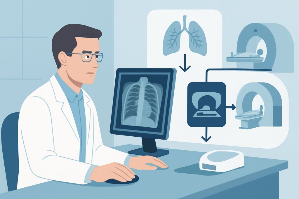
For the initial evaluation of suspected bronchiectasis, the ACR guidelines endorse a logical, stepwise approach, starting with a simple study and progressing to a more definitive one if needed. Both chest radiography and non-contrast chest CT are rated Usually appropriate, reflecting their distinct roles in the workup.
Step 1: Radiography chest
The chest radiograph is the recommended first-line imaging test. It is widely available, inexpensive, and delivers a very low radiation dose (☢ <0.1 mSv). While its sensitivity for mild bronchiectasis is limited, it can reveal classic findings in moderate to severe disease, such as “tram tracks” (parallel lines from thickened bronchial walls), ring shadows (dilated bronchi seen end-on), and evidence of mucus plugging or air-fluid levels. Critically, it also helps screen for alternative diagnoses that may mimic bronchiectasis, such as lung masses, significant parenchymal scarring, or overt pneumonia.
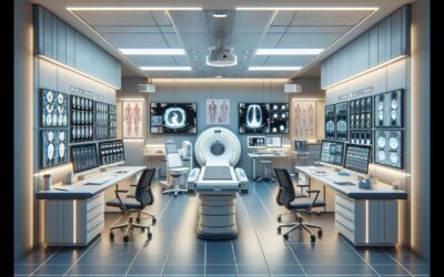
Step 2: CT chest without IV contrast
If the chest radiograph is negative or equivocal but clinical suspicion remains high, a non-contrast chest CT is the definitive next step. It is considered the gold standard for diagnosing bronchiectasis. A high-resolution CT (HRCT) protocol, which uses thin-slice images and a sharp reconstruction algorithm, is essential for optimal visualization of the airways. HRCT can precisely characterize the type (cylindrical, varicose, or cystic), extent, and distribution of bronchiectasis, which is vital for management and for investigating the underlying cause. The radiation dose is higher than a radiograph (☢☢☢ 1-10 mSv), which is why it is typically reserved as a second-line test or for cases with high pre-test probability.
Why Alternatives Are Rated Lower
- CT chest with IV contrast is rated Usually not appropriate for the initial diagnosis. Intravenous contrast does not improve the visualization of bronchial wall thickening or luminal dilation. It adds cost, the risk of allergic reaction, and the potential for contrast-induced nephropathy without providing additional diagnostic information for this specific question. Contrast is reserved for evaluating complications like hemoptysis, where a CTA may be needed to look for a bleeding source.
- MRI chest without IV contrast is rated May be appropriate. While it avoids ionizing radiation, its spatial resolution for the small airways is inferior to HRCT. It is also more susceptible to motion artifact from breathing and cardiac motion. MRI is generally considered a problem-solving tool in specific populations, such as young patients with cystic fibrosis requiring frequent follow-up, rather than a primary diagnostic test for new-onset suspicion in adults.
What’s Next After Initial Imaging? Downstream Workflow
The results of the initial imaging study will guide your next steps, which typically involve confirming the diagnosis, assessing severity, and searching for an underlying etiology.
If the chest radiograph is positive for bronchiectasis:
The next step is almost always a `CT chest without IV contrast` (using an HRCT protocol). The CT confirms the diagnosis and provides a detailed anatomical map of the disease, which is essential for long-term management. Following CT confirmation, the workup should focus on identifying the cause. This includes laboratory tests (e.g., quantitative immunoglobulins, sweat chloride or genetic testing for cystic fibrosis, sputum culture for bacteria and NTM) and consultation with a pulmonologist.
If the chest radiograph is negative or nonspecific:
A normal chest radiograph does not rule out bronchiectasis, especially in milder cases. If your clinical suspicion remains high based on the patient’s history of chronic productive cough and recurrent infections, you should proceed to a `CT chest without IV contrast`. If the CT is also negative, the diagnosis of bronchiectasis is unlikely, and you should pursue alternative causes for the patient’s chronic cough.
If imaging suggests a complication or alternative diagnosis:
If the initial study reveals an acute pneumonia, a lung mass, or significant interstitial changes, the clinical workflow pivots to address that finding. If the patient has known bronchiectasis and presents with an acute change (e.g., worsening symptoms, new consolidation on imaging), they now fit the “Bronchiectasis. Assessment of complications or treatment response” scenario, which may involve different imaging choices, sometimes including contrast-enhanced CT.
Pitfalls to Avoid (and When to Get Help)
Navigating the workup for suspected bronchiectasis requires careful interpretation of both clinical and imaging findings. Be mindful of these common pitfalls:
- Stopping the workup too early: Do not dismiss the diagnosis based on a normal chest radiograph if the patient’s symptoms are highly suggestive. Bronchiectasis is an HRCT diagnosis.
- Ordering the wrong CT protocol: For initial diagnosis, a non-contrast HRCT is required. Ordering a standard-thickness CT or a CT with IV contrast is suboptimal; the former may miss the findings, and the latter adds unnecessary risk and cost.
- Overlooking mimics: Remember that conditions like chronic bronchitis and asthma can cause bronchial wall thickening, but true bronchiectasis requires irreversible bronchial dilation (best seen as a broncho-arterial ratio >1).
If a patient presents with massive hemoptysis (e.g., >100 mL of blood in 24 hours), this is a medical emergency. The standard diagnostic workflow should be bypassed in favor of immediate stabilization and consultation with interventional radiology and thoracic surgery, often preceded by a `CTA chest with IV contrast` to identify the bleeding source.
Related ACR Topics and Tools
For a comprehensive overview of imaging for all related clinical presentations, please refer to the parent topic hub article. For additional tools to help with ordering decisions and patient communication, see the resources below.
- Parent Topic Hub: For breadth across all scenarios in Tracheobronchial Disease, see our parent guide: Tracheobronchial Disease: ACR Appropriateness Decoded.
- ACR Criteria Lookup: To explore imaging recommendations for adjacent or alternative clinical scenarios, visit the ACR Appropriateness Criteria Lookup tool.
- Imaging Protocols: For detailed technical parameters of the recommended studies, consult the Imaging Protocol Library.
- Radiation Dose: To discuss cumulative radiation exposure with your patients, use the Radiation Dose Calculator.
Frequently Asked Questions
Why start with a chest X-ray if a non-contrast CT is the gold standard for bronchiectasis?
Starting with a chest radiograph is a cost-effective, low-radiation strategy. It can confirm moderate-to-severe disease, suggest alternative diagnoses, and establish a baseline. A CT is reserved for cases where the radiograph is negative or equivocal but clinical suspicion remains high, avoiding unnecessary radiation and cost for all patients.
Is a CT with intravenous contrast ever appropriate for evaluating bronchiectasis?
Yes, but not for the initial diagnosis. A contrast-enhanced CT (specifically, a CTA) is indicated when evaluating complications, most notably moderate to severe hemoptysis, to identify hypertrophied bronchial arteries or other vascular abnormalities that may be the source of bleeding and potential targets for embolization.
What is the difference between a standard chest CT and a high-resolution CT (HRCT)?
An HRCT protocol uses much thinner slices (typically 1-1.5 mm) compared to a standard CT (3-5 mm) and employs a special high-spatial-frequency reconstruction algorithm. This technique provides exquisite detail of the lung parenchyma and airways, making it far superior for detecting the subtle bronchial wall thickening and dilation characteristic of bronchiectasis.
Can a normal high-resolution chest CT definitively rule out bronchiectasis?
A technically adequate HRCT is extremely sensitive for bronchiectasis. If a high-resolution study is interpreted as normal by an experienced radiologist, clinically significant bronchiectasis is highly unlikely, and other causes for the patient’s chronic respiratory symptoms should be prioritized.
Does this imaging workflow change if the patient has a known diagnosis like cystic fibrosis (CF)?
The fundamental principles are the same, but the threshold to proceed to CT may be lower. In patients with known CF, bronchiectasis is an expected finding. Imaging is often used to assess for changes from baseline or to evaluate for acute exacerbations. While a chest radiograph may still be used for routine monitoring, clinicians often proceed directly to non-contrast HRCT for a more detailed assessment when symptoms change.
Reviewed by Pouyan Golshani, MD, Interventional Radiologist — May 30, 2026