What Is the Best Initial Imaging for Suspected Chronic Superior Vena Cava Syndrome?
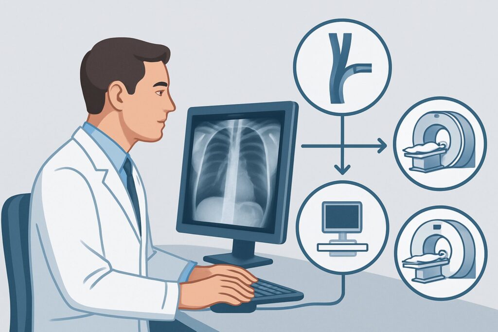
A 64-year-old man with a history of a remote mediastinal malignancy treated with radiation presents to your clinic with several months of progressive facial puffiness, a sensation of head fullness when bending over, and newly prominent veins across his chest. His symptoms are bothersome but have not acutely worsened. You suspect a chronic superior vena cava (SVC) or brachiocephalic vein occlusion, a slow-burning process that requires a detailed anatomical map for diagnosis and management. This article details the clinical workflow for choosing the right initial imaging study in this specific scenario, guided by the American College of Radiology (ACR) Appropriateness Criteria, which rate MRA and MRV chest without and with IV contrast as Usually Appropriate.
Who Fits This Clinical Scenario?
This guidance applies to adult patients with a subacute or chronic presentation (developing over weeks to months) suggestive of central thoracic venous obstruction. The classic patient has a constellation of symptoms known as Superior Vena Cava Syndrome, but in a low-grade, compensated form.
Inclusion criteria for this workflow:
- Insidious Onset: Symptoms like facial or arm edema, plethora (a ruddy complexion), dyspnea, and the development of visible chest wall venous collaterals have appeared gradually.
- Stable Condition: The patient is not in acute respiratory distress.
- Underlying Risk Factors: There is often a known risk factor, such as a history of thoracic malignancy, prior chest radiation, or the presence of an indwelling central venous catheter, pacemaker, or implantable cardioverter-defibrillator (ICD).
Exclusion criteria (route to a different workflow):
- Acute Presentation: Patients with rapidly progressing symptoms over hours to days, especially with stridor, severe dyspnea, or altered mental status, fit the acute SVC occlusion scenario. This is a potential emergency requiring a more rapid diagnostic and therapeutic pathway, often favoring CT.
- Isolated Unilateral Arm Swelling: If symptoms are confined to one arm without facial, neck, or chest wall signs, the primary suspicion shifts to a more peripheral process like subclavian or axillary vein thrombosis, which has a different diagnostic algorithm often starting with ultrasound.
What Diagnoses Are You Working Up in This Scenario?
The choice of imaging is driven by the need to differentiate between several potential causes of chronic central venous obstruction. The study must not only confirm the occlusion but also, if possible, identify the underlying etiology to guide subsequent management.
Malignancy (Extrinsic Compression or Invasion) This is the most common cause of SVC syndrome overall. A slow-growing tumor, such as non-small cell lung cancer, lymphoma, or metastatic disease (e.g., from breast or testicular cancer), can gradually compress or invade the SVC or brachiocephalic veins. The chronic nature of the presentation suggests the body has had time to develop collateral pathways.
Thrombosis (Intrinsic Occlusion) An organized, chronic thrombus is a frequent culprit, particularly in patients with indwelling central venous devices. The foreign body (catheter or lead) acts as a nidus for thrombus formation, which can slowly propagate and occlude the vessel. Over time, this bland thrombus becomes fibrotic and incorporated into the vessel wall.
Fibrosing Mediastinitis A less common but important consideration is fibrosing mediastinitis, an excessive fibrotic reaction in the mediastinum that encases and constricts the central veins. It can be idiopathic or a delayed reaction to infections like histoplasmosis or tuberculosis. Its infiltrative, non-mass-like nature can be challenging to diagnose without high-quality cross-sectional imaging.
Post-Radiation Fibrosis In patients with a history of radiation therapy to the chest, a late complication is radiation-induced fibrosis. This can cause direct stenosis of the great vessels, leading to a clinical picture identical to other forms of chronic SVC obstruction.
Why MRA and MRV chest without and with IV contrast Is the Recommended Study for This Presentation
For the non-emergent, chronic presentation, the ACR panel designates several imaging modalities as Usually Appropriate, including both CT and MR-based techniques. However, MRA and MRV chest without and with IV contrast offers distinct advantages that make it a premier choice for initial evaluation.
The primary strength of Magnetic Resonance Angiography (MRA) and Venography (MRV) is its superior soft-tissue contrast. This allows for detailed characterization of the occlusive process and the surrounding mediastinal anatomy without the use of ionizing radiation (Adult RRL: O 0 mSv). The study can often distinguish between bland thrombus, tumor invasion, and external compression from a mass or fibrotic tissue—information that is critical for determining the next step in management. Furthermore, MRV excels at mapping the intricate network of collateral veins (such as the azygos, hemiazygos, and internal mammary systems), which is vital for planning potential endovascular interventions.
Why are other studies rated differently for this specific scenario?
- CT Venography (CTV) chest with IV contrast: This is also rated Usually Appropriate and is an excellent alternative. It is typically faster and more widely available than MRI. CTV provides superb spatial resolution for delineating vessel anatomy and collateral pathways. However, it involves a significant dose of ionizing radiation (Adult RRL: ☢☢☢☢ 10-30 mSv) and may provide less definitive characterization of the soft tissue cause of the obstruction compared to MRI.
- US duplex Doppler upper extremity and chest: Rated May be appropriate (Disagreement), ultrasound is not a suitable initial study for this central problem. While it can effectively evaluate the peripheral subclavian and axillary veins, its acoustic window is limited by the sternum and clavicles, preventing reliable visualization of the brachiocephalic veins and the SVC itself. It cannot provide the comprehensive anatomical map needed.
- Catheter venography: Also rated May be appropriate (Disagreement) for initial diagnosis, this invasive procedure is now largely reserved for therapeutic intervention (e.g., stenting) after non-invasive imaging has defined the anatomy. Using it for primary diagnosis exposes the patient to the risks of an invasive procedure and radiation.
Ordering **MRA and MRV chest without and with IV contrast** is crucial. The pre-contrast sequences help identify vessel wall hematoma or calcification, while the dynamic post-contrast imaging provides the essential vascular map.
What’s Next After MRA and MRV chest without and with IV contrast? Downstream Workflow
The imaging results directly inform the subsequent clinical pathway, which is typically multidisciplinary.
- If the study is positive for malignant obstruction: The next step is to obtain a tissue diagnosis and complete staging. This may involve a CT-guided biopsy of a mediastinal mass, bronchoscopy, or mediastinoscopy. The patient should be referred to medical and radiation oncology to plan primary treatment of the cancer, which is the definitive therapy for the SVC obstruction. Interventional radiology may be consulted for palliative stenting if symptoms are severe.
- If the study is positive for benign stenosis or thrombosis: The patient should be referred to interventional radiology or vascular surgery. The primary treatment is often endovascular, involving balloon angioplasty and stenting to re-establish patency. Management of anticoagulation is also a key component of the treatment plan.
- If the study is negative for central venous occlusion: If a high clinical suspicion persists despite a normal MRA/MRV, the differential diagnosis must be broadened. Consider other causes of elevated central venous pressure, such as right-sided heart failure, constrictive pericarditis, or tricuspid regurgitation. An echocardiogram would be a logical next diagnostic step.
- If the study is indeterminate: In rare cases where MRI cannot definitively characterize the nature of the obstruction, a CTV may provide complementary information. In very complex cases, direct catheter venography may be required to clarify pressure gradients and anatomy before an intervention.
Pitfalls to Avoid (and When to Get Help)
- Mistaking Chronic for Acute: Do not apply this deliberate, MR-focused workup to a patient with acute stridor or severe respiratory distress. That is a medical emergency requiring a faster protocol, often centered on CT.
- Ordering a Non-Contrast Study: A non-contrast MRI or CT of the chest is rated Usually not appropriate for this indication. Intravenous contrast is essential to opacify the vessels and make a diagnosis.
- Forgetting Renal Function: Gadolinium-based contrast agents used for MRA/MRV require adequate renal function. Always check the patient’s eGFR before ordering the study.
- Ignoring Device Contraindications: Ensure the patient has no non-MRI-conditional pacemakers, defibrillators, or other metallic implants that would preclude an MRI.
If the patient’s symptoms are rapidly worsening or if imaging confirms a high-grade obstruction with minimal collateralization, escalate care immediately with consultations to interventional radiology and the relevant specialty (e.g., oncology) for urgent intervention.
Related ACR Topics and Tools
This article covers one specific scenario in depth. For a broader view of all clinical variants and imaging options for this condition, please consult the parent topic guide. For additional tools to help with imaging decisions, see the resources below.
- For breadth across all scenarios in Thoracic Venous Occlusions-Suspected Superior Vena Cava Syndrome, see our parent guide: Thoracic Venous Occlusions-Suspected Superior Vena Cava Syndrome: ACR Appropriateness Decoded.
- ACR Appropriateness Criteria Lookup — for adjacent scenarios
- Imaging Protocol Library — for technique on the recommended study
- Radiation Dose Calculator — for cumulative dose conversations
Frequently Asked Questions
Why is MRA/MRV preferred over CTV if both are ‘Usually Appropriate’ for chronic SVC occlusion?
While both are excellent, MRA/MRV is often preferred in the chronic, non-emergent setting because it avoids ionizing radiation and its superior soft-tissue contrast can better differentiate the cause of the occlusion (e.g., bland thrombus vs. tumor invasion). This can be crucial for planning the next steps in management.
What if my patient has an MRI-incompatible pacemaker?
If a patient has a contraindication to MRI, such as a non-conditional pacemaker, then CT Venography (CTV) of the chest with IV contrast is the best alternative. It is also rated ‘Usually Appropriate’ by the ACR and provides excellent anatomical detail of the central veins and collateral pathways.
Is a chest radiograph useful as a first step for suspected chronic SVC syndrome?
A chest radiograph is rated ‘May be appropriate (Disagreement)’. While it might show a widened mediastinum suggesting a mass, it cannot directly visualize the venous anatomy or confirm an occlusion. It is not sensitive or specific enough to serve as the primary diagnostic test and will almost always need to be followed by CT or MRI, so it is often better to proceed directly to cross-sectional imaging if clinical suspicion is high.
Does the ‘MRA’ component add value if I’m only suspecting a venous problem?
Yes. Including the MRA sequences is valuable because large mediastinal masses causing venous compression can also compress or involve adjacent arteries, such as the aorta or pulmonary arteries. Assessing the arterial system simultaneously provides a more complete picture of the mediastinal anatomy and can be important for surgical or radiation planning.
What is the difference in workup for a chronic versus an acute presentation of SVC syndrome?
The primary difference is urgency and the choice of modality. An acute presentation is a potential emergency, and the diagnostic goal is rapid confirmation and localization. CT is often favored in the acute setting due to its speed and availability. In the chronic setting, the workup is less urgent, allowing for a more detailed study like MRI to better characterize the underlying cause and plan definitive treatment.
Reviewed by Pouyan Golshani, MD, Interventional Radiologist — August 4, 2026