What Is the Best Initial Imaging for Suspected DCIS Recurrence After Lumpectomy?
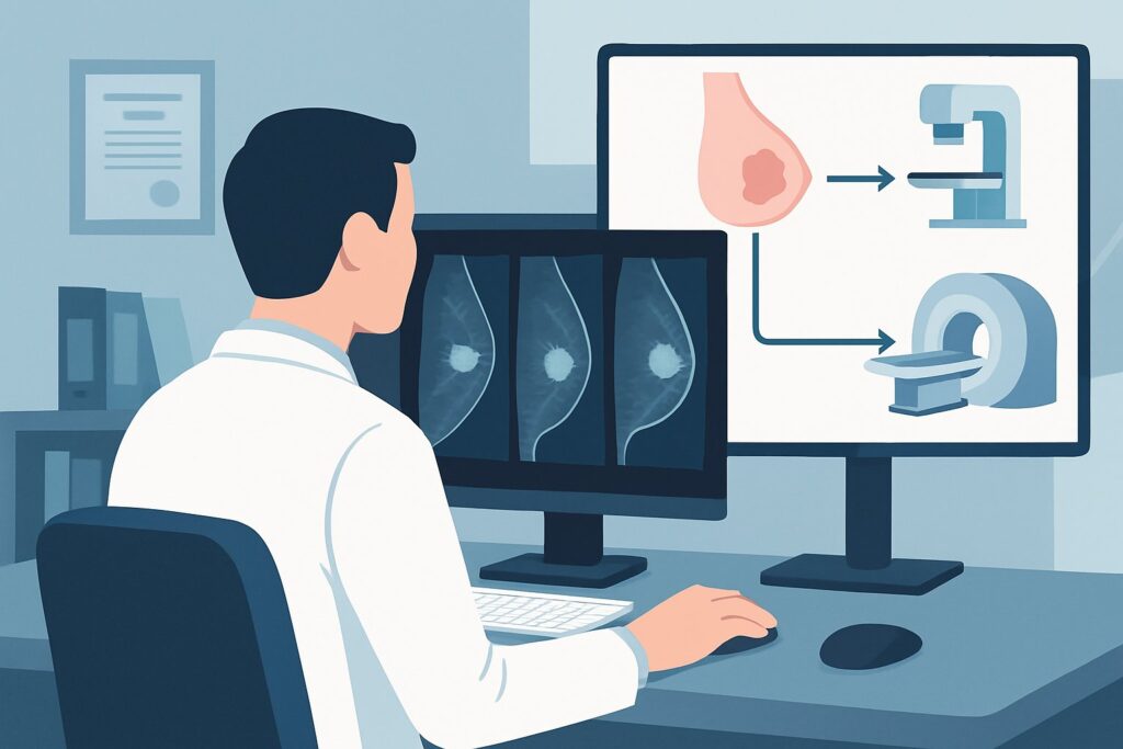
A 58-year-old woman, five years status post-lumpectomy and radiation for Ductal Carcinoma in Situ (DCIS), presents to your clinic for evaluation of a new, firm area she felt near her surgical scar. On examination, you confirm a 1-cm, non-tender, firm nodularity. The clinical question is immediate: what is the most appropriate initial imaging study to order to evaluate for a potential local recurrence? This scenario requires a targeted approach to differentiate between benign post-treatment changes and a new malignancy. According to the American College of Radiology (ACR) Appropriateness Criteria, both US breast and diagnostic mammography (including digital breast tomosynthesis) are rated Usually Appropriate as the initial imaging steps.
Who Fits This Clinical Scenario for Suspected DCIS Recurrence?
This guidance is specifically for an adult patient with a personal history of DCIS treated with breast conservation therapy (BCT), which typically includes lumpectomy followed by radiation. The key trigger for this workflow is the new onset of symptoms, a concerning finding on physical examination, or an abnormal laboratory value suggesting a potential local recurrence.
This clinical workflow applies if your patient:
- Has a history of DCIS treated with BCT (lumpectomy).
- Is now presenting with a new, focal symptom (e.g., a palpable lump, skin thickening, nipple discharge, focal pain).
- Has a new, concerning finding on clinical breast examination.
It is crucial to distinguish this situation from similar, but distinct, clinical scenarios that require different imaging pathways. This guidance does not apply to:
- Asymptomatic patients undergoing routine surveillance: A patient with a history of BCT for DCIS who has no new symptoms or findings would follow a routine surveillance protocol, which is a separate ACR variant.
- Patients with a history of mastectomy: The evaluation of the chest wall after mastectomy involves different imaging considerations and is covered under a different ACR scenario.
- Patients with a newly diagnosed, untreated DCIS: The initial workup for a new diagnosis focuses on defining the extent of disease, not evaluating for recurrence.
What Diagnoses Are You Working Up in This Scenario?
When a patient presents with a new finding in a previously treated breast, the differential diagnosis is focused but critical. The primary goal of imaging is to distinguish between benign post-treatment changes and a true recurrence of cancer.
Local Recurrence (Invasive or DCIS)
This is the most consequential diagnosis to exclude. A local recurrence after BCT for DCIS can manifest as either recurrent DCIS or, more commonly, as an invasive ductal carcinoma. Recurrences can present as palpable masses, new suspicious microcalcifications, or architectural distortion on imaging. Prompt and accurate diagnosis is essential for timely management.
Fat Necrosis and Post-Surgical Scarring
These are the most common benign mimickers of recurrence. Following surgery and radiation, the breast tissue undergoes significant healing and remodeling. Fat necrosis can form a firm, palpable mass that feels suspicious on physical exam. Similarly, scar tissue can mature and contract, creating a palpable nodularity. On imaging, these changes can sometimes appear complex, requiring careful correlation.
Benign Lesions
The treated breast is still functional breast tissue, and new benign lesions can develop independently of the prior cancer. This includes simple or complex cysts, which are common, as wellas fibroadenomas or other benign solid masses. Ultrasound is particularly effective at characterizing these types of lesions.
Post-Radiation Changes
Radiation therapy can cause long-term changes in the breast parenchyma and skin, including edema and fibrosis. While often diffuse, these changes can sometimes present focally and become symptomatic, warranting an imaging evaluation to rule out an underlying mass.
Why Are Ultrasound and Diagnostic Mammography the Recommended First Steps?
For a patient with a new palpable finding in a treated breast, the ACR designates US breast, Mammography diagnostic, and Digital breast tomosynthesis (DBT) diagnostic as Usually Appropriate. This multi-modal approach provides the most comprehensive initial evaluation.
Ultrasound is an indispensable tool for evaluating a palpable abnormality. It excels at characterizing masses as cystic or solid, defining margins, and assessing vascularity. In the post-treatment breast, where architectural distortion from scarring and radiation can limit mammographic sensitivity, ultrasound provides a direct, real-time assessment of the palpable area of concern. Furthermore, it involves no ionizing radiation (0 mSv) and is the ideal modality for guiding a potential biopsy.
Diagnostic mammography, preferably with DBT, is the essential counterpart to ultrasound. While ultrasound targets the palpable finding, mammography evaluates the entire breast for other signs of recurrence, such as suspicious microcalcifications—the classic sign of DCIS—which may not be visible on ultrasound. DBT, or 3D mammography, is particularly valuable in this setting. It reduces the effect of overlapping tissue and post-surgical scarring, improving the detection of subtle architectural distortion or new densities that could represent recurrence. The radiation dose for diagnostic mammography is low (ACR RRL®: ☢☢ 0.1-1 mSv).
Why are other advanced imaging modalities not recommended for initial workup?
- MRI breast without and with IV contrast: This is rated Usually Not Appropriate as a first-line test for a focal symptom. While breast MRI is highly sensitive, its specificity is lower in the post-treatment setting. Normal post-surgical and post-radiation changes, such as inflammation and scar tissue, can demonstrate enhancement, leading to a higher rate of false-positive findings. This can trigger unnecessary biopsies and patient anxiety. MRI is typically reserved as a problem-solving tool if mammography and ultrasound are inconclusive.
- Image-guided core biopsy: This is also rated Usually Not Appropriate as an initial step. A biopsy is a diagnostic procedure, not an initial imaging modality. It is the logical next step after a suspicious lesion has been identified and characterized by mammography or ultrasound. Ordering a biopsy without prior characterization is a workflow error.
What’s Next After Initial Breast Imaging? Downstream Workflow
The results of the diagnostic mammogram and targeted ultrasound, reported using the BI-RADS classification system, will dictate the next steps in the patient’s care.
- If the findings are suspicious (BI-RADS 4 or 5): A suspicious mass, cluster of microcalcifications, or area of architectural distortion requires tissue sampling for a definitive diagnosis. The next step is an image-guided core needle biopsy. The modality used for guidance (ultrasound, stereotactic/mammographic, or MRI) depends on which imaging test best visualizes the target lesion.
- If the findings are negative or benign (BI-RADS 1 or 2): When imaging identifies a clear benign cause for the palpable finding (e.g., a simple cyst, classic fat necrosis) and shows no other suspicious features, the patient can typically return to their standard annual surveillance schedule. Close clinical follow-up in 3-6 months may be considered to ensure stability of the palpable finding.
- If the findings are probably benign (BI-RADS 3): This category is used for findings that have a very low likelihood of malignancy (<2%). The standard recommendation is short-interval imaging follow-up, typically in six months, to ensure stability. Biopsy may be considered if the patient has a particularly high-risk history or expresses significant anxiety.
Pitfalls to Avoid (and When to Get Help)
Navigating the workup of a suspected breast cancer recurrence requires careful attention to detail to avoid common errors.
- Pitfall 1: Relying on a single modality. Do not order only an ultrasound for a palpable lump without a concurrent diagnostic mammogram. Mammography is essential for detecting non-palpable calcifications or other findings elsewhere in the breast.
- Pitfall 2: Ordering a screening mammogram. For a symptomatic patient, a diagnostic mammogram is required. This allows the radiologist to perform additional views and a targeted ultrasound during the same visit, providing a more immediate and comprehensive evaluation.
- Pitfall 3: Prematurely ordering an MRI. Jumping to MRI as the first test for a palpable lump often leads to indeterminate findings and unnecessary biopsies due to its high sensitivity but lower specificity in the post-treatment setting.
- Pitfall 4: Not correlating imaging with the physical exam. Ensure the radiologist is aware of the exact location of the palpable concern. The imaging report should explicitly state whether a sonographic finding corresponds to the palpable lump.
If imaging results are discordant with a highly suspicious clinical exam (e.g., a persistently growing, hard, fixed mass with negative imaging), escalation to a breast surgeon for consideration of an excisional biopsy is warranted.
Related ACR Topics and Tools
For a comprehensive overview of all clinical variants related to this topic, please consult the parent article. Additional GigHz tools can help you apply these criteria in your practice.
- For breadth across all scenarios in Imaging of Ductal Carcinoma in Situ (DCIS), see our parent guide: Imaging of Ductal Carcinoma in Situ (DCIS): ACR Appropriateness Decoded.
- ACR Appropriateness Criteria Lookup — for adjacent scenarios
- Imaging Protocol Library — for technique on the recommended study
- Radiation Dose Calculator — for cumulative dose conversations
Frequently Asked Questions
My patient has a new lump but her recent annual screening mammogram was normal. Should I still order imaging?
Yes. A screening mammogram is designed for asymptomatic patients. A new, palpable lump is a clinical change that requires a diagnostic workup, which includes a diagnostic mammogram (with special views of the area of concern) and a targeted ultrasound, even if a recent screening study was negative.
Is a breast MRI ever appropriate in this scenario?
Breast MRI is rated ‘Usually Not Appropriate’ for the *initial* evaluation of a new symptom after BCT for DCIS. However, it can be a valuable problem-solving tool if the findings on mammography and ultrasound are inconclusive or discordant with the clinical exam. It may also be used for extent-of-disease evaluation if a recurrence is confirmed by biopsy.
How do you differentiate scar tissue from recurrence on imaging?
This is a key challenge for the radiologist. Generally, post-surgical scars are stable or evolve predictably over time, often appearing as architectural distortion that retracts. A recurrence may present as a new, developing, or enlarging mass, or new suspicious microcalcifications. Comparison with all prior imaging studies is critical to assess for change, which is the most important indicator of a potential recurrence.
If the patient had a mastectomy instead of a lumpectomy, would the imaging workup be the same?
No. The workup would be different. A patient with a history of mastectomy who presents with a palpable lump on the chest wall would be evaluated under a different ACR Appropriateness Criteria variant. The primary imaging tool in that case is often ultrasound of the chest wall, as there is no remaining breast tissue to evaluate with mammography.
What if the ultrasound is negative but I can still feel a definite lump?
This is known as a ‘clinically occult’ but ‘palpably evident’ finding. If a skilled radiologist performs a targeted ultrasound and confirms no corresponding sonographic abnormality, the next step is careful clinical follow-up. If the lump persists or grows, a referral to a breast surgeon for consultation and potential excisional biopsy may be necessary, as a small percentage of cancers can be missed by both mammography and ultrasound.
Reviewed by Pouyan Golshani, MD, Interventional Radiologist — May 30, 2026