What Imaging Is Next for a Palpable Breast Mass with a Negative Mammogram?
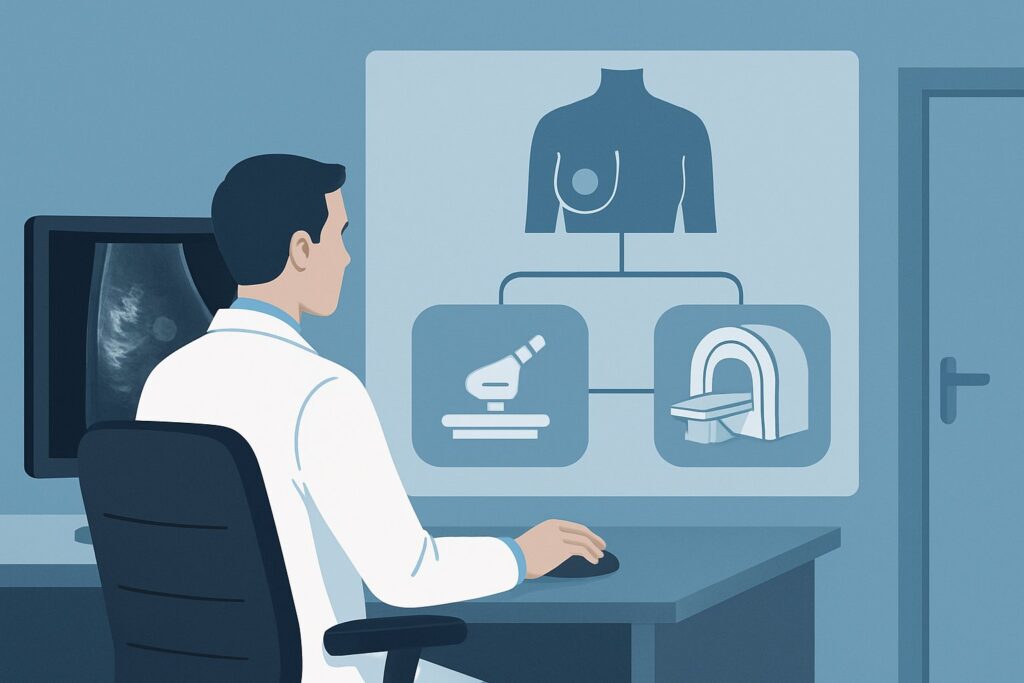
A 45-year-old female presents for follow-up. You saw her last week for a new, firm, non-tender lump she found in her left breast. You ordered a diagnostic mammogram, and the report is now in your inbox: BI-RADS 1 (Negative). The patient, however, is still concerned—she can still feel the lump, and so can you. This common clinical crossroads presents a critical question: when the initial, high-quality mammogram shows nothing, what is the appropriate next step to ensure a potential malignancy is not missed? This article provides a detailed workflow for this specific scenario, guiding you through the diagnostic rationale, downstream decisions, and potential pitfalls. Based on the American College of Radiology (ACR) Appropriateness Criteria, the next step, breast ultrasound, is rated Usually Appropriate.
Who Fits This Clinical Scenario?
This guidance is specifically for the workup of a palpable breast mass in an adult female, 40 years of age or older, whose diagnostic mammogram was interpreted as negative (BI-RADS 1). A BI-RADS 1 category signifies that there are no suspicious masses, calcifications, or areas of architectural distortion visible on the mammogram. The breast tissue is considered normal.
It is crucial to distinguish this situation from several similar, but distinct, clinical scenarios that require different management pathways:
- Women younger than 30: For these patients, ultrasound is typically the initial imaging modality of choice, not mammography.
- Initial imaging for a palpable mass: If this were the patient’s first presentation before any imaging was ordered, the initial recommendation would be diagnostic mammography (often with tomosynthesis) and a targeted ultrasound performed concurrently. This article addresses the sequential step after a negative mammogram has already been performed.
- Benign mammogram findings (BI-RADS 2): If the mammogram identified a clearly benign finding (e.g., a simple cyst, oil cyst, or stable fibroadenoma) that corresponds to the palpable lump, the workup is often complete. This scenario is for a completely negative study.
- Suspicious mammogram findings (BI-RADS 4 or 5): If the mammogram had revealed a suspicious finding, the next step would be image-guided biopsy, not another imaging modality.
This article focuses exclusively on the patient whose palpable physical exam finding is discordant with a negative mammographic evaluation.
What Diagnoses Are You Working Up in This Scenario?
When a palpable mass is not visible on a mammogram (termed “mammographically occult”), the differential diagnosis remains broad, but the primary goal is to exclude malignancy. The persistence of a palpable finding in the face of negative imaging necessitates further investigation.
Occult Breast Carcinoma: This is the most consequential diagnosis to exclude. Certain cancers, particularly invasive lobular carcinoma, can grow in an infiltrative pattern without forming a discrete, dense mass, making them difficult to detect on mammography. Furthermore, in women with dense breast tissue, a true mass can be obscured by the surrounding fibroglandular tissue, resulting in a false-negative mammogram.
Cyst: A simple or complex fluid-filled cyst is a very common cause of a palpable lump. Cysts are often not visible on mammography unless they have calcified rims or are large enough to displace surrounding tissue. They are, however, easily characterized by ultrasound.
Fibroadenoma or other Benign Solid Mass: A fibroadenoma is a common benign tumor that can be “iso-dense,” meaning it has the same radiographic density as the surrounding breast tissue. This can make it effectively invisible on a mammogram, yet it remains palpable and is readily identifiable on ultrasound.
Focal Fibrosis or Adenosis: These are benign, non-cancerous changes in the breast tissue that can create a palpable area of firmness or a lump. Like other benign entities, they may not have a distinct appearance on mammography but can be evaluated with ultrasound to confirm the absence of suspicious features.
Why Is Breast Ultrasound the Recommended Next Step After a Negative Mammogram?
For a woman aged 40 or older with a palpable mass and a negative mammogram, the ACR designates US breast as Usually Appropriate. This recommendation is rooted in the complementary strengths of ultrasound and mammography.
The primary rationale is that ultrasound excels where mammography can be limited. Its superior ability to evaluate dense breast tissue and to differentiate cystic from solid structures makes it the ideal problem-solving tool in this scenario. A targeted ultrasound, where the sonographer places the transducer directly over the palpable area of concern (often marked on the skin), provides a real-time, high-resolution assessment of the underlying tissue. This direct physical-to-imaging correlation is critical for determining if the palpable finding corresponds to a true lesion.
In contrast, other advanced imaging modalities are considered less appropriate at this stage:
- MRI breast without and with IV contrast is rated Usually not appropriate. While breast MRI has very high sensitivity for detecting invasive cancer, its specificity is lower. Using it at this early stage could lead to a higher rate of false positives, prompting unnecessary biopsies and patient anxiety. MRI is typically reserved for situations where both mammography and ultrasound are negative but clinical suspicion remains exceptionally high, or for high-risk screening.
- Image-guided core biopsy is also rated Usually not appropriate as the next step. A biopsy is a tissue sampling procedure, not an imaging study. It is only performed once an imaging target has been identified. Ordering a biopsy without an ultrasound or MRI target is illogical, as there is no lesion to guide the needle toward.
From a safety perspective, ultrasound involves no ionizing radiation (Relative Radiation Level: 0 mSv) and does not require intravenous contrast, making it a safe and repeatable examination. When ordering, it is essential to be specific: “Targeted ultrasound of the palpable left breast mass, approximately 2 o’clock, 4 cm from the nipple,” to ensure the radiologist focuses on the precise area of clinical concern.
What’s Next After Breast Ultrasound? Downstream Workflow
The results of the targeted breast ultrasound will dictate the subsequent steps in the patient’s management. The goal is to achieve concordance between the clinical exam and the imaging findings.
If the ultrasound is negative (BI-RADS 1): If a careful, targeted ultrasound of the palpable area reveals no abnormality, the lump likely represents normal, prominent fibroglandular tissue. In most cases, the patient can be reassured, and a plan for routine clinical follow-up (e.g., in 6 months) is appropriate. The workup is concordantly negative.
If the ultrasound identifies a simple cyst (BI-RADS 2): This is a definitive benign finding. The palpable lump is explained, and no further imaging workup is necessary. If the cyst is large or tender, ultrasound-guided aspiration can be performed for symptomatic relief.
If the ultrasound shows a probably benign solid mass (BI-RADS 3): This typically corresponds to a finding like a fibroadenoma. The standard recommendation is short-interval imaging follow-up, usually with a repeat targeted ultrasound in 6 months, to ensure stability. Biopsy is sometimes offered as an alternative to follow-up, depending on patient preference or specific lesion features.
If the ultrasound reveals a suspicious mass (BI-RADS 4 or 5): If the ultrasound identifies a solid mass with irregular margins, posterior acoustic shadowing, or other suspicious features, the palpable finding is now correlated with a targetable lesion. The next step is an ultrasound-guided core needle biopsy to obtain a pathologic diagnosis.
Pitfalls to Avoid (and When to Get Help)
Navigating this scenario requires careful clinical judgment to avoid common missteps. Here are key pitfalls to be aware of:
- False Reassurance from a Negative Mammogram: The most significant error is stopping the workup after a negative mammogram. A persistent palpable mass always warrants further investigation, typically with ultrasound. Remember the mantra: “We image what we can feel.”
- Non-Targeted Ultrasound: Ordering a “complete bilateral breast ultrasound” instead of a “targeted ultrasound of the palpable mass” can be a pitfall. The sonographer may not focus on the specific area of concern, potentially missing a subtle lesion. Clear communication on the order is vital.
- Ignoring Clinical-Radiologic Discordance: If both the mammogram and targeted ultrasound are negative, but the palpable mass is highly suspicious on clinical exam (e.g., hard, fixed, new, or growing), do not stop. This discordance is a red flag.
When to Escalate: If a palpable mass persists and is clinically suspicious despite negative findings on both mammography and targeted ultrasound, referral to a breast surgeon is the appropriate next step. The surgeon may recommend close clinical follow-up, a breast MRI, or even an excisional biopsy.
Related ACR Topics and Tools
For a comprehensive overview of all clinical variants related to palpable breast masses, from initial workup to post-biopsy management, please see our parent topic hub article. It provides a breadth of information that complements this in-depth guide.
- For breadth across all scenarios in Palpable Breast Masses, see our parent guide: Palpable Breast Masses: ACR Appropriateness Decoded.
- To look up other clinical scenarios, use the Imaging Appropriateness Selector tool.
- To review standard imaging techniques, consult the Imaging Protocol Library.
- For discussions about cumulative exposure with patients, the Radiation Dose Calculator can be a helpful resource.
Frequently Asked Questions
What does a BI-RADS 1 (Negative) result mean on a mammogram report?
BI-RADS 1, or ‘Negative,’ means the radiologist found no evidence of cancer or other significant abnormalities. There are no suspicious masses, calcifications, or areas of architectural distortion. It is a normal mammogram. However, it does not rule out the possibility of a mass that is not visible on mammography, which is why a palpable finding still requires further evaluation.
Why not just go straight to biopsy if I can feel a lump?
A biopsy is a procedure to sample tissue from a specific target. If a mammogram is negative, there is no visible target to guide a needle. Performing a ‘blind’ biopsy of a palpable area is less accurate and not the standard of care. Ultrasound is used first to identify a specific lesion that corresponds to the palpable lump, which can then be accurately targeted for biopsy if it appears suspicious.
Does the recommendation for ultrasound change if the patient has very dense breasts?
No, the recommendation for a targeted ultrasound is even stronger in women with dense breasts. Breast density can lower the sensitivity of mammography, making it more likely to obscure an underlying mass. Ultrasound is not limited by breast density and is an excellent tool for evaluating palpable findings in this context.
Is a 3D mammogram (tomosynthesis) enough, or is ultrasound still needed if it’s negative?
Even if the negative mammogram was performed with 3D tomosynthesis, which improves cancer detection in dense breasts, a targeted ultrasound is still the recommended next step for a palpable mass. Tomosynthesis is still an X-ray-based technology and may not visualize a mass that is iso-dense with surrounding tissue. The ACR guidelines support using ultrasound to evaluate the palpable finding regardless of the type of mammogram performed.
If the targeted ultrasound is also negative, what is the appropriate clinical follow-up interval?
When both a high-quality diagnostic mammogram and a targeted ultrasound are negative, the finding is highly likely to be benign. The standard recommendation is clinical follow-up. A common interval is 3 to 6 months to ensure the palpable finding is stable, resolving, or not changing in character. If the lump grows or becomes more suspicious on exam, the patient should return sooner for re-evaluation and potential referral to a breast surgeon.
Reviewed by Pouyan Golshani, MD, Interventional Radiologist — June 27, 2026