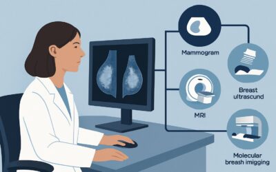
What Is the Right Supplemental Breast Screening for Intermediate-Risk, Nondense Breasts?

A 48-year-old woman with a family history of postmenopausal breast cancer in her mother is here for her annual wellness visit. Her lifetime risk, calculated using the Tyrer-Cuzick model, is 18%, placing her in the intermediate-risk category. Her prior mammograms have consistently shown nondense, scattered fibroglandular tissue. You are considering the most appropriate imaging strategy: is her standard annual mammogram sufficient, or is supplemental screening indicated given her risk profile? This article provides a detailed clinical workflow for this specific scenario, guiding the decision on supplemental breast cancer screening for an intermediate-risk woman with nondense breasts. According to the American College of Radiology (ACR) Appropriateness Criteria, the standard of care, Digital breast tomosynthesis screening, is rated Usually Appropriate.
Who Fits This Clinical Scenario for Supplemental Breast Screening?
This guidance applies to a specific patient population: asymptomatic adult women undergoing screening for breast cancer who are at an intermediate risk and have nondense breasts. Understanding each component is critical to applying this workflow correctly.
Inclusion Criteria:
- Intermediate Risk: This category is defined as a calculated lifetime risk of 15% to 20%. Common factors placing a woman in this group include a personal history of lobular carcinoma in situ (LCIS), atypical ductal hyperplasia (ADH), or atypical lobular hyperplasia (ALH). A significant family history that does not meet the criteria for high-risk status can also qualify.
- Nondense Breasts: This corresponds to Breast Imaging Reporting and Data System (BI-RADS) density categories A (the breasts are almost entirely fatty) or B (there are scattered areas of fibroglandular density). This determination is made by the radiologist on prior mammograms.
- Asymptomatic: The patient has no clinical signs or symptoms of breast cancer, such as a palpable lump, nipple discharge, or skin changes. This workflow is for screening, not diagnostic workup.
Exclusion Criteria:
This workflow should not be applied to patients who fall into different clinical scenarios. For example, a woman with a lifetime risk greater than 20%, a known pathogenic mutation (e.g., BRCA1/2), or a history of chest radiation before age 30 is considered high-risk and requires a different screening protocol, typically including annual MRI. Similarly, a patient with heterogeneously or extremely dense breasts (BI-RADS C or D) presents a different challenge where mammographic sensitivity is reduced, altering the recommendations for supplemental screening.
What Diagnoses Are You Working Up in This Scenario?
In a screening context, the “differential diagnosis” refers to the spectrum of potential occult malignancies we aim to detect at their earliest, most treatable stage. For an intermediate-risk woman, the goal is to find cancers that may arise due to her elevated risk profile while minimizing false positives in her mammographically favorable nondense breast tissue.
Invasive Ductal Carcinoma (IDC): As the most common type of invasive breast cancer, IDC is a primary target of any screening program. In nondense breasts, it often presents as a spiculated mass or suspicious calcifications, which are typically well-visualized on mammography. Digital breast tomosynthesis enhances the detection of these lesions by resolving overlapping tissue.
Ductal Carcinoma In Situ (DCIS): This is the most common form of non-invasive breast cancer. The classic presentation of DCIS is pleomorphic microcalcifications. Mammography is highly sensitive for detecting these calcifications, especially in nondense breasts, making it a critical tool for identifying this precursor to invasive disease.
Invasive Lobular Carcinoma (ILC): While less common than IDC, ILC is a key consideration because it can be mammographically occult. It often grows in a diffuse, single-file pattern without forming a discrete mass or calcifications. Instead, it may present as subtle architectural distortion. This is a primary reason why advanced mammographic techniques are valuable even in nondense breasts.
Benign Findings: A successful screening exam not only finds cancer but also confidently characterizes benign findings (e.g., cysts, fibroadenomas, intramammary lymph nodes) to prevent unnecessary patient anxiety and additional workup.
Why Is Digital Breast Tomosynthesis the Recommended Study for This Patient?
For an intermediate-risk woman with nondense breasts, the ACR rates Digital breast tomosynthesis (DBT) screening as Usually Appropriate. This recommendation is based on a careful balance of cancer detection efficacy, radiation exposure, and the potential for false positives.
The primary advantage of DBT, or 3D mammography, over conventional 2D mammography is its ability to minimize the effect of overlapping breast tissue. By acquiring a series of low-dose images from different angles, DBT reconstructs a quasi-three-dimensional view of the breast. Even in nondense tissue, this can unmask subtle lesions, particularly the architectural distortion associated with invasive lobular carcinoma, which might otherwise be obscured. Studies have consistently shown that DBT increases the cancer detection rate while simultaneously reducing the recall rate for false alarms compared to 2D mammography alone.
The radiation dose for DBT is low (ACR Relative Radiation Level ☢☢, 0.1-1 mSv), comparable to that of standard 2D mammography and well within accepted safety limits for an annual screening examination.
Why Alternatives Are Rated Lower for This Scenario:
- Breast Ultrasound (US): Rated Usually Not Appropriate for screening in this context. The main value of supplemental screening US is to find mammographically occult cancers in dense breasts. In nondense breasts, where mammography is highly sensitive, adding screening ultrasound provides minimal additional cancer detection and is associated with a substantial increase in false-positive findings, leading to unnecessary biopsies and patient anxiety.
- Breast MRI with IV Contrast: Rated May Be Appropriate (with panel disagreement). While MRI is the most sensitive imaging modality for breast cancer, its role in this specific population is debated. For intermediate-risk women with nondense breasts, the incremental cancer yield of MRI over DBT is modest. This small benefit must be weighed against MRI’s significant disadvantages: higher cost, the need for intravenous gadolinium contrast, longer examination time, and a higher rate of false-positive findings. The “Disagreement” among the ACR panel highlights the lack of consensus on whether these trade-offs are justified for this group.
When ordering the recommended study, specifying “Screening Mammogram with Tomosynthesis” is sufficient. For detailed technical parameters used by technologists and radiologists, see our comprehensive guide on the Screening Mammography (with DBT) protocol.
What Is the Downstream Workflow After a DBT Screening?
The results of a screening DBT are categorized using the BI-RADS system, which dictates the next steps in the clinical workflow.
Negative or Benign Result (BI-RADS 1 or 2): If the examination is negative or shows clearly benign findings, the patient has a normal screening result. The recommendation is to continue with routine annual screening. No further immediate imaging is required.
Incomplete Result (BI-RADS 0): This is a common outcome, indicating that the radiologist needs additional imaging to make a final assessment. This is not necessarily a suspicious finding; often, it is to clarify an area of overlapping tissue or better characterize a potential lesion. The patient will be called back for a diagnostic workup, which may include additional mammographic views (like spot compression or magnification) and/or a targeted breast ultrasound.
Probably Benign Result (BI-RADS 3): This category is used for a finding that has a very high probability ( >98%) of being benign, but its stability has not been previously documented. The standard recommendation is a short-interval follow-up examination, typically a diagnostic mammogram or ultrasound in six months, to ensure the finding is stable. Biopsy is generally avoided for these lesions unless they change over the follow-up period.
Suspicious or Highly Suggestive of Malignancy (BI-RADS 4 or 5): These findings warrant a recommendation for tissue sampling. The patient should be referred for a biopsy. The modality used to guide the biopsy (e.g., stereotactic, ultrasound-guided) will depend on which imaging technique best visualizes the suspicious lesion.
Common Pitfalls to Avoid in This Screening Scenario
Navigating supplemental screening requires careful attention to risk assessment and adherence to evidence-based guidelines. Here are several common pitfalls to avoid for this specific patient presentation:
- Miscalculating Lifetime Risk: Inaccurately assessing a patient’s risk is the most critical error. Underestimating risk (e.g., missing a second-degree relative with premenopausal cancer) may lead to under-screening, while overestimating it may lead to unnecessary and costly tests like MRI. Use a validated model like Tyrer-Cuzick or Claus.
- Reflexively Ordering Screening Ultrasound: Do not add screening breast ultrasound for an intermediate-risk patient with nondense breasts. The ACR guidelines are clear that this is Usually Not Appropriate due to a high false-positive rate with little proven benefit in this population.
- Confusing Screening with Surveillance: This workflow is for primary screening in women never diagnosed with breast cancer. A patient with a personal history of breast cancer is in a surveillance population, which is considered high-risk and follows a different guideline, often involving annual MRI.
If a patient’s risk calculation is borderline or their family history is complex, a referral to a high-risk breast clinic or a genetic counselor is the appropriate escalation to ensure the correct screening strategy is implemented.
Related ACR Topics and Tools
For a comprehensive overview of all clinical variants related to supplemental screening and breast density, please see our parent topic hub article. Additionally, the following GigHz tools can help you apply these guidelines in your practice.
- For breadth across all scenarios in Supplemental Breast Cancer Screening Based on Breast Density, see our parent guide: Supplemental Breast Cancer Screening Based on Breast Density: ACR Appropriateness Decoded.
- ACR Appropriateness Criteria Lookup — for quickly checking recommendations for adjacent or different clinical scenarios.
- Imaging Protocol Library — for detailed technical specifications on how recommended studies are performed.
- Radiation Dose Calculator — for discussing cumulative radiation exposure with patients undergoing regular screening.
Frequently Asked Questions
What exactly defines ‘intermediate risk’ for breast cancer screening?
Intermediate risk for breast cancer is generally defined as a lifetime risk of 15% to 20%, as calculated by a validated risk assessment model like the Tyrer-Cuzick (IBIS) or Claus models. Clinical factors that often place a woman in this category include a personal history of lobular carcinoma in situ (LCIS), atypical ductal hyperplasia (ADH), or a significant family history that does not meet the criteria for high-risk status (e.g., one first-degree relative with postmenopausal breast cancer).
If my patient has nondense breasts, why is 3D mammography (DBT) better than standard 2D?
Even in nondense breasts (BI-RADS A or B), there can be overlapping fibroglandular tissue that obscures underlying cancers on a standard 2D mammogram. Digital breast tomosynthesis (DBT), or 3D mammography, creates a series of thin slices through the breast, effectively reducing this overlap. This improves the detection of subtle findings like architectural distortion, which is a common sign of invasive lobular cancer, and has been shown to decrease the number of patients called back for false alarms.
Why isn’t screening breast MRI recommended for this patient group?
While breast MRI is the most sensitive test for detecting breast cancer, it is rated as ‘May Be Appropriate’ with disagreement for this specific group. The rationale is based on a risk-benefit analysis. For intermediate-risk women with nondense breasts, the additional number of cancers found by MRI compared to DBT is relatively small. This modest benefit is weighed against MRI’s significant downsides, including higher cost, the need for IV gadolinium contrast, and a much higher rate of false-positive findings that can lead to unnecessary biopsies and patient anxiety.
My patient had a personal history of breast cancer. Does this workflow still apply?
No. A personal history of invasive breast cancer or DCIS places a patient in a high-risk surveillance category, not a screening category. Surveillance protocols are different and typically involve annual mammography (with DBT) and annual supplemental breast MRI, often staggered at six-month intervals. This article’s workflow is strictly for women without a personal history of breast cancer.
What if a patient’s breast density changes from dense to nondense over time?
This is a common physiological change, particularly after menopause as glandular tissue involutes and is replaced by fat. The screening strategy should always be based on the patient’s current breast density and risk factors. If a patient was previously receiving supplemental screening (e.g., ultrasound) because she had dense breasts, that supplemental imaging may no longer be appropriate now that her breasts are nondense, assuming her risk category has not changed.
Reviewed by Pouyan Golshani, MD, Interventional Radiologist — May 29, 2026