What Is the Best Initial Imaging for a Palpable Lump After Mastectomy?

A 62-year-old woman, four years post-left mastectomy for invasive ductal carcinoma, presents to your clinic for a follow-up. During her self-exam, she noticed a new, firm, non-tender nodule along the medial aspect of her mastectomy scar. She has had no breast reconstruction. You are now faced with a critical decision: what is the most appropriate initial imaging study to evaluate this new finding and differentiate a benign post-surgical change from a potential tumor recurrence? This article provides a focused, evidence-based workflow for this specific clinical scenario, explaining why the American College of Radiology (ACR) rates breast ultrasound as Usually Appropriate as the first-line imaging modality.
Who Fits This Clinical Scenario?
This guidance applies specifically to female patients with a history of mastectomy for breast cancer or prophylactic reasons, who have not undergone any form of breast reconstruction (neither implant-based nor autologous tissue flap). The key presenting symptom is a new, palpable lump or an area of clinically significant, focal pain on the chest wall of the mastectomy side. The purpose of imaging is to characterize this new finding.
This workflow is distinct from other post-mastectomy scenarios. It does not apply to:
- Asymptomatic Screening: This article is for diagnostic workup of a new symptom, not for routine cancer screening in an asymptomatic patient after mastectomy. That situation is covered in a separate ACR Appropriateness Criteria variant.
- Patients with Reconstruction: The presence of breast implants or autologous tissue flaps (like a TRAM or DIEP flap) fundamentally changes the anatomy and imaging approach. Evaluating a lump in a reconstructed breast requires a different diagnostic algorithm.
- Diffuse or Non-Focal Pain: While this scenario includes “clinically significant pain,” it generally refers to pain that can be localized to a specific area, often corresponding to a palpable abnormality. Diffuse, non-focal chest wall pain may warrant a different workup.
Correctly identifying that your patient fits this specific variant—a new focal finding in a non-reconstructed mastectomy site—is the first step to ordering the right test.
What Diagnoses Are You Working Up in This Scenario?
When a patient presents with a new lump at a mastectomy site, the differential diagnosis is focused, with one critical possibility driving the workup. The goal of imaging is to distinguish benign post-surgical changes from this primary concern.
Local Tumor Recurrence
This is the most consequential diagnosis to exclude. Recurrence can manifest as a nodule in the skin, subcutaneous tissue, pectoralis muscle, or deeper chest wall structures. Early detection is crucial for treatment planning and prognosis. Imaging aims to identify any solid, vascular, or suspicious-appearing mass that would warrant a biopsy.
Fat Necrosis
A very common benign entity, fat necrosis is scar tissue that forms after surgery or radiation. It can present as a firm, irregular, and sometimes painful mass, clinically mimicking a recurrence. On imaging, it has a variable appearance but often shows characteristic features that can suggest a benign diagnosis and prevent unnecessary intervention.
Suture Granuloma
This is a benign inflammatory reaction to retained suture material from the mastectomy. It typically presents as a firm, palpable nodule directly along or deep to the surgical scar. Ultrasound can often identify the suture material at the center of the inflammatory mass, confirming the diagnosis.
Post-Surgical Fluid Collection (Seroma/Hematoma)
While most common in the early post-operative period, seromas or hematomas can persist, become organized, and develop a thick fibrous capsule, making them feel like a firm mass months or even years later. Ultrasound is excellent at identifying the fluid nature of these collections, even when complex.
Why Is Breast Ultrasound the Recommended Initial Study for This Presentation?
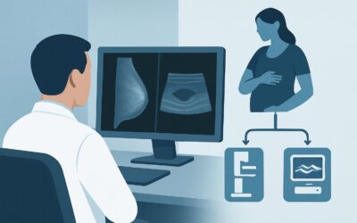
The ACR designates breast ultrasound (US) as Usually Appropriate for the initial evaluation of a palpable lump or focal pain at a mastectomy site without reconstruction. The rationale is based on its high diagnostic yield, safety profile, and ability to guide subsequent management effectively.
Ultrasound provides excellent spatial resolution of the superficial tissues of the chest wall, including the skin, subcutaneous fat, and underlying musculature. This allows for precise characterization of a palpable finding. It can reliably differentiate simple fluid collections (cysts, seromas) from solid masses. For solid nodules, sonographic features such as shape, margins, echotexture, and orientation can help distinguish benign findings like fat necrosis from suspicious lesions concerning for recurrence. The addition of color Doppler imaging assesses vascularity, which can further increase suspicion for malignancy.
Perhaps most importantly, ultrasound is the ideal modality for guiding a percutaneous core needle biopsy if a suspicious solid lesion is identified, allowing for a definitive pathologic diagnosis with minimal invasiveness.
Radiation and Contrast Considerations
A key advantage of ultrasound is the complete absence of ionizing radiation (Adult RRL=O 0 mSv). This is particularly important for cancer survivors who may have had prior radiation therapy and will likely undergo future surveillance imaging. Furthermore, ultrasound does not require intravenous contrast, avoiding any risk of allergic reaction or contrast-induced nephropathy.
Why Other Modalities Are Rated Lower
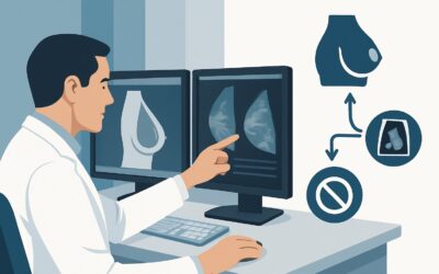
- Mammography or Digital Breast Tomosynthesis (DBT): These are rated as May be appropriate. Without breast tissue to compress, their utility is limited. While a tangential view of the palpable lump can sometimes be performed, it is often technically challenging and provides less information about the soft tissues than ultrasound. Mammography’s primary strength is detecting suspicious microcalcifications, which are a less common presentation of recurrence in this setting.
- Breast MRI with and without IV Contrast: This is rated as Usually not appropriate for the initial workup. While MRI is very sensitive for detecting recurrence, it is less specific and can show enhancement in benign post-surgical tissues like scar and granulation tissue, leading to false-positive results and unnecessary anxiety and biopsies. Its role is reserved for problem-solving if ultrasound is inconclusive or for assessing the deep extent of a biopsy-proven recurrence.
What’s Next After Breast Ultrasound? The Downstream Workflow
The results of the initial breast ultrasound will direct the next steps in a clear, logical sequence. The goal is to achieve a definitive diagnosis efficiently while avoiding unnecessary procedures.
If the Result is a Suspicious Solid Mass (BI-RADS 4 or 5)
The immediate next step is an ultrasound-guided core needle biopsy. This is the standard of care to obtain tissue for histopathologic analysis. The pathology results will confirm whether the lesion is a recurrence, allowing for prompt consultation with surgical and medical oncology to plan further treatment.
If the Result is Clearly Benign (BI-RADS 2)
If the ultrasound identifies features classic for a benign entity, such as a simple seroma, a suture granuloma with a visible suture, or typical fat necrosis, no further imaging or intervention is needed. The patient can be reassured, and routine clinical follow-up is sufficient. The imaging report provides a confident, non-malignant explanation for the palpable finding.
If the Result is Negative or Indeterminate (BI-RADS 0 or 3)
If the ultrasound shows no correlating abnormality for the palpable lump (a negative study), clinical correlation is key. If the palpable finding is subtle and the ultrasound is reassuring, a short-term clinical follow-up in 3-6 months may be appropriate. If there is a high degree of clinical suspicion despite a negative ultrasound, or if the finding is indeterminate (e.g., a complex fluid collection or a probably benign solid nodule), options include a short-interval follow-up ultrasound to ensure stability or proceeding to a problem-solving modality like MRI, though this is less common.
Pitfalls to Avoid (and When to Get Help)
Navigating this clinical scenario is generally straightforward, but a few common pitfalls can lead to diagnostic delays or unnecessary tests.
- Attributing all lumps to scar tissue: Never assume a new, persistent, or growing lump is “just scar tissue” without an imaging evaluation. While benign causes are common, the risk of recurrence necessitates a formal workup.
- Ordering the wrong initial test: Jumping directly to an MRI or PET/CT is contrary to evidence-based guidelines. It increases costs, can lead to false positives, and delays the most direct path to diagnosis, which is typically an ultrasound-guided biopsy.
- Accepting an incomplete ultrasound: Ensure the radiology report confirms that the sonographic finding corresponds to the exact location of the patient’s palpable concern. A “limited” or “non-targeted” scan may miss the relevant pathology.
If an initial ultrasound confirms a deeply infiltrating mass that appears to invade the pectoralis muscle or ribs, this is a red flag for chest wall invasion. In this case, escalation to a contrast-enhanced chest MRI or CT is appropriate to fully stage the extent of the disease before any surgical planning.
Related ACR Topics and Tools
For a comprehensive overview of all clinical variants related to imaging after mastectomy, including scenarios with breast reconstruction, please refer to our parent topic guide. For additional tools to assist in your clinical practice, see the resources below.
- For breadth across all scenarios in Imaging after Mastectomy and Breast Reconstruction, see our parent guide: Imaging after Mastectomy and Breast Reconstruction: ACR Appropriateness Decoded.
- To look up other clinical scenarios, visit the ACR Appropriateness Criteria Lookup.
- For detailed procedural techniques, explore the Imaging Protocol Library.
- To discuss radiation exposure with patients, use the Radiation Dose Calculator.
Frequently Asked Questions
Why not order a mammogram for a lump after mastectomy, since that was the original screening tool?
A mammogram requires compressing breast tissue to spread it out for imaging. After a mastectomy without reconstruction, there is no breast tissue to compress. While a special view can sometimes image the skin and chest wall, ultrasound is far superior for evaluating a focal, palpable soft tissue lump in this setting, as it provides detailed characterization without radiation or compression.
My patient has focal pain but no distinct lump. Does this workflow still apply?
Yes. The ACR scenario includes ‘clinically significant pain.’ A targeted ultrasound of the painful area is the appropriate first step. It can identify non-palpable causes of pain, such as a small recurrence, a suture granuloma, Mondor’s disease (superficial thrombophlebitis), or a neuroma in the scar.
If the ultrasound is negative, can I confidently rule out recurrence?
A high-quality ultrasound that finds no suspicious correlate for a palpable finding is very reassuring. However, no imaging test is perfect. If there is a strong clinical suspicion for recurrence despite a negative ultrasound (e.g., a growing or persistent, hard mass), close clinical follow-up is essential. In rare cases, a problem-solving MRI or even a clinical biopsy of the palpable area might be considered after discussion with a breast specialist.
What if the palpable lump is in the axilla (armpit) on the side of the mastectomy?
This workflow still applies. The axilla is a common site for nodal recurrence. An axillary ultrasound is the appropriate initial imaging test to evaluate the lymph nodes and surrounding soft tissues for any suspicious findings. If a suspicious node is found, an ultrasound-guided biopsy would be the next step.
Is a biopsy always necessary for a solid-appearing lump found on ultrasound?
Not always, but often. If a solid lump has features that are classic for a benign entity, like a suture granuloma with a visible suture or typical fat necrosis, the radiologist may confidently label it as benign (BI-RADS 2) and recommend no biopsy. However, any new solid mass that does not have unequivocally benign features will be categorized as suspicious (BI-RADS 4 or 5), and a biopsy will be recommended to achieve a definitive diagnosis.
Reviewed by Pouyan Golshani, MD, Interventional Radiologist — May 29, 2026