What Is the Correct Screening After Mastectomy Without Reconstruction? An ACR Guide

A 58-year-old female with a history of left-sided breast cancer, treated with mastectomy ten years ago, presents for her annual wellness visit. She has had no breast reconstruction and is asymptomatic, with no new lumps, pain, or skin changes on her chest wall. During the exam, she asks, “Do I still need a mammogram on that side, or some other kind of scan to check for cancer?” You are now faced with determining the appropriate surveillance strategy for her mastectomy site. This article details the evidence-based clinical workflow for this specific scenario, explaining why the American College of Radiology (ACR) Appropriateness Criteria rate routine screening imaging, including ultrasound, as Usually not appropriate.
Who Fits This Clinical Scenario for Post-Mastectomy Screening?
This guidance applies specifically to female patients undergoing routine cancer screening who meet all the following criteria:
- A personal history of breast cancer.
- Treated with unilateral or bilateral mastectomy.
- No subsequent breast reconstruction (i.e., a flat chest wall).
- Currently asymptomatic, with no new palpable lumps, skin changes, nipple discharge, or focal pain at the mastectomy site.
It is critical to distinguish this screening scenario from a diagnostic workup. This advice does not apply to patients who present with a new clinical finding. A patient with a palpable lump or clinically significant pain on the side of a mastectomy requires a different, diagnostic imaging pathway. Similarly, patients who have undergone breast reconstruction with implants or autologous tissue flaps follow separate surveillance guidelines, as the presence of reconstructed tissue alters the imaging approach.
What Are We Screening For After Mastectomy Without Reconstruction?
In a patient who has undergone mastectomy without reconstruction, surveillance is focused on the early detection of locoregional recurrence. While imaging is not the primary screening tool, understanding the potential targets of surveillance clarifies the role of the clinical exam.
Chest Wall Recurrence: This is the primary concern. Recurrence can manifest as nodules within the skin, subcutaneous tissue, or deeper, involving the pectoralis muscle or ribs. These are often superficial enough to be detected by careful palpation during a physical exam before they would be incidentally found on a non-targeted imaging study.
Regional Nodal Recurrence: The cancer can also recur in the nearby lymph node basins, including the axilla (armpit), supraclavicular (above the collarbone), and internal mammary nodes. A thorough physical exam includes palpation of these areas to detect any new or enlarged lymph nodes.
Benign Post-Surgical Changes: It is also important to recognize benign findings that can mimic recurrence. These include suture granulomas, seromas (fluid collections), and fat necrosis. These entities can present as palpable lumps, underscoring why any new finding requires a diagnostic workup, even though most will be benign.
Why Is Routine Screening Imaging ‘Usually Not Appropriate’ After Mastectomy?
The central principle guiding surveillance in this scenario is that a structured, high-quality physical examination is the most effective screening tool. The ACR designates all routine imaging modalities as “Usually not appropriate” for screening asymptomatic patients after mastectomy without reconstruction due to a lack of evidence supporting their use and the potential for harm.
The rationale is multifactorial. First, there is no high-quality data demonstrating that routine screening with ultrasound, MRI, or any other modality improves long-term survival or other clinically meaningful outcomes compared to surveillance with regular clinical breast exams (CBE) and patient self-awareness.
Second, the yield of screening imaging on a flat chest wall is very low. The absence of breast parenchyma and the presence of post-surgical scar tissue make interpretation difficult and can lead to a high rate of false-positive findings. These false positives can trigger a cascade of unnecessary anxiety, further imaging, and invasive biopsies for what often turn out to be benign post-operative changes like scar tissue or fat necrosis.
Let’s examine why specific, commonly considered modalities are not recommended for screening:
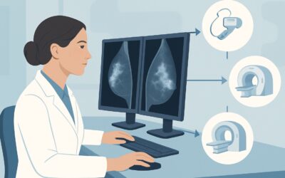
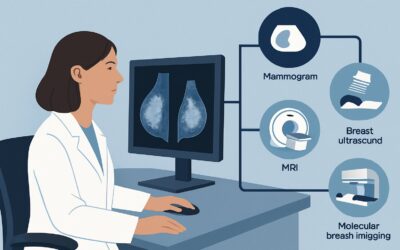
- Mammography and Digital Breast Tomosynthesis: Rated Usually not appropriate. Without breast tissue to compress, mammography is technically challenging and provides little to no useful information about the chest wall. It also involves a small amount of radiation (ACR Relative Radiation Level ☢☢).
- MRI Breast Without and With IV Contrast: Rated Usually not appropriate. While MRI is a very sensitive imaging test, its use for routine screening in this population is not supported. Post-surgical scarring and inflammation can enhance after contrast administration, mimicking recurrence and leading to low specificity. Its role is reserved for problem-solving in a diagnostic setting, not for screening.
The consensus is that the potential harms of routine screening imaging—including false positives, patient anxiety, and healthcare costs—outweigh the unproven benefits in this specific asymptomatic population. The standard of care remains a diligent physical exam.
What’s Next? The Correct Post-Mastectomy Surveillance Workflow
The appropriate workflow for a patient after mastectomy without reconstruction is centered on clinical surveillance, shifting to a diagnostic pathway only when an abnormality is detected.
For the Asymptomatic Patient (Screening Pathway):
The recommended surveillance consists of:
- Provider-Performed Clinical Breast Exam (CBE): A thorough physical examination of the chest wall, mastectomy scars, and regional lymph node basins (axillary, supraclavicular) should be performed every 6 to 12 months for the first five years, and annually thereafter.
- Patient Self-Examination: Patients should be educated on breast self-awareness to recognize and report any new or changing lumps, bumps, or skin changes between clinical visits.
- Contralateral Breast Screening: If the patient has a remaining contralateral breast, it should undergo standard annual screening with mammography or tomosynthesis.
No routine imaging of the mastectomy site is indicated if the exam is normal.
For the Symptomatic Patient (Diagnostic Pathway):
If a new abnormality is detected on a clinical exam or reported by the patient (e.g., a palpable nodule), the workflow immediately transitions to a diagnostic workup:
- Targeted Ultrasound: This is the initial imaging modality of choice. Ultrasound is excellent for characterizing a palpable finding on the chest wall, determining if it is solid or cystic, and guiding a biopsy if necessary.
- Biopsy: Any sonographically suspicious solid lesion requires a biopsy for definitive histopathologic diagnosis.
- Further Imaging (e.g., MRI): If ultrasound is inconclusive or if there is concern for deeper chest wall invasion, a contrast-enhanced MRI may be considered for further evaluation and pre-operative planning.
This two-tiered approach ensures that imaging is used judiciously and effectively when a clinical question needs to be answered, avoiding the pitfalls of low-yield screening.
Common Pitfalls in Post-Mastectomy Surveillance
Ordering “Reassurance” Scans: A common pitfall is ordering a routine annual ultrasound of the chest wall for an asymptomatic patient, often at their request. This practice is not supported by evidence and can lead to a cascade of unnecessary interventions. Patient education on the value of a physical exam is the preferred approach.
Incomplete Physical Exam: The clinical exam must be comprehensive. Failing to carefully palpate the entire length of the mastectomy scar, the full chest wall, the axilla, and the supraclavicular fossa can lead to a missed recurrence.
Dismissing Patient-Reported Findings: Never dismiss a lump or change reported by the patient. Even if an initial clinical exam feels normal, a patient’s report of a new, persistent finding warrants a low threshold for a targeted diagnostic ultrasound.
If a new, persistent, or enlarging palpable abnormality is found on the chest wall or in the regional nodal basins, the situation requires immediate escalation to diagnostic imaging, beginning with a targeted ultrasound of the specific area of concern.
Related ACR Topics and Tools
This article focuses on a single clinical scenario. For a comprehensive overview of all related presentations and imaging guidelines, or to explore the tools used to make these recommendations, please refer to the following resources.
- For breadth across all scenarios in this topic, see our parent guide: Imaging after Mastectomy and Breast Reconstruction: ACR Appropriateness Decoded.
- Explore other clinical situations with the ACR Appropriateness Criteria Lookup.
- Review imaging techniques in the Imaging Protocol Library.
- Discuss radiation exposure with patients using the Radiation Dose Calculator.
Frequently Asked Questions
Does this ‘no imaging’ recommendation apply if the patient had a skin-sparing or nipple-sparing mastectomy?
This is a nuanced area. While the ACR criteria do not differentiate, these techniques intentionally leave more residual tissue at risk compared to a traditional mastectomy. Some cancer centers may have specific protocols that include screening MRI for these patients, particularly if they are at high genetic risk. This decision should be made in a multidisciplinary setting, but routine imaging is not the universal standard of care.
What is the correct screening for the contralateral, intact breast?
This article’s recommendations apply only to the mastectomy side. The contralateral breast is considered at higher risk, and the patient should continue with standard annual screening, typically with digital breast tomosynthesis (3D mammography), as per high-risk screening guidelines.
If a patient is very anxious and requests screening imaging, should I order it?
The recommended approach is to provide thorough patient education. Explain that a high-quality physical exam is the evidence-based standard of care and that imaging has a high risk of false positives, which can lead to unnecessary anxiety and biopsies without a proven survival benefit. Ordering imaging solely for reassurance is generally discouraged by clinical guidelines.
How does this surveillance change for patients with a high-risk genetic mutation like BRCA1/2?
Even for patients with high-risk genetic mutations, the standard of care for surveillance after a risk-reducing mastectomy is a thorough annual clinical exam. The amount of residual tissue is minimal, and the risk-benefit profile still does not favor routine screening imaging of the chest wall. The focus of imaging surveillance would be on any other at-risk tissues, such as the ovaries or a contralateral breast if present.
Is a chest CT scan a good screening tool for chest wall recurrence?
No, a chest CT is not a recommended screening tool for locoregional breast cancer recurrence. It is not sensitive for detecting small, superficial nodules on the chest wall and delivers a significantly higher radiation dose than other modalities. Its use is reserved for staging known cancer or when there is clinical suspicion of deep chest wall invasion or distant metastatic disease, not for screening an asymptomatic patient.
Reviewed by Pouyan Golshani, MD, Interventional Radiologist — May 29, 2026