What Is the Best Initial Imaging for Suspected Dementia with Lewy Bodies?
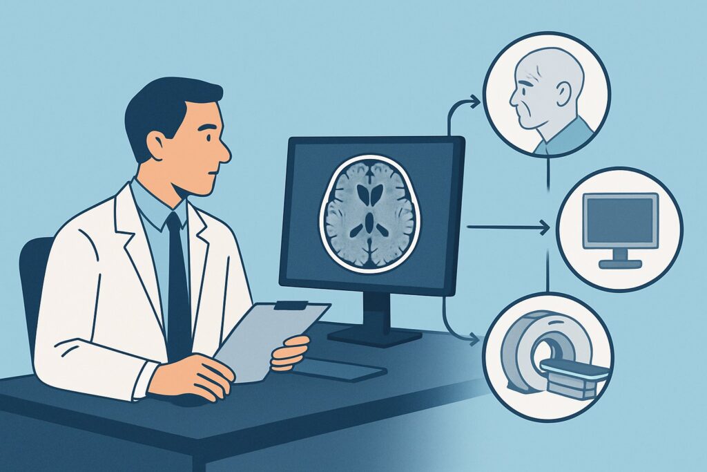
A 72-year-old man is brought to your clinic by his family due to a six-month history of progressive cognitive decline, marked by fluctuating attention and recurrent, well-formed visual hallucinations of small animals in the house. On examination, you note mild bradykinesia and a subtle resting tremor in his right hand. You suspect Dementia with Lewy Bodies (DLB), a diagnosis that carries significant implications for management, particularly regarding neuroleptic sensitivity. The immediate clinical question is which imaging study to order first to support the diagnosis and, critically, to exclude other pathologies. This article details the American College of Radiology (ACR) guided workflow for this specific presentation. For this scenario, the ACR rates MRI head without IV contrast as Usually appropriate.
Who Fits This Clinical Scenario?
This guidance applies to adult patients presenting with a progressive cognitive impairment where the clinical suspicion points toward Dementia with Lewy Bodies. The key features that place a patient in this specific workflow are the core clinical signs of DLB:
- Cognitive Decline: A progressive dementia that interferes with social or occupational function.
- Core Features: The presence of at least one of the following is essential:
- Recurrent, well-formed, and detailed visual hallucinations.
- Spontaneous parkinsonian motor signs (bradykinesia, rest tremor, or rigidity).
- Fluctuating cognition with pronounced variations in attention and alertness.
This workflow is distinct from other dementia workups. It is crucial to differentiate this presentation from nearby scenarios:
- Exclusion 1: Predominant Memory Loss. If the patient’s primary and most prominent symptom is amnestic memory loss without early or significant parkinsonism or hallucinations, the workup for Suspected Alzheimer Disease is more suitable.
- Exclusion 2: Behavioral or Language Changes. If the dominant features are profound changes in personality, behavior (e.g., disinhibition, apathy), or a progressive aphasia, the evaluation should follow the pathway for Suspected Frontotemporal Dementia.
- Exclusion 3: Mild, Non-Disabling Symptoms. If cognitive symptoms are present but do not yet meet the criteria for dementia (i.e., they do not significantly impair daily functioning), the patient fits the Mild Cognitive Impairment scenario, which has a distinct diagnostic approach.
What Diagnoses Are You Working Up in This Scenario?
The imaging choice is driven by a differential diagnosis that extends beyond just confirming DLB. The goal is to find supportive evidence while rigorously excluding mimics that require different management.
Dementia with Lewy Bodies (DLB) is the leading consideration. It is the second most common cause of neurodegenerative dementia after Alzheimer disease. Imaging in DLB is often aimed at identifying patterns of atrophy and, most importantly, ruling out other structural causes. While structural MRI can be non-specific, the relative preservation of the medial temporal lobes is a key supportive feature that helps distinguish it from typical Alzheimer’s.
Alzheimer Disease (AD) remains a critical differential. Atypical presentations of AD can include visual symptoms, though well-formed hallucinations are less common. Structural MRI is highly valuable here, as it may reveal the characteristic medial temporal lobe and hippocampal atrophy strongly associated with AD, steering the diagnosis away from DLB.
Parkinson Disease Dementia (PDD) is clinically and pathologically very similar to DLB. The primary distinction is timing: in PDD, dementia develops at least one year after the onset of well-established motor symptoms of Parkinson’s disease. In DLB, cognitive symptoms appear before or concurrently with motor symptoms. Their imaging findings on structural MRI are often indistinguishable.
Vascular Dementia must be considered, especially in patients with cardiovascular risk factors. A stepwise cognitive decline or focal neurological signs would increase suspicion. MRI is superior to other modalities for detecting the burden of chronic small vessel ischemic disease, lacunar infarcts, or strategic single infarcts that could explain the patient’s symptoms.
Structural Mimics are less common but must be excluded. These include Normal Pressure Hydrocephalus (NPH), chronic subdural hematomas, primary or metastatic brain tumors, and the sequelae of prior strokes. Anatomic imaging is essential for ruling out these treatable or otherwise consequential conditions.
Why Is MRI Head without IV Contrast the Recommended Study for This Presentation?
For an adult with cognitive impairment, visual hallucinations, and parkinsonism, the ACR designates MRI head without IV contrast as Usually appropriate. This recommendation is based on its superior ability to evaluate brain structure, differentiate between dementia subtypes, and rule out mimics, all without using ionizing radiation.
The primary role of the initial MRI is twofold. First, it serves to exclude other pathologies. Its high soft-tissue contrast is unmatched for identifying tumors, inflammation, evidence of prior strokes, or signs of Normal Pressure Hydrocephalus. Second, it provides crucial information about patterns of cerebral atrophy. In suspected DLB, a key finding is the relative preservation of hippocampal and medial temporal lobe volume, especially when compared to the significant atrophy seen in these regions in typical Alzheimer disease. While generalized cortical atrophy may be present, the absence of focal hippocampal volume loss is an important clue.
This study is performed without intravenous contrast, as there is no initial suspicion for a pathology (like tumor or active inflammation) that would require it. This avoids the risks and costs associated with gadolinium-based contrast agents. The radiation level is O (0 mSv), making it a safe choice for initial evaluation.
Why Other Studies Are Rated Differently
- CT head without IV contrast is also rated Usually appropriate and is a valid alternative, particularly when MRI is contraindicated (e.g., incompatible implanted devices) or not readily available. However, its soft-tissue resolution is significantly lower than MRI, making it less sensitive for detecting subtle atrophy patterns or small vessel ischemic changes. It is excellent for ruling out acute hemorrhage, large tumors, or hydrocephalus but provides less diagnostic nuance. It involves a moderate radiation dose (ACR RRL=☢☢☢, 1-10 mSv).
- Amyloid PET/CT brain is rated Usually not appropriate for this initial workup. While amyloid pathology is a feature of DLB (often co-occurring with synucleinopathy), it is also the defining feature of Alzheimer disease. Therefore, a positive amyloid PET scan does not reliably differentiate between the two conditions and is not recommended as a first-line diagnostic tool in this scenario.
- SPECT or SPECT/CT brain striatal (e.g., DaTscan) is also rated Usually appropriate. However, it is typically considered a second-line or problem-solving study rather than the initial imaging test. It assesses the integrity of the dopamine transporter system, which is impaired in DLB but normal in Alzheimer disease. While highly specific, structural imaging with MRI is prioritized to first rule out anatomic causes for the patient’s symptoms.
What’s Next After MRI Head without IV Contrast? Downstream Workflow
The results of the initial MRI will guide the subsequent diagnostic and management pathway. The workflow is not linear and depends on integrating the imaging findings with the clinical picture.
- If the MRI shows non-specific findings or is consistent with DLB: If the scan reveals generalized atrophy but relative preservation of the medial temporal lobes, and it excludes other structural causes, the findings support a clinical diagnosis of DLB. Management can proceed based on this working diagnosis.
- If diagnostic uncertainty persists: If the MRI is non-diagnostic but clinical suspicion for DLB remains high, the next step is often functional imaging. A SPECT or SPECT/CT brain striatal (DaTscan) is an excellent choice to confirm dopaminergic deficit, which would strongly support DLB over AD. Alternatively, an FDG-PET/CT brain, also rated Usually appropriate, can be performed to look for the “cingulate island sign”—a characteristic pattern of preserved metabolism in the posterior cingulate cortex that is highly suggestive of DLB.
- If the MRI suggests an alternative diagnosis: If the scan reveals significant, focal medial temporal lobe atrophy, the diagnosis shifts toward Alzheimer disease. If extensive white matter changes or strategic infarcts are found, Vascular Dementia becomes more likely. Findings of enlarged ventricles out of proportion to sulcal widening would prompt a workup for Normal Pressure Hydrocephalus. Each of these findings directs the clinical team down a different management pathway.
Pitfalls to Avoid (and When to Get Help)
Navigating the workup for suspected DLB requires careful attention to clinical and imaging nuances. Here are common pitfalls to avoid:
- Over-reliance on a “normal” MRI: In early DLB, the structural MRI is often reported as normal or showing only age-related atrophy. This does not rule out the diagnosis. The key is the pattern of atrophy (or lack thereof) relative to other dementia types.
- Misinterpreting parkinsonism: Not all parkinsonism in the elderly is Parkinson’s disease. The concurrent onset with cognitive symptoms is a red flag for DLB.
- Forgetting functional imaging: If the structural MRI is unrevealing but the diagnosis is uncertain, failing to proceed to functional imaging (like a DaTscan) can lead to a missed or delayed diagnosis.
- Ordering the wrong PET scan: An Amyloid PET is Usually not appropriate as a first step here because it doesn’t differentiate DLB from AD. A DaTscan or FDG-PET is more diagnostically useful in this specific context.
If the clinical picture and initial imaging results are conflicting or unclear, escalation to a neurologist or geriatrician with expertise in cognitive disorders is the appropriate next step.
Related ACR Topics and Tools
This article focuses on a single clinical scenario. For a comprehensive overview of imaging for all forms of dementia and to explore related clinical presentations, please consult our parent guide and the tools below.
- For breadth across all scenarios in Dementia, see our parent guide: Dementia: ACR Appropriateness Decoded
- ACR Appropriateness Criteria Lookup: Look up ratings for thousands of clinical scenarios.
- Imaging Protocol Library: Review detailed imaging techniques for recommended studies.
- Radiation Dose Calculator: Estimate cumulative radiation exposure for your patients.
Frequently Asked Questions
Why is MRI without contrast preferred over MRI with contrast for suspected DLB?
Intravenous contrast is used to highlight areas of inflammation, infection, or tumors by identifying breakdowns in the blood-brain barrier. In the initial workup for a neurodegenerative condition like Dementia with Lewy Bodies (DLB), these processes are not suspected. The primary goal is to assess brain structure and atrophy patterns, for which non-contrast sequences are sufficient and optimal. Omitting contrast avoids the potential risks, costs, and time associated with gadolinium-based agents. The ACR rates MRI with contrast as ‘May be appropriate (Disagreement)’ for this reason.
My patient has a pacemaker and cannot get an MRI. What is the best alternative?
If MRI is contraindicated, the ACR rates ‘CT head without IV contrast’ as ‘Usually appropriate’. It is the best alternative for initial structural imaging. While it has lower soft-tissue resolution than MRI, it is effective at ruling out major structural causes like tumors, chronic subdural hematomas, and hydrocephalus. If the CT is non-diagnostic and clinical uncertainty remains, functional imaging like a DaTscan (SPECT/CT brain striatal) can be a valuable next step to assess for dopaminergic deficits.
What is a DaTscan and when should I order it in this workup?
A DaTscan (Dopamine Transporter Scan) is a nuclear medicine imaging test (SPECT/CT brain striatal) that visualizes the dopamine transporters in the brain. In neurodegenerative parkinsonian syndromes like DLB and Parkinson’s disease, there is a loss of these transporters. A DaTscan is typically abnormal in DLB but normal in Alzheimer disease, essential tremor, or drug-induced parkinsonism. While rated ‘Usually appropriate,’ it is best used as a second-line test after an initial structural MRI has ruled out other causes, or when there is high diagnostic uncertainty between DLB and Alzheimer disease.
Can an MRI definitively diagnose Dementia with Lewy Bodies?
No, a structural MRI cannot definitively diagnose DLB on its own. The diagnosis of DLB is primarily clinical, based on a combination of symptoms and examination findings. The role of the MRI is to provide supportive evidence and, more importantly, to exclude other conditions that can mimic DLB. A finding of generalized atrophy with relative sparing of the medial temporal lobes supports the diagnosis, but a ‘normal’ MRI does not rule it out.
What specific MRI sequences should I request from the radiologist?
When ordering the MRI, it is helpful to request a ‘dementia protocol’ if your institution has one. This typically includes high-resolution 3D T1-weighted sequences (e.g., MPRAGE, SPGR) for assessing atrophy and volumetric analysis, FLAIR and T2-weighted sequences to evaluate for white matter disease and vascular pathology, and a susceptibility-weighted sequence (e.g., SWI, GRE) to look for microhemorrhages. This combination provides a comprehensive structural evaluation for a dementia workup.
Reviewed by Pouyan Golshani, MD, Interventional Radiologist — May 29, 2026