What Is the Best Initial Imaging for Suspected Pelvic Endometriosis in an Adult?
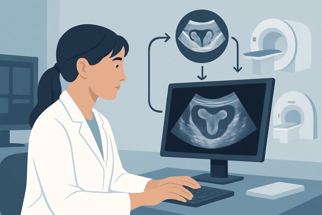
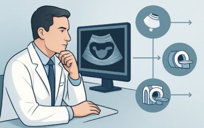
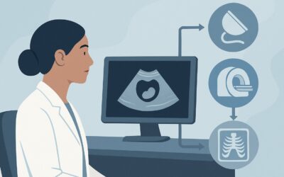
A 34-year-old woman presents to your clinic with a multi-year history of worsening dysmenorrhea and deep dyspareunia. Her symptoms are cyclical and significantly impact her quality of life. On examination, she has uterine tenderness and nodularity along the uterosacral ligaments. You suspect pelvic endometriosis, but the diagnosis needs to be supported before considering laparoscopy or empiric hormonal therapy. The immediate clinical question is which imaging study provides the most diagnostic value as the first step in her workup. This article details the American College of Radiology (ACR) Appropriateness Criteria for this exact scenario, explaining why a specific imaging modality is the recommended starting point. For the initial evaluation of clinically suspected pelvic endometriosis in an adult, the ACR rates a combined ‘US pelvis transabdominal and US pelvis transvaginal’ as Usually Appropriate.
Who Fits This Clinical Scenario?
This guidance applies specifically to adult patients for whom there is a clinical suspicion of pelvic endometriosis, and this is the initial imaging evaluation. The typical presentation includes one or more of the “three D’s”: dysmenorrhea (painful periods), dyspareunia (painful intercourse), and dyschezia (painful bowel movements), often accompanied by chronic pelvic pain or infertility. The key element is that no prior advanced imaging (like MRI or a dedicated endometriosis ultrasound) has been performed for this indication.
This workflow is distinct from several related clinical situations. This article does not apply if:
- A prior ultrasound was performed but was negative or indeterminate. That patient falls into a different clinical variant requiring a decision on the next best imaging study, often involving Magnetic Resonance Imaging (MRI).
- Symptoms strongly suggest rectosigmoid involvement. If a patient presents with prominent cyclic dyschezia, hematochezia, or a palpable posterior cul-de-sac mass, the imaging strategy may be tailored differently from the outset to better evaluate the bowel.
- The patient has an established postoperative diagnosis. A patient with a history of surgical confirmation of endometriosis who presents with new or recurrent symptoms requires a follow-up imaging approach, which has its own set of considerations.
Correctly identifying the patient’s place in the diagnostic journey ensures the most efficient and highest-yield imaging is selected first.
What Diagnoses Are You Working Up in This Scenario?
When ordering initial imaging for suspected endometriosis, the primary goal is to identify endometriotic implants and assess the extent of disease. However, the patient’s symptoms overlap with several other gynecologic and non-gynecologic conditions, making a broad differential essential.
Endometriosis is the primary diagnosis under consideration. The imaging study aims to detect endometriomas (so-called “chocolate cysts” of the ovary), deep infiltrating endometriosis (DIE) affecting the uterosacral ligaments, posterior cul-de-sac, bladder, or bowel, and signs of adenomyosis, a common co-existing condition. Identifying these findings can guide medical therapy or surgical planning.
Adenomyosis is a frequent mimicker and concurrent diagnosis, where endometrial tissue exists within the myometrium. It often presents with similar symptoms of heavy menstrual bleeding and pelvic pain. Ultrasound can often distinguish its characteristic features, such as a bulky, globular uterus with myometrial cysts or striations, from endometriosis.
Pelvic Inflammatory Disease (PID), particularly in its chronic form, can lead to adhesions and tubo-ovarian abscesses that cause chronic pelvic pain. Ultrasound is highly effective at identifying hydrosalpinx, pyosalpinx, and complex adnexal masses characteristic of PID, differentiating it from the solid, infiltrating lesions of endometriosis.
Ovarian Neoplasms, both benign (like hemorrhagic cysts or dermoids) and malignant, must be considered in any patient with an adnexal mass or pelvic pain. Ultrasound is the frontline modality for characterizing ovarian lesions, assessing features like septations, solid components, and vascularity to stratify risk and determine the need for further evaluation with MRI or surgical consultation.
Fibroids (Leiomyomas) are an extremely common cause of pelvic pain, pressure, and abnormal bleeding. Ultrasound readily identifies the location, size, and number of fibroids, helping to determine if they are the primary cause of the patient’s symptoms.
Why Is a Combined Transabdominal and Transvaginal Ultrasound the Recommended Study?
The ACR designates ‘US pelvis transabdominal and US pelvis transvaginal’ as Usually Appropriate for the initial imaging of suspected pelvic endometriosis. This recommendation is based on the modality’s high accessibility, lack of ionizing radiation, and excellent diagnostic capability for key findings when performed meticulously.
The two-part approach is critical. The transabdominal (TA) portion provides a broad overview of the entire pelvis, which is essential for identifying large masses, assessing the upper extent of fibroids, or detecting pathology outside the reach of the transvaginal probe. The transvaginal (TV) portion provides high-resolution images of the uterus, endometrium, ovaries, and adnexa. For endometriosis, the TV ultrasound is particularly crucial for identifying the classic ground-glass appearance of endometriomas and detecting the subtle signs of deep infiltrating endometriosis, such as uterosacral ligament thickening or obliteration of the posterior cul-de-sac.
While MRI is also rated as Usually Appropriate, ultrasound is typically the preferred first-line study due to its lower cost and wider availability. MRI is often reserved for cases where ultrasound is inconclusive or when there is a high suspicion for deep infiltrating endometriosis that requires preoperative surgical planning.
Alternative studies receive lower ratings for specific reasons in this initial workup:
- CT of the pelvis (with or without IV contrast) is rated Usually Not Appropriate. CT has poor soft-tissue resolution for identifying endometrial implants and endometriomas, which can be mistaken for simple or hemorrhagic cysts. Furthermore, it exposes the patient to ionizing radiation (ACR Relative Radiation Level ☢☢☢ to ☢☢☢☢) without providing superior diagnostic information for this specific indication.
- US pelvis transabdominal alone is also Usually Not Appropriate. Without the high-resolution detail from the transvaginal component, small endometriomas, ovarian adhesions, and signs of deep infiltrating endometriosis are frequently missed, leading to a high rate of false-negative studies.
What’s Next After Ultrasound? Downstream Workflow
The results of the initial pelvic ultrasound will guide the subsequent clinical pathway. The decision tree branches based on whether the findings are positive, negative, or indeterminate for endometriosis.
If the ultrasound is positive for endometriosis: A definitive finding of an endometrioma or clear signs of deep infiltrating endometriosis can confirm the clinical suspicion. The next steps are typically clinical, involving a discussion with the patient about management options. This may include initiating medical therapy (e.g., hormonal suppression) or referral to a gynecologic surgeon for consultation, especially if symptoms are severe, fertility is a concern, or extensive disease is identified.
If the ultrasound is negative: A negative ultrasound does not definitively rule out endometriosis, as superficial peritoneal implants are below the resolution of any imaging modality. If clinical suspicion remains high despite a negative initial ultrasound, the patient may fit the criteria for the next ACR scenario: ‘Adult. Clinically suspected pelvic endometriosis. Indeterminate or negative ultrasound.’ In this case, an MRI of the pelvis without and with IV contrast becomes a Usually Appropriate next step to search for more subtle findings of DIE that may have been missed on ultrasound.
If the ultrasound is indeterminate: The study may reveal ambiguous findings, such as a complex adnexal cyst that is not classic for an endometrioma or non-specific pelvic adhesions. In this situation, similar to a negative study with high clinical suspicion, proceeding to pelvic MRI is the logical next step. MRI provides superior soft-tissue characterization and can often resolve the ambiguity, differentiating an endometrioma from a hemorrhagic cyst or neoplasm and better defining the extent of adhesive disease.
Pitfalls to Avoid (and When to Get Help)
In the initial workup of suspected endometriosis, several common pitfalls can delay diagnosis or lead to unnecessary testing. First, avoid ordering a transabdominal-only ultrasound; the transvaginal component is essential for evaluating the key pelvic structures with sufficient detail. Second, do not default to CT; it offers poor diagnostic yield for this condition and involves needless radiation exposure. A third pitfall is accepting a “normal” ultrasound report as definitive exclusion of the disease when clinical suspicion is high. Remember that superficial endometriosis is not visible on imaging. If the patient’s symptoms are classic and debilitating, a negative ultrasound should prompt consideration of the next diagnostic step, such as MRI or referral to a specialist for consideration of diagnostic laparoscopy, which remains the gold standard. If you encounter complex adnexal masses or findings suggestive of malignancy on the initial ultrasound, immediate escalation to a gynecologic oncologist is warranted.
Related ACR Topics and Tools
This article focuses on a single clinical scenario. For a comprehensive overview of imaging for all related presentations, from rectosigmoid disease to postoperative follow-up, please consult our parent guide. To explore other scenarios or find technical details on imaging protocols, the following GigHz resources are available.
- For breadth across all scenarios in Endometriosis, see our parent guide: Endometriosis: ACR Appropriateness Decoded.
- To explore adjacent scenarios and their ACR ratings: ACR Appropriateness Criteria Lookup
- For technical specifications on the recommended study: Imaging Protocol Library
- For discussing radiation exposure with patients for other studies: Radiation Dose Calculator
Frequently Asked Questions
Why is MRI also rated ‘Usually Appropriate’ if ultrasound is the recommended first step?
While both are rated ‘Usually Appropriate,’ they serve different roles. Ultrasound is recommended as the first-line study due to its lower cost, wider availability, and lack of contraindications. MRI is an excellent problem-solving tool and is often used as the next step if the ultrasound is negative or inconclusive in a patient with high clinical suspicion, or for detailed preoperative mapping of deep infiltrating endometriosis.
Does the patient need to have a full bladder for the initial endometriosis ultrasound?
Yes, for the transabdominal portion of the exam, a full bladder is necessary. It acts as an acoustic window, pushing the bowel out of the way and providing a clearer view of the uterus and adnexa. The patient will be asked to empty her bladder before the transvaginal portion of the study.
Can ultrasound reliably diagnose deep infiltrating endometriosis (DIE)?
In experienced hands, transvaginal ultrasound can be very effective at identifying DIE, a technique sometimes referred to as ‘sonovaginography’ or a ‘dynamic’ endometriosis scan. The sonographer specifically looks for nodules in the posterior cul-de-sac, uterosacral ligaments, and rectovaginal septum, and assesses for organ mobility (the ‘sliding sign’). However, operator experience is a significant factor, and MRI is often considered more reproducible for mapping the full extent of DIE.
If the ultrasound shows a simple cyst, does that rule out endometriosis?
Not necessarily. A simple cyst is not a feature of endometriosis, but its presence doesn’t exclude endometriosis elsewhere. The patient could have a functional ovarian cyst concurrently with superficial peritoneal endometriosis that is not visible on ultrasound. The key is to look for specific signs like endometriomas (which have a characteristic ‘ground-glass’ appearance, not a simple anechoic appearance) and signs of DIE.
Should I order a CA-125 level along with the initial ultrasound?
A CA-125 level has limited utility in the initial diagnosis of endometriosis. While it can be elevated in moderate to severe endometriosis, it is a non-specific marker that can also be elevated in many other conditions, including fibroids, adenomyosis, pelvic inflammatory disease, and even normal menstruation. It is not recommended as a routine screening or diagnostic tool for this initial presentation.
Reviewed by Pouyan Golshani, MD, Interventional Radiologist — May 30, 2026