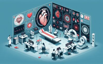
What Is the Best Next Imaging Study for a Known Cardiac Mass of Unknown Etiology?
A 68-year-old patient with new-onset atrial fibrillation undergoes a transthoracic echocardiogram (TTE) as part of their initial workup. The study reveals an incidental, mobile, 2-cm mass in the left atrium. The mass is well-circumscribed, but its attachment point and tissue characteristics are unclear. You, the ordering clinician, are now faced with a critical decision: the mass is confirmed, but its etiology is unknown. Is it a benign myxoma, a dangerous thrombus, or something more sinister? This article provides a clinical workflow for selecting the next imaging study for an adult patient with a known cardiac mass of unknown etiology, guided by the American College of Radiology (ACR) Appropriateness Criteria. For this specific scenario, multiple advanced imaging modalities, including transesophageal echocardiography, cardiac MRI, and cardiac CT, are rated as Usually Appropriate.
Who Fits This Clinical Scenario for Cardiac Mass Evaluation?
This guidance is designed for a specific clinical situation: an adult patient in whom a cardiac mass has already been detected on a prior imaging study, most commonly a transthoracic echocardiogram (TTE), but the nature of the mass remains undetermined.
Inclusion criteria for this workflow:
- Patient is an adult.
- A cardiac mass has been definitively identified on prior imaging.
- The etiology, or specific diagnosis, of the mass is unknown.
- The clinical question is which next imaging study to order for further characterization.
This workflow is not intended for patients who fall into closely related but distinct clinical scenarios. You should seek different guidance if your patient:
- Is suspected of having a cardiac mass but has not yet had initial imaging. This situation falls under the ACR variant for Initial imaging of a suspected cardiac mass.
- Has a cardiac mass with an established diagnosis (e.g., a known myxoma or thrombus) and requires follow-up imaging. This is a separate surveillance scenario.
- Is a pediatric patient. While some ACR ratings include pediatric radiation levels, the differential diagnosis and typical workflow can differ significantly in children.
Correctly identifying your patient’s scenario is the crucial first step to ensuring the most appropriate and highest-yield imaging is performed.
What Diagnoses Are You Working Up When a Cardiac Mass Is Found?
When an intracardiac mass of unknown etiology is discovered, the differential diagnosis is broad, ranging from benign and incidental findings to life-threatening malignancies. The goal of subsequent imaging is to narrow this differential to guide management, which could involve anticoagulation, surgical resection, or systemic therapy.
The most common primary cardiac tumor in adults is a benign myxoma. These typically arise in the left atrium from the interatrial septum and can present with embolic events, obstruction, or constitutional symptoms. Their characteristic mobile nature and stalk-like attachment are key features to identify on imaging. Other benign tumors include papillary fibroelastomas, which are small, frond-like masses typically on valve leaflets, and lipomas, which are composed of fatty tissue.
Malignant primary cardiac tumors are rare but aggressive. Sarcomas, particularly angiosarcomas, are the most frequent type and often present as large, infiltrative masses in the right atrium, commonly associated with a pericardial effusion. Primary cardiac lymphoma is another consideration, especially in immunocompromised patients.
Metastatic disease to the heart is significantly more common than primary cardiac malignancy. Cancers that frequently metastasize to the heart include lung cancer, breast cancer, melanoma, and renal cell carcinoma. These can appear as discrete nodules or diffuse infiltration of the myocardium or pericardium.
Finally, non-neoplastic masses are critical to differentiate. A thrombus is a frequent finding, especially in patients with atrial fibrillation, ventricular dysfunction, or a history of myocardial infarction. Vegetations from endocarditis are another key consideration, particularly in patients with fever, new-onset murmur, or risk factors like intravenous drug use.
Why Advanced Imaging Is Usually Appropriate for Characterizing a Known Cardiac Mass
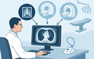
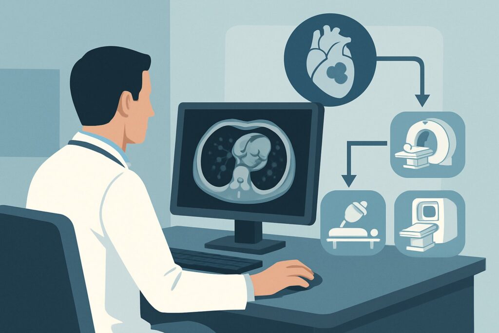
After a mass is detected on an initial study like TTE, the ACR designates several advanced imaging modalities as Usually Appropriate. The choice among them depends on the initial findings, institutional expertise, and the specific clinical question. No single test is perfect for all scenarios; they offer complementary information.
US echocardiography transesophageal (TEE) is often the immediate next step. By placing the ultrasound probe in the esophagus, TEE provides superior spatial resolution compared to TTE, especially for structures in the posterior heart like the left atrium, left atrial appendage, and mitral valve. It is excellent for precisely defining a mass’s size, shape, mobility, and point of attachment—critical details for surgical planning or differentiating a pedunculated myxoma from a broad-based thrombus. As an ultrasound-based modality, it involves no ionizing radiation (0 mSv).
MRI heart function and morphology without and with IV contrast (Cardiac MRI) is the premier modality for non-invasive tissue characterization. Its multi-parametric approach can distinguish different tissue types. For example, specific imaging sequences can suppress the signal from fat to confirm a lipoma. The pattern of gadolinium contrast enhancement helps differentiate an avascular thrombus (no enhancement) from a vascularized tumor (variable enhancement). Late gadolinium enhancement (LGE) sequences are particularly powerful for identifying infiltration, fibrosis, and distinguishing tumor from scar or thrombus. Like TEE, it uses no ionizing radiation (0 mSv).
CT heart function and morphology with IV contrast (Cardiac CT) is another Usually Appropriate option. Its primary strengths are speed, excellent spatial resolution, and the ability to assess for calcification, coronary artery involvement, and extracardiac extension. ECG-gating is essential to minimize motion artifact. While it provides less soft-tissue detail than MRI, it is a valuable alternative for patients with contraindications to MRI (e.g., certain implanted devices) or in urgent settings. It does involve a significant radiation dose (☢☢☢☢ 10-30 mSv).
For cases where malignancy is a high concern, FDG-PET/CT heart or FDG-PET/MRI heart are also Usually Appropriate. These hybrid imaging techniques combine anatomic detail with functional information about metabolic activity. Malignant and inflammatory masses typically show high FDG uptake, while benign tumors and thrombi do not. This can be invaluable for differentiating benign from malignant processes and for systemic staging.
An MRI of the heart without contrast is rated as May be appropriate because it lacks the crucial tissue characterization information provided by gadolinium enhancement, limiting its diagnostic utility. Similarly, a standard, non-gated CT chest with IV contrast is Usually not appropriate for this indication, as cardiac motion will render the mass poorly defined.
What Is the Downstream Workflow After Characterizing a Cardiac Mass?
The results of advanced cardiac imaging will dictate the subsequent clinical pathway, which typically involves a multidisciplinary heart team including cardiology, cardiac surgery, and oncology.
- If the mass has features classic for a benign tumor (e.g., myxoma): The next step is typically a consultation with a cardiac surgeon. Surgical resection is often recommended for myxomas and other mobile tumors due to the high risk of systemic embolization or valvular obstruction, and the surgery is often curative.
- If the mass is highly suspicious for a thrombus: Management focuses on therapeutic anticoagulation. Follow-up imaging is performed after a course of treatment to confirm resolution or reduction in size. If the mass persists despite adequate anticoagulation, the diagnosis should be reconsidered.
- If the mass is suspicious for malignancy (primary or metastatic): The workflow becomes more complex. An endomyocardial biopsy may be considered, though this carries risks and may have a low diagnostic yield. More often, the focus shifts to a systemic search for a primary tumor (if metastasis is suspected) using whole-body imaging like PET/CT. Management is coordinated with an oncologist and may involve chemotherapy, radiation, or palliative care. Surgical resection is rarely curative for metastatic disease but may be considered for palliation of symptoms.
- If the findings are indeterminate: The case requires careful multidisciplinary discussion. A combination of modalities (e.g., both Cardiac MRI and PET/CT) may be needed to gather sufficient evidence. In some cases, where the risk of malignancy is low but the risk of embolization is high, empiric surgical resection for a definitive histopathologic diagnosis may be the most prudent course.
Pitfalls to Avoid (and When to Get Help)
Navigating the workup of a cardiac mass requires careful attention to detail to avoid common diagnostic errors.
- Pitfall 1: Mistaking a normal variant for a mass. Prominent structures like the crista terminalis, Chiari network, or eustachian valve can sometimes be misinterpreted as a mass on initial imaging. Advanced imaging like TEE or Cardiac MRI is crucial for clarification.
- Pitfall 2: Ordering a non-cardiac protocol. A standard “CT Chest” is not a substitute for a dedicated, ECG-gated “Cardiac CT.” The lack of gating will result in a non-diagnostic study due to motion artifact, delaying diagnosis and exposing the patient to unnecessary radiation.
- Pitfall 3: Overlooking contraindications. Before ordering Cardiac MRI, screen for contraindications to gadolinium (e.g., severe renal insufficiency) and to the magnetic field itself (e.g., incompatible pacemakers or metallic implants).
- Pitfall 4: Not considering the full clinical context. A mass in a patient with a known primary malignancy is a metastasis until proven otherwise. A mass in a patient with fever and positive blood cultures is highly suspicious for a vegetation. The imaging findings must always be interpreted alongside the patient’s history.
If the diagnosis remains unclear after appropriate advanced imaging, or if the mass has features concerning for malignancy, escalation to a multidisciplinary heart team is essential for collaborative decision-making.
Related ACR Topics and Tools
For a comprehensive overview of all clinical variants related to the evaluation of cardiac masses, from initial suspicion to post-treatment follow-up, please see our parent guide. Additional GigHz resources can help you apply these criteria in your daily practice.
- For breadth across all scenarios in Evaluation of Cardiac Masses, see our parent guide: Evaluation of Cardiac Masses: ACR Appropriateness Decoded.
- To look up appropriateness criteria for other clinical presentations, use the ACR Appropriateness Criteria Lookup.
- For detailed procedural techniques on the recommended studies, explore the Imaging Protocol Library.
- To discuss radiation exposure with your patients, consult the Radiation Dose Calculator.
Frequently Asked Questions
My patient’s TTE showed a possible mass, but the images were poor. Should I still follow this workflow?
If the initial TTE was technically limited and a mass is only ‘suspected’ but not definitively seen, you may be in a slightly different scenario: ‘Initial imaging for a suspected cardiac mass.’ In that case, repeating the TTE with contrast or proceeding directly to TEE is often the first step to simply confirm or refute the presence of a mass before moving to more advanced modalities like MRI or CT.
Why are both Cardiac MRI and Cardiac CT rated ‘Usually Appropriate’? How do I choose between them?
They provide complementary information. Cardiac MRI is superior for soft-tissue characterization—differentiating thrombus from tumor, or fat from fibrous tissue. Cardiac CT is faster, better for assessing calcification and coronary artery involvement, and is a strong alternative if a patient has contraindications to MRI. The choice often depends on the specific question you’re asking after the initial echo and patient-specific factors.
The mass is in the right atrium. Does that change the imaging choice?
Location is a key factor in the differential diagnosis. While the imaging choices remain the same, the pre-test probability of certain diagnoses changes. For example, angiosarcomas and metastatic disease have a predilection for the right atrium, whereas myxomas are most common in the left atrium. This clinical suspicion might increase the utility of a PET/CT earlier in the workup to assess for metabolic activity and systemic disease.
Is a biopsy always necessary if malignancy is suspected?
Not always. Endomyocardial biopsy is an invasive procedure with significant risks (e.g., perforation, arrhythmia) and can have a low diagnostic yield (sampling error). Often, a presumptive diagnosis is made based on a combination of imaging features (e.g., an infiltrative, FDG-avid mass) and the overall clinical picture, especially if a primary extra-cardiac malignancy is found. The decision to biopsy is made by a multidisciplinary team.
My patient has a pacemaker. Can they still get a Cardiac MRI?
It depends. Many modern pacemakers and implantable cardioverter-defibrillators (ICDs) are now ‘MR-conditional,’ meaning they are safe for MRI under specific protocols and with cardiology supervision. However, older devices may be an absolute contraindication. This must be verified carefully before ordering the study. If the patient has a non-conditional device, Cardiac CT is the preferred cross-sectional imaging alternative.
Reviewed by Pouyan Golshani, MD, Interventional Radiologist — May 30, 2026