What Is the Best Next Imaging Study for Suspected Acalculous Cholecystitis After a Negative Ultrasound?
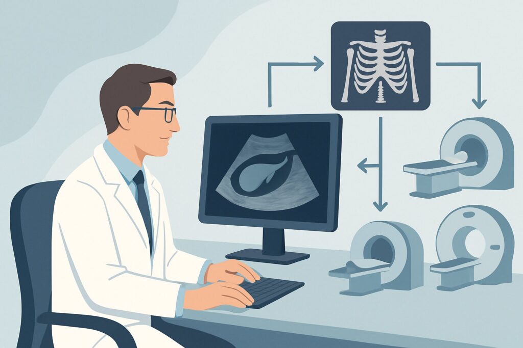
It’s 2 AM in the intensive care unit. Your patient, who is five days post-cardiac surgery, develops a new fever, leukocytosis, and right upper quadrant tenderness. A bedside ultrasound is performed, showing a distended gallbladder without stones, sludge, or definitive sonographic signs of cholecystitis. The clinical suspicion for acalculous cholecystitis is high, but the initial imaging is equivocal. This scenario presents a critical diagnostic challenge: what is the most appropriate next step to confirm or exclude a life-threatening condition? This article provides a detailed clinical workflow for this specific situation, guiding your choice for the next imaging study. Based on the American College of Radiology (ACR) Appropriateness Criteria, a Nuclear medicine scan gallbladder is rated Usually appropriate.
Who Fits This Clinical Scenario?
This guidance is specifically for patients with right upper quadrant (RUQ) pain where there is a strong clinical suspicion for acalculous cholecystitis, and an initial ultrasound has returned negative or equivocal results. The typical patient profile is often a critically ill individual who is unable to provide a clear history. Key inclusion criteria include:
- Significant systemic illness (e.g., sepsis, major trauma, burns, recent major surgery).
- Prolonged fasting or receiving total parenteral nutrition (TPN).
- New onset of fever, elevated white blood cell (WBC) count, and RUQ tenderness.
- An initial RUQ ultrasound that fails to show gallstones or specific signs of acute cholecystitis (like sonographic Murphy sign, significant wall thickening, or pericholecystic fluid).
This workflow is distinct from other clinical presentations. This article does not apply if:
- Gallstones are clearly identified on ultrasound. This would be suspected calculous cholecystitis, a different diagnostic pathway.
- The patient is ambulatory with no fever or elevated WBC count. This presentation points toward a different sibling scenario, often involving biliary dyskinesia or chronic cholecystitis.
- The ultrasound is definitively positive for acute cholecystitis. In that case, further diagnostic imaging is often unnecessary, and the focus shifts to treatment.
What Diagnoses Are You Working Up in This Scenario?
When an ultrasound is unrevealing in a critically ill patient with RUQ pain, the differential diagnosis remains broad but is centered on urgent conditions. The choice of the next imaging study is intended to differentiate between these possibilities.
Acalculous Cholecystitis: This is the primary diagnosis of concern. It represents gallbladder inflammation in the absence of gallstones, typically caused by biliary stasis and gallbladder ischemia. It accounts for a fraction of all acute cholecystitis cases but carries a much higher morbidity and mortality rate, making timely diagnosis essential. The lack of stones on ultrasound makes this a top consideration in this specific patient population.
Gangrenous Cholecystitis: A severe complication of acute cholecystitis (either calculous or acalculous) where the gallbladder wall becomes ischemic and necrotic. Ultrasound findings can be subtle or absent in early stages, and the condition can progress rapidly to perforation and peritonitis.
Biliary Sludge or Functional Obstruction: While not true stones, thick biliary sludge can obstruct the cystic duct, leading to a similar clinical picture. Ultrasound is not perfectly sensitive for detecting obstructing sludge, and a functional study may be needed to assess for impaired gallbladder emptying.
Non-Biliary Causes: An equivocal ultrasound means other diagnoses must be considered. These include a subhepatic or intrahepatic abscess, pancreatitis with peripancreatic fluid tracking superiorly, or even a right lower lobe pneumonia with diaphragmatic irritation. The next imaging study should ideally help evaluate for these alternatives if the primary diagnosis is excluded.
Why Is a Nuclear Medicine Gallbladder Scan the Recommended Study for This Presentation?
For a patient with suspected acalculous cholecystitis and a non-diagnostic ultrasound, the ACR designates a Nuclear medicine scan gallbladder (also known as cholescintigraphy or a HIDA scan) as Usually appropriate. The rationale is based on its ability to assess function rather than just anatomy.
A HIDA scan uses a radiotracer (e.g., 99mTc-mebrofenin) that is taken up by hepatocytes and excreted into the biliary system. The core diagnostic question in acalculous cholecystitis is whether the cystic duct is patent. If the radiotracer is seen in the common bile duct but the gallbladder fails to visualize within 60 minutes (or after 3-4 hours on delayed imaging), it signifies a functional obstruction of the cystic duct. This finding is highly sensitive and specific for acute cholecystitis in this clinical context.
In contrast, other imaging modalities are rated lower for this specific scenario:
- CT abdomen with IV contrast is rated May be appropriate. While excellent for identifying alternative pathologies like abscesses or pancreatitis, its findings for acalculous cholecystitis (e.g., gallbladder wall thickening, pericholecystic fluid) are often nonspecific in critically ill patients who may have generalized edema or ascites. It also carries a higher radiation dose (☢☢☢ 1-10 mSv) compared to cholescintigraphy (☢☢ 0.1-1mSv).
- MRI abdomen without and with IV contrast with MRCP is also rated May be appropriate. MRI provides outstanding soft-tissue detail and can diagnose cholecystitis and its complications without ionizing radiation (O 0 mSv). However, it is often logistically challenging for unstable, ventilated patients in the ICU, requiring transport and longer acquisition times. Its primary role is often reserved for complex cases or when the diagnosis remains unclear after other tests.
The HIDA scan directly answers the key pathophysiologic question, making it the most direct and efficient next step. When ordering, ensure the patient has been fasting for at least 4-6 hours to promote gallbladder filling, and be aware that patients on TPN or who have fasted for over 24 hours may require pre-treatment with sincalide (CCK) to empty a sludge-filled gallbladder before the scan.
What’s Next After a Nuclear Medicine Gallbladder Scan? Downstream Workflow
The results of the cholescintigraphy scan will guide your next clinical actions. The workflow typically branches into three paths based on the findings.
If the study is positive (non-visualization of the gallbladder): This result confirms cystic duct obstruction and, in this clinical setting, establishes the diagnosis of acute acalculous cholecystitis. The immediate next step is an urgent consultation with general surgery or interventional radiology. Treatment options include emergent cholecystectomy (if the patient is a surgical candidate) or, more commonly in critically ill patients, percutaneous cholecystostomy (image-guided drainage of the gallbladder) to control the source of sepsis.
If the study is negative (visualization of the gallbladder): A normal HIDA scan, where the gallbladder fills with the radiotracer, effectively rules out acute acalculous cholecystitis with a very high negative predictive value. This result should prompt you to aggressively search for an alternative diagnosis. The workflow now pivots to investigating other causes of sepsis or RUQ pain. This is an appropriate time to consider a CT of the abdomen and pelvis to look for an abscess, pancreatitis, bowel pathology, or other non-biliary sources that may have been missed on the initial ultrasound.
If the study is indeterminate: Occasionally, the scan may be equivocal. This can happen in patients with severe liver dysfunction, leading to poor tracer excretion. In some cases, morphine augmentation can be used; morphine causes sphincter of Oddi contraction, which increases pressure in the common bile duct and promotes tracer flow into the gallbladder if the cystic duct is patent. If the diagnosis remains uncertain after cholescintigraphy, a discussion between the clinical team, radiology, and surgery is warranted to decide between proceeding with a different imaging modality like CT/MRI or moving directly to empiric treatment like cholecystostomy if the patient is deteriorating.
Pitfalls to Avoid (and When to Get Help)
Navigating this diagnostic pathway requires careful attention to detail to avoid common errors. Be mindful of these potential pitfalls:
- Improper Patient Preparation: A HIDA scan can yield a false-positive result if the patient has eaten recently (gallbladder is already contracted) or has been fasting for too long (gallbladder is full of viscous bile). Ensure NPO status is appropriate (4-6 hours) and consider CCK pre-treatment for prolonged fasting.
- Ignoring Clinical Deterioration: While cholescintigraphy is the best diagnostic test, it should not unduly delay treatment in a rapidly decompensating septic patient. If the patient is in shock, it may be more appropriate to proceed directly to empiric image-guided cholecystostomy.
- Over-reliance on a Single Negative Study: While a negative ultrasound and a negative HIDA scan make acalculous cholecystitis highly unlikely, they do not exclude gangrenous cholecystitis, which can occur with a patent cystic duct. If clinical suspicion remains high despite negative functional imaging, escalate to CT or MRI to evaluate the gallbladder wall for signs of necrosis.
If the clinical picture and imaging results are discordant, or if the patient continues to decline, escalate immediately by involving surgical and interventional radiology colleagues for a multidisciplinary discussion on the next steps.
Related ACR Topics and Tools
This article focuses on a single, specific clinical scenario. For a comprehensive overview of all variants and initial imaging strategies for right upper quadrant pain, or to explore the technical details of the recommended studies, the following resources are valuable.
- For breadth across all scenarios in Right Upper Quadrant Pain, see our parent guide: Right Upper Quadrant Pain: ACR Appropriateness Decoded.
- To look up appropriateness ratings for adjacent or different clinical scenarios, use the ACR Appropriateness Criteria Lookup.
- For detailed procedural steps and technical parameters for various imaging studies, consult the Imaging Protocol Library.
- To discuss radiation exposure with patients and track cumulative dose, the Radiation Dose Calculator is a helpful tool.
Frequently Asked Questions
Why not just order a CT scan first after a negative ultrasound?
While a CT scan is rated ‘May be appropriate,’ a nuclear medicine gallbladder scan (HIDA) is ‘Usually appropriate’ because it directly assesses cystic duct patency—the key functional deficit in acalculous cholecystitis. CT findings like wall thickening can be nonspecific in critically ill patients. The HIDA scan provides a more definitive yes/no answer for the primary diagnosis in question and involves less radiation.
What is morphine augmentation during a HIDA scan?
If the gallbladder does not visualize after 60 minutes but tracer is seen in the small bowel, a small dose of intravenous morphine can be administered. Morphine causes the sphincter of Oddi to contract, increasing pressure within the common bile duct. This pressure gradient promotes the flow of the radiotracer into the gallbladder if the cystic duct is patent, shortening the scan time and increasing specificity.
What if my patient cannot be NPO (nothing by mouth) for the required time?
This is a significant challenge. If a patient has eaten within 4 hours of the planned scan, the test will likely be non-diagnostic (false positive) as the gallbladder will be contracted. The scan must be delayed until an adequate fasting period is achieved. In an emergent situation where this is not possible, another modality like CT or MRI may need to be considered, with the understanding that they are less specific for this particular diagnosis.
Does this guidance apply to pediatric patients?
Acalculous cholecystitis is much less common in children but can occur, particularly in those with critical illness, congenital heart disease, or on long-term TPN. The diagnostic principles are similar, and cholescintigraphy remains the most accurate non-invasive test. However, all imaging decisions in pediatric patients should be made in consultation with a pediatric radiologist, and radiation dose should be carefully managed using ‘As Low As Reasonably Achievable’ (ALARA) principles.
Can a HIDA scan be performed at the bedside for an unstable ICU patient?
Yes, portable nuclear medicine cameras are available and can perform cholescintigraphy at the bedside in the ICU. This avoids the risks associated with transporting a critically ill, ventilated patient to the nuclear medicine department, making it a practical choice for this specific patient population.
Reviewed by Pouyan Golshani, MD, Interventional Radiologist — May 29, 2026