What Is the Best Next Imaging Study for Suspected Inflammatory Arthritis When Radiographs Are Normal?
A 42-year-old patient presents with six months of progressive, symmetric pain and swelling in her metacarpophalangeal (MCP) and proximal interphalangeal (PIP) joints. Morning stiffness lasts over an hour. Her initial workup reveals an elevated C-reactive protein (CRP) and positive anti-cyclic citrullinated peptide (anti-CCP) antibodies, but her hand radiographs are reported as normal. You strongly suspect early rheumatoid arthritis, but the inconclusive initial imaging leaves you at a clinical decision point. How do you confirm active inflammation and detect early changes to guide treatment initiation? This article provides a detailed workflow for this specific scenario, grounded in the American College of Radiology (ACR) Appropriateness Criteria, which rates ultrasound of the area of interest as Usually Appropriate for this presentation.
Who Fits This Clinical Scenario?
This guidance is for clinicians evaluating a patient with chronic extremity joint pain where inflammatory arthritis is the leading suspicion, but initial radiographs are unrevealing. The key inclusion criteria are:
- Symptom Duration: The joint pain is chronic, typically lasting six weeks or longer.
- Clinical Suspicion: The history and physical exam suggest an inflammatory process (e.g., morning stiffness, joint swelling, symmetric involvement) over a mechanical or degenerative one. This may be supported by elevated inflammatory markers or positive serologies (e.g., rheumatoid factor, anti-CCP), but also applies to seronegative presentations.
- Initial Imaging: Conventional radiographs of the symptomatic joints have been performed and are either normal or show non-specific findings, such as soft tissue swelling without erosions or definitive joint space narrowing.
This workflow is not intended for patients where the primary suspicion is for crystalline arthropathy or erosive osteoarthritis. If you suspect gout based on a classic presentation (e.g., acute monoarthritis of the first metatarsophalangeal joint), a different imaging pathway may be more appropriate. Similarly, if the clinical picture points toward erosive osteoarthritis (e.g., “gull-wing” deformities in a patient without inflammatory markers), the diagnostic considerations and imaging rationale differ.
What Diagnoses Are You Working Up in This Scenario?
When initial radiographs are negative in a patient with suspected inflammatory arthritis, advanced imaging aims to detect early pathological changes that precede the bone destruction visible on X-ray. The primary goal is to confirm inflammation, identify its specific features, and establish a baseline for monitoring treatment response.
Rheumatoid Arthritis (RA): This is often the primary consideration, especially in cases of symmetric polyarthritis of the small joints of the hands and feet with positive RF or anti-CCP antibodies. Advanced imaging is tasked with identifying synovitis (inflammation of the joint lining), tenosynovitis (inflammation of tendon sheaths), and subtle bone erosions or bone marrow edema that are invisible on radiographs. Detecting these features early is critical for initiating disease-modifying antirheumatic drugs (DMADs) to prevent irreversible joint damage.
Psoriatic Arthritis (PsA): A key seronegative spondyloarthropathy, PsA can present with a variety of patterns, including a symmetric polyarthritis mimicking RA. Imaging in suspected PsA focuses on identifying not only synovitis but also characteristic features like enthesitis (inflammation where tendons and ligaments attach to bone), dactylitis (“sausage digit”), and tenosynovitis. These findings can help differentiate it from RA.
Other Seronegative Spondyloarthropathies: Conditions like reactive arthritis or enteropathic arthritis can also manifest with peripheral joint inflammation. While the clinical history is paramount, imaging can confirm the presence of active synovitis or enthesitis, supporting the diagnosis when systemic features are ambiguous.
Undifferentiated Inflammatory Arthritis: In some patients, symptoms and signs are suggestive of an inflammatory process, but they do not meet the full classification criteria for a specific disease. In this context, imaging serves to confirm the presence of objective inflammation (e.g., synovitis with hyperemia on power Doppler ultrasound), justifying a provisional diagnosis and potential trial of therapy while monitoring for evolution into a defined condition.
Why Is Ultrasound of the Area of Interest the Recommended Study for This Presentation?
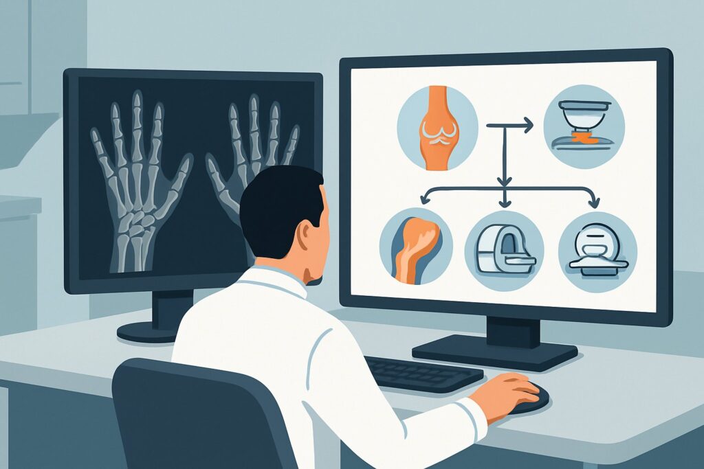
For a patient with suspected inflammatory arthritis and normal radiographs, both ultrasound (US) and magnetic resonance imaging (MRI) are rated as Usually Appropriate by the ACR. However, ultrasound is often the preferred initial advanced imaging modality due to its unique advantages in this clinical context.
Ultrasound provides high-resolution, real-time visualization of soft tissues and bone surfaces. Its primary strength is the direct assessment of synovitis, which appears as synovial hypertrophy and joint effusion. Critically, the use of power Doppler with US can detect synovial hyperemia, a direct indicator of active inflammation. This capability is highly sensitive for active disease and can help guide treatment decisions. US is also superior to radiographs in detecting very early bone erosions, which appear as distinct cortical breaks. Furthermore, it excels at identifying tenosynovitis and enthesitis, key features of spondyloarthropathies.
MRI without or with IV contrast is also Usually Appropriate and offers excellent sensitivity for synovitis, tenosynovitis, and bone marrow edema—a potential precursor to erosions. It provides a more comprehensive, multi-planar view of the entire joint and surrounding structures. However, US is often more accessible, less expensive, and does not involve exposure to gadolinium-based contrast agents. It also allows for a dynamic examination and easy comparison with the contralateral, asymptomatic side in real time.
Alternative modalities are rated lower for specific reasons:
- Bone Scan (Whole Body or with SPECT/CT): This is rated Usually Not Appropriate. While sensitive to bone turnover, it is highly non-specific. Increased radiotracer uptake can be caused by inflammation, degeneration, trauma, or infection, and the modality cannot differentiate between these causes or visualize the specific soft-tissue findings of inflammatory arthritis. It also involves significant radiation exposure (☢☢☢ 1-10 mSv).
- Computed Tomography (CT): CT (with or without contrast) is also Usually Not Appropriate. While excellent for evaluating cortical bone and established erosions, it has poor soft-tissue contrast resolution compared to US and MRI, making it insensitive for detecting early synovitis or bone marrow edema.
Both US and MRI are radiation-free (0 mSv), a significant advantage for a patient population that may require serial imaging over time to monitor disease activity and treatment response.
What’s Next After Ultrasound? Downstream Workflow
The results of the ultrasound will guide your next steps, moving you from suspicion to diagnosis and management.
If the study is positive for inflammatory arthritis: Findings of definite synovitis (especially with a positive power Doppler signal), tenosynovitis, or erosions confirm an active inflammatory process. In a patient with a strong clinical and serologic picture for RA, these findings are often sufficient to establish the diagnosis and initiate DMARD therapy. The imaging results provide an objective baseline to assess treatment efficacy on follow-up examinations. Consultation with a rheumatologist is the standard next step to formalize the diagnosis and management plan.
If the study is negative: A technically adequate ultrasound that shows no evidence of synovitis, tenosynovitis, or erosions in the most symptomatic joints makes active inflammatory arthritis in those specific locations less likely. The next step is to reconsider the differential diagnosis. Could the symptoms be due to a non-inflammatory condition like fibromyalgia or early, non-erosive osteoarthritis? If clinical suspicion for inflammatory disease remains high despite the negative scan, consider imaging a different, clinically active joint region. In some cases, an MRI may be considered to look for bone marrow edema, which can be missed on US.
If the study is indeterminate: Ambiguous findings, such as minimal synovial thickening without a power Doppler signal or non-specific fluid, can be challenging. In these cases, correlation with the clinical picture is essential. A follow-up US in several weeks could clarify the situation, or an MRI could be pursued for its high sensitivity and comprehensive view. Consultation with a musculoskeletal radiologist or rheumatologist can help interpret borderline findings in the correct clinical context.
Pitfalls to Avoid (and When to Get Help)
Navigating the workup for early inflammatory arthritis requires careful attention to detail to avoid common missteps.
- Technique and Operator Dependence: The quality of a musculoskeletal ultrasound is highly dependent on the skill of the sonographer and the quality of the equipment. Ensure the study is performed at a center with expertise in musculoskeletal US.
- Incomplete Examination: When ordering, be specific about the joints to be evaluated. A comprehensive scan should assess for synovitis, tenosynovitis, and erosions in a standardized manner. Failure to use power Doppler is a critical omission, as it is essential for assessing active inflammation.
- Imaging Asymptomatic Joints: The diagnostic yield is highest when imaging the most clinically active and symptomatic joints. A negative scan of a non-tender joint does not rule out disease elsewhere.
- Over-interpreting Degenerative Changes: Osteoarthritic changes can sometimes be confused with inflammatory findings. Differentiating small osteophytes from erosions requires experience.
If the imaging findings are discordant with the clinical picture, or if they suggest an alternative serious diagnosis like infection or malignancy, prompt escalation to a rheumatologist is warranted for further evaluation and management.
Related ACR Topics and Tools
The ACR Appropriateness Criteria are a powerful resource for evidence-based imaging decisions. For a broader overview of related scenarios, please consult our parent topic guide. For additional tools to refine your imaging orders and discuss them with patients, see the resources below.
- For breadth across all scenarios in Chronic Extremity Joint Pain-Suspected Inflammatory Arthritis, Crystalline Arthritis, or Erosive Osteoarthritis, see our parent guide: Chronic Extremity Joint Pain-Suspected Inflammatory Arthritis, Crystalline Arthritis, or Erosive Osteoarthritis: ACR Appropriateness Decoded.
- To explore other clinical scenarios, use the ACR Appropriateness Criteria Lookup.
- For details on imaging techniques, visit the Imaging Protocol Library.
- To help with patient conversations about medical radiation, use the Radiation Dose Calculator.
Frequently Asked Questions
Why not just order an MRI, since it is also rated ‘Usually Appropriate’?
While MRI is an excellent modality, ultrasound is often preferred as the first advanced imaging step because it is typically less expensive, more widely available, and can be performed more quickly. It also allows for a dynamic assessment of joints and tendons and a real-time comparison with the patient’s asymptomatic side. MRI is a superb problem-solving tool, especially if US is negative or equivocal and clinical suspicion remains high.
What if my patient has a pacemaker or other contraindication to MRI?
In cases where a patient has a contraindication to MRI, such as an incompatible pacemaker, cochlear implant, or certain metallic foreign bodies, ultrasound becomes the definitive advanced imaging modality of choice. It provides outstanding soft tissue and cortical bone detail without the risks associated with strong magnetic fields.
Does the choice of imaging change if the patient is seronegative (RF and anti-CCP negative)?
No, the imaging recommendation remains the same. In seronegative patients, objective evidence of inflammation on imaging is even more critical to support the diagnosis. Ultrasound or MRI can confirm the presence of synovitis or enthesitis, helping to diagnose conditions like seronegative RA or psoriatic arthritis.
How specific should my order for ultrasound be?
Your order should be as specific as possible to maximize diagnostic yield. Instead of ordering a generic ‘ultrasound of hands,’ specify the clinical concern and the joints of interest (e.g., ‘Ultrasound of bilateral hands and wrists to evaluate for synovitis and erosions; clinical concern for early inflammatory arthritis. Please evaluate MCPs, PIPs, and wrists with power Doppler.’).
Can ultrasound be used to monitor response to treatment?
Yes, ultrasound is an excellent tool for monitoring treatment response. A decrease in synovial hypertrophy and, most importantly, a reduction or resolution of the power Doppler signal can provide objective evidence that a treatment (like a DMARD or biologic) is working, often before the patient reports significant symptomatic improvement.
Reviewed by Pouyan Golshani, MD, Interventional Radiologist — May 30, 2026