What Is the First Imaging Study for Knee Trauma with Patellar Tenderness?
A 14-year-old presents to urgent care after an awkward fall during a basketball game. The exact mechanism is unclear, but he complains of direct pain over his kneecap. He was able to limp off the court and can bear weight, but your exam reveals focal tenderness directly over the patella and a moderate joint effusion. You suspect a possible fracture, but what is the most appropriate first step to confirm or exclude this diagnosis without ordering unnecessary, higher-cost studies? This article provides a detailed clinical workflow for this specific scenario, guided by the American College of Radiology (ACR) Appropriateness Criteria. For this presentation, the ACR designates Radiography knee as Usually appropriate for initial imaging.
Who Fits This Clinical Scenario for Acute Knee Trauma?
This imaging pathway is designed for a specific subset of patients with acute knee injuries. The key inclusion criteria are:
- The patient is an adult or a child aged 5 years or older.
- There has been an acute traumatic event, but the precise mechanism is unknown or poorly described.
- The physical exam reveals focal patellar tenderness and a knee effusion.
- Crucially, the patient is able to walk or bear weight, even if with a limp.
It is critical to distinguish this presentation from similar but distinct clinical scenarios that follow different imaging guidelines. This workflow does not apply if:
- The patient cannot bear weight: An inability to bear weight for four steps is a key component of the Ottawa Knee Rules and suggests a higher likelihood of a significant fracture, potentially altering the imaging workup.
- Tenderness is located elsewhere: If tenderness is isolated to the fibular head or tibial plateau without patellar involvement, the differential diagnosis shifts, and different ACR variants may apply.
- There was a high-energy mechanism: Patients injured in a motor vehicle accident or who have suffered a knee dislocation require a more comprehensive evaluation, often involving advanced imaging like Computed Tomography (CT) to assess for complex fractures or vascular injury.
What Diagnoses Are You Working Up in This Scenario?
With focal patellar tenderness and an effusion after trauma, the clinical question centers on ruling out specific, consequential injuries. The initial imaging choice is tailored to identify or exclude these primary concerns.
Patellar Fracture
This is the most direct and common concern given the exam findings. A direct blow or forceful quadriceps contraction can cause a transverse, vertical, or comminuted fracture of the patella. Standard radiographs are highly effective at visualizing these fractures and are the cornerstone of diagnosis.
Osteochondral Fracture
An acute patellar dislocation or subluxation event, even if it spontaneously reduced and was not witnessed, can shear off a fragment of articular cartilage and underlying bone from the patella or lateral femoral condyle. These intra-articular bodies can cause mechanical symptoms and require intervention. Radiographs can often detect the bony fragment.
Quadriceps or Patellar Tendon Rupture
While Magnetic Resonance Imaging (MRI) is the definitive study for tendon evaluation, a complete rupture of the extensor mechanism can be suggested on radiographs. A high-riding patella (patella alta) suggests a patellar tendon rupture, while a low-riding patella (patella baja) suggests a quadriceps tendon rupture. These are critical diagnoses not to miss.
Traumatic Hemarthrosis from Internal Derangement
The presence of an effusion, which is likely a hemarthrosis in the setting of acute trauma, indicates a significant intra-articular injury. While radiographs cannot directly visualize ligaments or menisci, they are the essential first step to rule out an associated fracture (e.g., a Segond fracture with an Anterior Cruciate Ligament tear) before considering further imaging like MRI.
Why Is Knee Radiography the Recommended First Study for This Presentation?
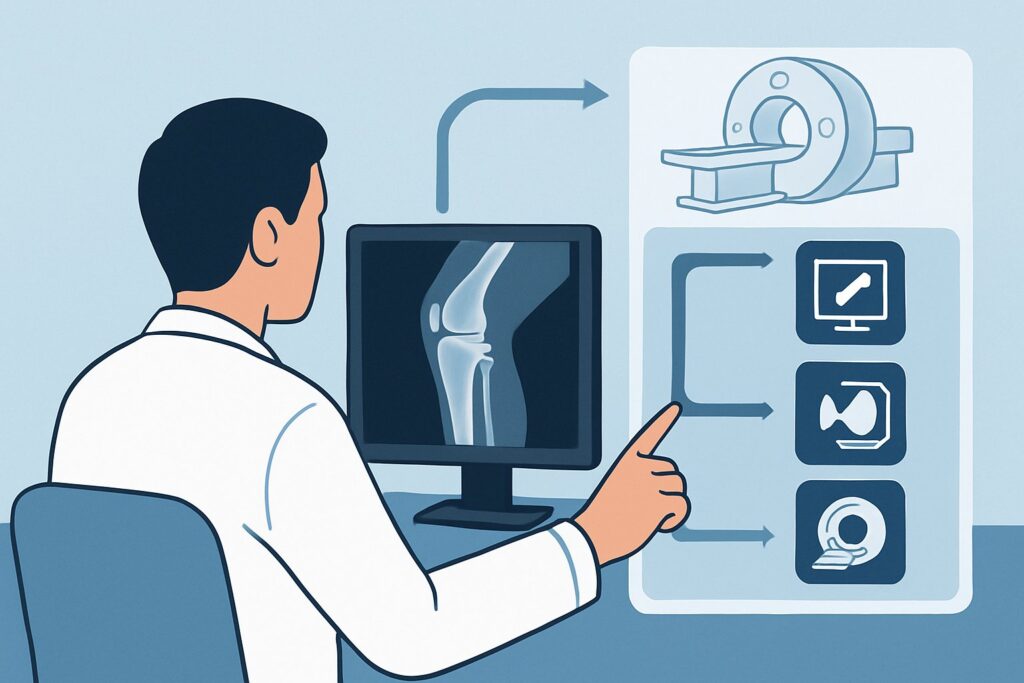
The ACR designates Radiography knee as Usually appropriate for this clinical scenario because it directly, efficiently, and safely addresses the most pressing diagnostic questions. A standard knee series, which typically includes anteroposterior (AP), lateral, and often sunrise (or merchant) views, provides a wealth of information.
The primary rationale is the high diagnostic yield for fractures, particularly of the patella, which is the main concern based on the physical exam. Radiographs are widely available, inexpensive, and quick to perform, making them the ideal initial screening tool. The lateral view is particularly valuable for assessing joint effusion and evaluating the position of the patella to screen for major extensor mechanism injuries. The sunrise view provides an unimpeded look at the patella and the patellofemoral joint, crucial for identifying subtle vertical fractures or osteochondral defects.
In contrast, more advanced imaging modalities are rated lower for this initial evaluation:
- MRI knee without IV contrast is rated Usually not appropriate as the first study. While MRI provides exquisite detail of soft tissues like ligaments, menisci, and cartilage, it is not necessary to rule out a simple patellar fracture. It is more costly, takes longer to acquire, and has limited availability in many acute care settings. MRI is reserved for cases where radiographs are negative but high clinical suspicion for a significant soft tissue injury persists.
- CT knee without IV contrast is also rated Usually not appropriate for initial imaging. CT offers superior bony detail compared to radiographs but exposes the patient to a higher radiation dose. Its role is typically in the post-radiograph setting to better characterize complex, already-diagnosed fractures for surgical planning, not as a first-line screening tool for this presentation.
The radiation dose from a knee radiograph series is minimal. For an adult, the relative radiation level (RRL) is ☢ (<0.1 mSv), and for a child, it is even lower at ☢ (<0.03 mSv), far below the level of routine background radiation exposure over a few days.
What’s Next After Radiography knee? Downstream Workflow
The results of the initial knee radiographs will dictate the subsequent clinical pathway. The decision tree branches based on whether the study is positive, negative, or indeterminate.
If Radiographs Are Positive for Fracture
A definitive fracture of the patella or other bone requires orthopedic consultation. The management will depend on the fracture pattern, specifically the amount of displacement and the integrity of the extensor mechanism. A non-displaced fracture may be managed non-operatively with immobilization, while a displaced or comminuted fracture often requires surgical fixation.
If Radiographs Are Negative
If no fracture is identified, the focus shifts to managing a presumed soft tissue injury. The patient can typically be managed with rest, ice, compression, and elevation (RICE), along with analgesics. The key next step is follow-up. If symptoms of instability, locking, or persistent pain and effusion develop despite conservative management, the patient may fit a different clinical scenario: Suspect occult fracture or internal derangement after negative radiographs. At that point, an MRI of the knee without contrast often becomes the appropriate next step to evaluate for ligamentous, meniscal, or chondral injury.
If Radiographs Are Indeterminate
Occasionally, radiographs may be equivocal, perhaps showing a questionable cortical irregularity or an unusual ossicle. If clinical suspicion for a fracture remains high despite unclear radiographs (e.g., the patient develops an inability to perform a straight leg raise), further imaging may be warranted. Depending on the specific question, this could involve a CT scan for better bony characterization or an MRI to assess both bone and soft tissues.
Pitfalls to Avoid (and When to Get Help)
In this specific scenario, several common pitfalls can lead to diagnostic delays or errors. Be mindful of the following:
- Incomplete Radiographic Series: Failing to obtain a sunrise (patellar) view can cause you to miss a non-displaced vertical patellar fracture, which is often invisible on AP and lateral views.
- Misinterpreting a Bipartite Patella: A bipartite patella is a common congenital variant where the patella exists in two pieces. It is typically located superolaterally, has well-corticated (smooth) edges, and is often bilateral. Mistaking this for an acute fracture can lead to unnecessary immobilization and consultation.
- Ignoring Patellar Position: Overlooking a high-riding (patella alta) or low-riding (patella baja) patella on the lateral view can lead to a missed diagnosis of a functionally devastating quadriceps or patellar tendon rupture.
- Dismissing a Lipohomarthrosis: On a cross-table lateral radiograph, a fat-fluid level within the effusion (lipohomarthrosis) is a specific sign of an intra-articular fracture, even if the fracture line itself is not visible.
If the patient has a suspected open fracture, neurovascular compromise, or develops signs of a compartment syndrome, this constitutes a surgical emergency requiring immediate orthopedic consultation.
Related ACR Topics and Tools
The ACR Appropriateness Criteria are extensive, and patient presentations often fall between defined scenarios. For a comprehensive overview of all knee trauma variants, from minor sprains to complex dislocations, please consult our parent guide. For tools to help with ordering, protocoling, and patient communication, see the resources below.
- For breadth across all scenarios in Acute Trauma to the Knee, see our parent guide: Acute Trauma to the Knee: ACR Appropriateness Decoded.
- To explore other clinical scenarios, use the Imaging Appropriateness Selector.
- For detailed technical parameters of imaging studies, visit the Imaging Protocol Library.
- To discuss radiation exposure with patients, use the Radiation Dose Calculator.
Frequently Asked Questions
Why not order an MRI first if I suspect a ligament or meniscus tear along with the patellar injury?
While an MRI is the best test for ligaments and menisci, the American College of Radiology recommends radiographs first in this scenario to rule out a fracture. A fracture is a more immediate concern that can alter management significantly. If radiographs are negative and clinical suspicion for a soft tissue injury remains high after a period of conservative care, an MRI then becomes the appropriate next step.
Is a sunrise (patellar) view always necessary for a knee radiograph series?
In the setting of focal patellar tenderness, a sunrise (or merchant) view is highly recommended. It provides the best visualization of the patella in the axial plane and is crucial for identifying vertical patellar fractures and assessing the patellofemoral articulation, which are often missed on standard AP and lateral views.
What if the patient is a child under 5 years old?
This specific ACR guideline applies to children 5 years of age and older. Younger children have different injury patterns and considerations, such as physeal (growth plate) injuries and plastic deformities of bone. Imaging decisions in very young children often require consultation with pediatric orthopedic or radiology specialists.
If the knee radiograph is negative, when should I have the patient follow up?
If initial radiographs are negative, the patient is typically treated for a soft tissue sprain or contusion. A follow-up appointment in 5-7 days is reasonable to reassess pain, swelling, and function. If there is no improvement or if new mechanical symptoms like locking or instability develop, further evaluation, potentially including an MRI, is warranted.
Does the presence of a large effusion alone justify an MRI if the radiograph is negative?
A large effusion (especially a hemarthrosis) after trauma indicates a significant intra-articular injury, but it does not automatically mean an MRI is needed immediately. The standard workflow is still to start with radiographs to rule out a fracture. If radiographs are negative, the effusion is managed symptomatically, and the decision for a subsequent MRI is based on the evolution of clinical symptoms like instability or persistent pain.
Reviewed by Pouyan Golshani, MD, Interventional Radiologist — June 27, 2026