What Is the Next Imaging Step for a Child with Back Pain and a Suspicious Radiograph?

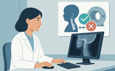
It’s late in the afternoon clinic when you see a 9-year-old with two weeks of worsening, non-traumatic back pain, now associated with a low-grade fever and refusal to bear weight. The initial spine radiographs you ordered are concerning, showing subtle vertebral endplate irregularity and disc space narrowing. You suspect an infectious, inflammatory, or neoplastic process. The immediate question is which advanced imaging study will provide a definitive diagnosis and guide urgent management. This article details the American College of Radiology (ACR) workflow for this specific scenario: a child with back pain, clinical red flags, and radiographic findings suspicious for a serious underlying pathology. For this presentation, the ACR rates MRI complete spine without and with IV contrast as Usually appropriate.
Who Fits This Clinical Scenario?
This guidance applies specifically to a pediatric patient presenting with back pain accompanied by at least one clinical “red flag” and abnormal or suspicious initial radiographs. The key inclusion criteria are:
- Patient Age: Child or adolescent.
- Symptoms: Back pain with one or more red flags, such as fever, night pain, weight loss, neurologic deficits (weakness, bowel/bladder dysfunction), constant or progressive pain, or elevated inflammatory markers (ESR, CRP).
- Prior Imaging: Radiographs (x-rays) have been performed and show findings suspicious for infection, inflammation, or malignancy. This could include vertebral body destruction, endplate changes, disc space narrowing, or a soft tissue mass.
This workflow is distinct from other common pediatric back pain presentations. This guidance does not apply if:
- Radiographs are negative: If the child has red flags but the initial x-rays are completely normal, the next imaging step is covered in a different ACR variant.
- There are no red flags: For a child with mechanical-sounding back pain and no systemic symptoms, the initial imaging approach is much more conservative.
- A specific skin lesion is present: Back pain associated with a palpable lump, hairy patch, or draining sinus suggests a congenital spinal anomaly, which has its own dedicated imaging pathway.
What Diagnoses Are You Working Up in This Scenario?
When a child presents with back pain, red flags, and suspicious radiographs, the differential diagnosis narrows to serious conditions that require prompt identification. The primary goal of advanced imaging is to differentiate among these possibilities and define the extent of disease.
Discitis and Vertebral Osteomyelitis: This is a leading concern in children with fever, focal back pain, and refusal to walk. Infection of the intervertebral disc (discitis) and adjacent vertebral bodies (osteomyelitis) can progress to epidural abscess formation and neurologic compromise. Imaging must clearly delineate the disc, vertebral marrow, and surrounding soft tissues.
Primary Bone Tumors: Malignant primary bone tumors, while not common, can present this way. Ewing sarcoma and osteosarcoma are key considerations. Benign but aggressive tumors like osteoid osteoma or aneurysmal bone cysts are also on the differential. Imaging is critical for characterizing the lesion, assessing for soft tissue extension, and guiding biopsy.
Metastatic Disease or Leukemia: The spine is a common site for metastases from other childhood cancers, particularly neuroblastoma and rhabdomyosarcoma. Leukemia can also infiltrate the vertebral marrow, causing pain and pathologic fractures. Imaging must be sensitive enough to detect marrow replacement and screen for additional lesions.
Inflammatory Spondyloarthropathy: Less commonly, a severe inflammatory process like juvenile idiopathic arthritis can manifest with significant back pain and radiographic changes. Imaging helps identify active inflammation (e.g., sacroiliitis, synovitis) that confirms a rheumatologic diagnosis.
Why Is MRI of the Complete Spine Without and With IV Contrast the Recommended Study?
The ACR designates MRI complete spine without and with IV contrast as Usually appropriate because it offers the highest diagnostic utility for the key differential diagnoses in this scenario, all without using ionizing radiation.
The rationale is multi-faceted. MRI provides unparalleled soft tissue and bone marrow contrast. Pre-contrast sequences (T1, T2, STIR/fat-suppressed T2) are highly sensitive for detecting edema in the bone marrow, disc spaces, and paraspinal soft tissues—the hallmark of infection and inflammation. They can also delineate the anatomic extent of a tumor. The addition of intravenous gadolinium-based contrast is crucial for differentiating a phlegmon from a drainable fluid collection in an abscess, characterizing tumor enhancement patterns, and assessing for leptomeningeal disease spread.
Evaluating the “complete spine” is often recommended over a focused “area of interest” because infectious and neoplastic processes in children can be multifocal. A complete spine survey prevents missing skip lesions or identifying an unsuspected primary site of disease.
Why are other studies rated lower?
- CT of the spine, rated May be appropriate, provides excellent bony detail but has poor soft tissue contrast, making it difficult to evaluate early osteomyelitis, discitis, or epidural abscess. It also involves significant ionizing radiation (Pediatric RRL: ☢☢☢☢ 3-10 mSv), a major consideration in children.
- Technetium-99m bone scan, also rated May be appropriate, is sensitive for detecting areas of increased bone turnover but is not specific. It cannot distinguish between infection, tumor, trauma, or inflammation. Furthermore, it provides poor anatomic detail and also involves radiation (Pediatric RRL: ☢☢☢☢ 3-10 mSv).
Ultimately, MRI provides the most comprehensive, safest, and diagnostically powerful information in a single study. Once you’ve decided on an MRI, understanding the specific sequences is key. For a detailed look at the foundational non-contrast sequences, our protocol guide provides a helpful overview of the technique and reading principles: MRI Lumbar Spine Without Contrast.
What’s Next After MRI of the Complete Spine? Downstream Workflow
The results of the contrast-enhanced spine MRI will dictate the subsequent clinical pathway. The goal is to move from diagnosis to definitive management, which often requires a multidisciplinary team.
- If the MRI is positive for infection (discitis/osteomyelitis/abscess): This is a medical emergency. The next steps involve urgent consultation with pediatric infectious disease and potentially pediatric neurosurgery or interventional radiology. Blood cultures should be drawn, and empiric intravenous antibiotics are typically initiated. If a drainable epidural or paraspinal abscess is identified, surgical or percutaneous drainage is required to prevent permanent neurologic injury.
- If the MRI is positive for a tumor: An urgent referral to pediatric oncology is paramount. The imaging characteristics will help narrow the differential, but a biopsy is almost always required for definitive histopathologic diagnosis. The MRI findings are crucial for staging and surgical planning.
- If the MRI is negative: A negative, high-quality MRI makes a serious underlying infection or tumor highly unlikely. The workup may then pivot toward rheumatologic or mechanical causes. Consultation with pediatric rheumatology may be appropriate if inflammatory markers remain elevated. If the pain persists without a clear cause, the patient may fit the criteria for the ACR scenario concerning back pain with red flags but negative advanced imaging.
- If the MRI is indeterminate: In rare cases, findings may be equivocal. This may prompt a discussion with the pediatric radiologist about the utility of a follow-up MRI, a different imaging modality like a bone scan (rated May be appropriate), or proceeding directly to biopsy if clinical suspicion remains high.
Pitfalls to Avoid (and When to Get Help)
In this high-stakes clinical scenario, several common pitfalls can delay diagnosis or lead to suboptimal outcomes. Be mindful of the following:
- Ordering a limited MRI: Requesting an MRI of only the lumbar spine when the pathology could be thoracic or cervical can miss the diagnosis. Unless pain is exquisitely localized, a complete spine survey is safer.
- Omitting IV contrast: Forgetting to order the MRI “without and with contrast” severely limits its ability to characterize infection and tumors. An abscess cannot be reliably distinguished from a phlegmon without contrast.
- Delaying the scan: In the presence of neurologic deficits, fever, or severe, unremitting pain, MRI should be obtained emergently. Delay can lead to irreversible spinal cord or nerve root damage.
- Misinterpreting normal developmental variants: Normal pediatric spinal anatomy, such as red marrow patterns and vertebral body shape, can sometimes mimic pathology. Always review the images with an experienced pediatric radiologist.
If there are any signs of rapidly progressing neurologic deficit (e.g., new-onset weakness, loss of bowel/bladder control), escalate immediately to a pediatric neurosurgeon for emergent evaluation, which should not be delayed for imaging.
Related ACR Topics and Tools
This article covers a single, specific clinical workflow. For a comprehensive overview of all pediatric back pain scenarios and to explore adjacent clinical questions, refer to the resources below.
- For breadth across all scenarios in Back Pain-Child, see our parent guide: Back Pain-Child: ACR Appropriateness Decoded.
- To explore other ACR guidelines, use the ACR Appropriateness Criteria Lookup.
- To review technical details for this and other studies, see the Imaging Protocol Library.
- To discuss radiation exposure with families for alternative studies, the Radiation Dose Calculator can help frame the conversation.
Frequently Asked Questions
Why is a complete spine MRI recommended instead of just imaging the area of pain?
In children, serious conditions like infection (osteomyelitis) and tumors (leukemia, neuroblastoma) can be multifocal, meaning they appear in multiple locations. A ‘skip lesion’ could be missed if only the most painful area is imaged. A complete spine MRI provides a comprehensive survey to ensure no other sites of disease are overlooked, which is critical for accurate staging and treatment planning.
Is an MRI safe for a young child who may not be able to hold still?
Yes, but it often requires sedation or general anesthesia to ensure a high-quality, motion-free study. This is a standard procedure coordinated between the radiology department and pediatric anesthesiology. The benefits of obtaining a definitive diagnosis in this high-risk scenario far outweigh the risks associated with sedation for most patients.
If the initial radiograph is suspicious, can I skip straight to biopsy instead of getting an MRI?
No, an MRI is a critical pre-biopsy step. It provides the ‘roadmap’ for the surgeon or interventional radiologist, identifying the safest and most diagnostically fruitful location to sample. It also defines the full extent of the disease, which a biopsy cannot do, and helps differentiate between solid tumor, abscess, and inflammatory tissue, guiding whether a biopsy or a drainage procedure is needed.
What if my hospital doesn’t have immediate access to an MRI scanner?
If MRI is not immediately available and the child has signs of neurologic compromise or sepsis, urgent transfer to a tertiary care pediatric center is the most appropriate next step. If transfer is not possible and the clinical situation is deteriorating, a contrast-enhanced CT scan may be considered as a temporizing measure to look for gross abnormalities like bone destruction or a large epidural abscess, but it is a suboptimal study and should not delay transfer.
Are there any contraindications to a contrast-enhanced MRI in this scenario?
The primary contraindication is a severe allergy to gadolinium-based contrast agents, which is very rare. Additionally, caution is used in patients with severe renal dysfunction (though this is uncommon in otherwise healthy children). The ordering physician should always screen for these conditions before the study.
Reviewed by Pouyan Golshani, MD, Interventional Radiologist — May 30, 2026