What Is the Next Imaging Study for Suspected Biliary Disease After an Equivocal Ultrasound?
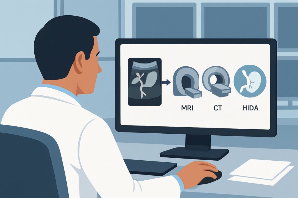
A 48-year-old woman presents to the emergency department at 2 a.m. with sharp right upper quadrant pain, a fever of 101.8°F, and a white blood cell count of 17,000/μL. You suspect an acute biliary process. The initial bedside ultrasound is equivocal—the gallbladder wall appears slightly thickened, but there are no definitive stones, no sonographic Murphy’s sign, and the common bile duct is non-dilated. The patient’s pain is worsening, and you need a definitive diagnosis to guide management. What is the next, most appropriate imaging study? This article provides a step-by-step clinical workflow for this exact scenario, grounded in the American College of Radiology (ACR) Appropriateness Criteria. For this presentation, the ACR rates MRI abdomen without and with IV contrast with MRCP as Usually Appropriate.
Who Fits This Clinical Scenario for Right Upper Quadrant Pain?
This guidance is specifically for patients who meet a distinct set of clinical and imaging criteria. The workflow applies when your patient presents with a clinical picture highly suggestive of an acute, infectious biliary process, but the initial, first-line imaging study has failed to provide a clear answer.
Inclusion Criteria (all must be met):
- Focal right upper quadrant (RUQ) pain.
- Systemic signs of inflammation or infection, such as fever and an elevated white blood cell (WBC) count (leukocytosis).
- A prior abdominal ultrasound that was either negative (showed no clear pathology) or equivocal (findings were indeterminate or non-diagnostic).
Exclusion Criteria (this guidance does not apply if):
- This is the initial imaging workup. For patients with suspected biliary disease who have not yet had an ultrasound, the initial imaging choice is different.
- The patient has no fever or leukocytosis. A patient with RUQ pain but no systemic signs of infection after a negative ultrasound falls into a different diagnostic algorithm, where the urgency and differential diagnosis shift.
- The initial ultrasound was definitively positive. If ultrasound clearly demonstrates gallstones, gallbladder wall thickening, pericholecystic fluid, and a sonographic Murphy’s sign, further diagnostic imaging is often unnecessary. The next step is typically a surgical consultation.
- Acalculous cholecystitis is the primary suspicion. While there is overlap, the workup for critically ill patients with suspected acalculous cholecystitis follows a more specific ACR pathway.
What Diagnoses Are You Working Up in This Scenario?
With an equivocal ultrasound in a febrile patient, your differential diagnosis must remain broad but focused on conditions that require timely intervention. The goal of the next imaging study is to differentiate between these possibilities, as management varies significantly.
Acute Cholecystitis
This remains the leading diagnosis. An ultrasound can be falsely negative or equivocal, especially in cases of a small, contracted gallbladder, obesity limiting visualization, or a stone impacted in the cystic duct that is not directly seen. The next imaging study must be highly sensitive for secondary signs of inflammation, such as wall edema, enhancement, and pericholecystic fluid.
Choledocholithiasis with or without Cholangitis
A stone in the common bile duct (CBD) is a crucial diagnosis to make. Ultrasound sensitivity for CBD stones can be low. If a stone is causing obstruction and stasis, it can lead to ascending cholangitis, a potentially life-threatening infection. The clinical triad of fever, RUQ pain, and jaundice (Charcot’s triad) is classic but not always present.
Hepatic Abscess
A pyogenic liver abscess can present identically to acute cholecystitis with fever, leukocytosis, and RUQ pain. Early abscesses or those located deep within the hepatic dome can be difficult to characterize on ultrasound. Misdiagnosing a liver abscess as simple cholecystitis could lead to a dangerous delay in percutaneous drainage or appropriate antibiotic therapy.
Complicated Cholecystitis
The initial ultrasound may not reveal the full extent of severe inflammation. Conditions like gangrenous cholecystitis (ischemia and necrosis of the gallbladder wall) or a contained perforation can be subtle on sonography but require more urgent surgical intervention.
Why Is MRI with MRCP the Recommended Study After an Equivocal Ultrasound?
When ultrasound is inconclusive, you need a high-resolution, multi-parametric imaging modality to solve the diagnostic puzzle. The ACR designates MRI abdomen without and with IV contrast with MRCP as a Usually Appropriate study, making it a top recommendation for this specific clinical challenge.
The power of this study lies in its combination of techniques. The Magnetic Resonance Cholangiopancreatography (MRCP) sequences are heavily T2-weighted, fluid-sensitive images that create a non-invasive map of the entire biliary tree. This provides exquisite detail for detecting even small, non-obstructing stones in the common bile duct or cystic duct that are frequently missed by both ultrasound and CT.
Following the MRCP, the administration of intravenous contrast allows for a comprehensive evaluation of the gallbladder and surrounding structures. Contrast-enhanced MRI sequences are highly sensitive for detecting gallbladder wall inflammation, edema, and enhancement patterns suggestive of acute cholecystitis. Furthermore, MRI excels at characterizing soft tissues, making it the superior modality for identifying complications like a hepatic abscess, gangrenous changes in the gallbladder wall, or subtle fluid collections from a perforation. This ability to confidently diagnose or exclude alternative pathologies is a key advantage.
Finally, MRI achieves this high diagnostic accuracy with no ionizing radiation (adult relative radiation level: O, 0 mSv), a significant benefit, particularly in younger patients or those who may require future imaging.
How do alternative studies compare?
- Nuclear medicine scan gallbladder (HIDA scan): This is also rated Usually Appropriate. A HIDA scan is a functional study that is excellent for answering one specific question: Is the cystic duct obstructed? Non-visualization of the gallbladder is highly specific for acute cholecystitis. However, it provides no anatomical information about alternative diagnoses like a liver abscess or choledocholithiasis. If the pre-test probability for cholecystitis is very high and other diagnoses are unlikely, HIDA is a strong choice. If the differential is broader, MRI is more comprehensive.
- CT abdomen with IV contrast: This is also rated Usually Appropriate and is often chosen for its speed and wide availability. CT is excellent for detecting complications like abscess formation or gallbladder perforation. However, its sensitivity for detecting gallstones (especially non-calcified ones) and subtle gallbladder wall inflammation is lower than that of MRI. It also involves significant ionizing radiation (adult relative radiation level: ☢☢☢, 1-10 mSv).
What’s Next After the MRI? Downstream Workflow
The results of the MRI will guide your next steps, providing a clear path for patient management. The downstream workflow depends directly on the findings.
- Positive for Acute Cholecystitis: If the MRI confirms acute cholecystitis (e.g., gallbladder wall thickening and enhancement, pericholecystic fluid, an impacted cystic duct stone), the next step is a prompt surgical consultation for cholecystectomy. If complications like gangrene or perforation are identified, the consultation becomes more urgent.
- Positive for Choledocholithiasis: If the MRCP identifies a stone in the common bile duct, the patient will require therapeutic intervention. This typically involves a gastroenterology consultation for Endoscopic Retrograde Cholangiopancreatography (ERCP) with sphincterotomy and stone extraction. This is especially urgent if there are signs of cholangitis.
- Positive for Hepatic Abscess: A confirmed liver abscess requires consultation with interventional radiology for percutaneous drain placement, along with tailored intravenous antibiotic therapy based on culture results.
- Negative or Non-diagnostic Study: If this high-level imaging study is negative for any acute pathology, it is time to reconsider the differential diagnosis. The focus should shift to non-biliary causes of RUQ pain, such as right-sided pyelonephritis, a penetrating duodenal ulcer, or musculoskeletal pain. Further workup would be guided by the evolving clinical picture.
Pitfalls to Avoid (and When to Get Help)
Navigating this clinical scenario requires avoiding common diagnostic traps that can delay appropriate care.
- Over-reliance on the initial ultrasound: Do not let a negative or equivocal ultrasound talk you out of a strong clinical suspicion. In a patient with fever, leukocytosis, and RUQ pain, an indeterminate ultrasound is an indication for further imaging, not for discharge.
- Forgetting the alternatives: While cholecystitis is common, fixating on it can cause you to miss a liver abscess or cholangitis, both of which have different and time-sensitive treatments.
- Ordering the wrong MRI: Simply ordering an “MRI of the abdomen” is not sufficient. You must specifically request “with and without IV contrast” and “with MRCP” to ensure the full, dedicated biliary protocol is performed.
- Delaying the second-line study: In a patient with signs of sepsis or severe pain, waiting too long for the definitive imaging study can lead to worsening infection or complications like gallbladder perforation.
If the patient develops signs of shock, hypotension, or altered mental status, this suggests severe sepsis or cholangitis. Escalate care immediately to the appropriate critical care setting and obtain surgical and/or gastroenterology consultations emergently, often in parallel with ordering the definitive imaging study.
Related ACR Topics and Tools
For a comprehensive overview of imaging for all presentations of right upper quadrant pain, and for tools to help with ordering and patient communication, the following resources are available.
- For breadth across all scenarios in Right Upper Quadrant Pain, see our parent guide: Right Upper Quadrant Pain: ACR Appropriateness Decoded.
- ACR Appropriateness Criteria Lookup — for adjacent scenarios
- Imaging Protocol Library — for technique on the recommended study
- Radiation Dose Calculator — for cumulative dose conversations
Frequently Asked Questions
Why not just go straight to a HIDA scan if I suspect acute cholecystitis?
A HIDA scan is an excellent and highly specific test for acute cholecystitis and is also rated ‘Usually Appropriate’ by the ACR in this scenario. However, it is a purely functional study. If your differential includes other possibilities like a liver abscess or choledocholithiasis (a stone in the common bile duct), the HIDA scan will not provide any anatomical information to diagnose or rule out these conditions. MRI with MRCP offers a more comprehensive evaluation when the diagnosis is uncertain.
Is CT a reasonable alternative if MRI is not immediately available?
Yes, CT abdomen with IV contrast is also rated ‘Usually Appropriate’ and is a very reasonable alternative, especially if MRI is unavailable or contraindicated, or if there is a high suspicion for a complication like perforation. CT is fast and excellent at detecting abscesses and free air. Its main limitations are lower sensitivity for gallstones and subtle gallbladder inflammation compared to MRI, and the use of ionizing radiation.
What if my patient has a contraindication to MRI, like an incompatible pacemaker?
In cases of an absolute contraindication to MRI, the other ‘Usually Appropriate’ options become the primary choices. You would typically choose between a CT abdomen with IV contrast and a HIDA scan. The choice depends on the most likely diagnosis. If you are concerned about complications like an abscess, CT is superior. If your suspicion is almost exclusively for uncomplicated acute cholecystitis, a HIDA scan is a very strong option.
Does the patient need to be NPO for an MRI with MRCP?
Yes, for an optimal MRCP study, the patient should ideally be NPO (nothing by mouth) for at least 4 to 6 hours. This minimizes bowel peristalsis, which can create motion artifact, and promotes gallbladder distention, which makes it easier to evaluate. For an emergent study, this requirement may be weighed against the clinical urgency.
What if the MRI shows a dilated common bile duct but no stone is seen on MRCP?
This can be a challenging situation. A dilated common bile duct in the setting of biliary symptoms is highly suggestive of a downstream obstruction, even if a stone isn’t visualized. This could be due to a small stone that has already passed (passage cholangitis), biliary sludge, or, less commonly, a stricture or small ampullary mass. This finding typically warrants a consultation with gastroenterology for consideration of ERCP for both diagnostic and therapeutic purposes.
Reviewed by Pouyan Golshani, MD, Interventional Radiologist — May 29, 2026