What Is the Next Step for a Small (<6 mm) Incidental Pulmonary Nodule on CT?
A 52-year-old man undergoes a computed tomography (CT) scan of the abdomen and pelvis for flank pain, which incidentally includes the lung bases. The radiology report is negative for acute abdominal pathology but notes a “4 mm solid, non-calcified, indeterminate pulmonary nodule in the right lower lobe.” As the ordering clinician, you now face a common but critical decision: What is the appropriate next step? This scenario triggers frequent questions about balancing the very low risk of malignancy with the risks of over-investigation, including radiation exposure and patient anxiety. For this specific presentation, the American College of Radiology (ACR) Appropriateness Criteria rate a follow-up `CT chest without IV contrast` as `May be appropriate`, reflecting a nuanced approach that often favors conservative management over immediate re-imaging.
## Who Fits This Clinical Scenario?
This workflow applies to a specific and common patient population: adults aged 35 and older who have an indeterminate solid pulmonary nodule measuring less than 6 mm in its greatest dimension, discovered incidentally on a chest CT performed for another reason.
Inclusion Criteria for This Guideline:
- Age: 35 years or older.
- Finding: An indeterminate, solid (not ground-glass or part-solid) pulmonary nodule.
- Size: Less than 6 mm in average or longest diameter.
- Detection: Found incidentally on a CT scan (not a screening or surveillance scan).
It is crucial to distinguish this scenario from similar presentations that require different management pathways. This guidance does not apply if:
- The patient has a known primary malignancy: In this case, any new pulmonary nodule is considered a potential metastasis until proven otherwise, triggering a more aggressive workup.
- The patient is severely immunocompromised: The differential for pulmonary nodules is much broader and includes opportunistic infections, requiring a different diagnostic algorithm.
- The nodule was found on a chest radiograph: A nodule seen on an X-ray requires a diagnostic chest CT for characterization first. This is a separate ACR scenario.
- The nodule is larger (≥ 6 mm): Larger nodules carry a higher pre-test probability of malignancy and follow more intensive surveillance or workup protocols, as detailed in a neighboring ACR variant.
## What Diagnoses Are You Working Up in This Scenario?
When an indeterminate pulmonary nodule under 6 mm is found, the primary goal is to differentiate between benign and potentially malignant causes without subjecting the patient to unnecessary procedures. The vast majority of these nodules are benign.
The most common cause is a benign granuloma or an intrapulmonary lymph node. Granulomas are small, organized collections of immune cells, often resulting from a healed infection like histoplasmosis or tuberculosis. They are extremely common, especially in certain geographic regions, and pose no threat. Intrapulmonary lymph nodes are also a frequent and normal finding.
A less common but consequential consideration is a very early-stage primary lung cancer, typically an adenocarcinoma. However, the probability of malignancy in a solid nodule of this size is exceedingly low—estimated to be less than 1% even in high-risk smokers. This low probability is the fundamental reason for a conservative, surveillance-based approach.
Other benign possibilities include a focal scar from prior inflammation or injury, or a small area of rounded atelectasis. These etiologies are clinically insignificant and are often indistinguishable from granulomas on a single CT scan, but their stability over time confirms their benign nature. The entire workflow is designed to confirm this stability with minimal intervention.
## Why Is Follow-Up CT Chest Without Contrast Only ‘May Be Appropriate’?
The ACR rating of `May be appropriate` for a follow-up `CT chest without IV contrast` is a deliberate choice reflecting evidence from large-scale studies and consensus guidelines, most notably from the Fleischner Society. The core principle is that for nodules this small, the risk of malignancy is so low that the potential harms of routine follow-up imaging—radiation exposure, cost, and patient anxiety—often outweigh the benefits.
Management is stratified by patient risk. For a low-risk patient (e.g., minimal or no smoking history, no other risk factors), guidelines suggest that no routine follow-up imaging is necessary for a single solid nodule < 6 mm. For a high-risk patient (e.g., heavy smoking history, family history of lung cancer), a single optional follow-up CT at 12 months may be considered to ensure stability. The `May be appropriate` rating captures this optionality; the decision to re-image is based on clinical judgment and shared decision-making.
When follow-up is elected, a non-contrast chest CT is the correct tool. It provides the necessary high-resolution detail to accurately measure the nodule and assess its morphology. The primary goal is to detect growth, which is the most reliable indicator of potential malignancy. Intravenous contrast is not needed because enhancement patterns are not reliable for nodules this small and do not add diagnostic value. Omitting contrast also avoids the associated risks of allergic reaction and contrast-induced nephropathy.
Alternatives are rated `Usually not appropriate` for clear reasons:
- FDG-PET/CT: This modality has poor spatial resolution and is notoriously insensitive for nodules smaller than 8-10 mm. It also has a high rate of false positives from inflammatory or infectious causes, making it the wrong tool for characterizing a small, indeterminate nodule.
- Image-guided transthoracic needle biopsy: Attempting to biopsy a sub-6 mm nodule is technically challenging, has a low diagnostic yield, and carries a significant risk of complications like pneumothorax and hemorrhage. The procedural risk is substantially higher than the risk of the nodule being malignant.
The radiation dose for a non-contrast chest CT is categorized as moderate (☢☢☢ 1-10 mSv). While this is not trivial, the decision to perform a single 12-month follow-up in a high-risk individual is a calculated trade-off. The goal is to avoid multiple, unnecessary scans.
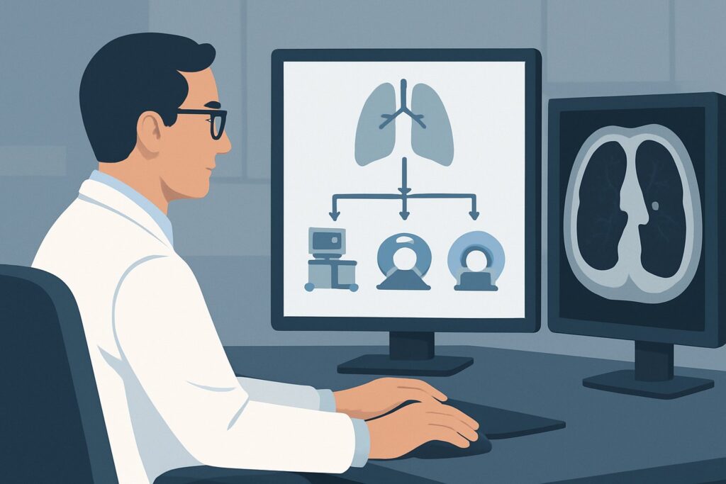
Once you’ve decided on a follow-up `CT chest without IV contrast`, our protocol guide covers the technique, contrast, and reading principles: CT Chest Without Contrast.
## What’s Next After CT chest without IV contrast? Downstream Workflow
The downstream workflow depends on two factors: the initial decision to perform follow-up imaging and the results of that scan if it is performed.
Scenario 1: No Follow-Up Imaging (Low-Risk Patient)
- Action: The finding is documented in the medical record. The patient should be informed of the incidental finding, the rationale for not pursuing follow-up (very low risk), and the importance of routine health maintenance, including smoking cessation if applicable. No further action is required regarding the nodule.
Scenario 2: Optional 12-Month Follow-Up CT Performed (High-Risk Patient)
- If the nodule is stable or has decreased in size: This confirms a benign etiology. No further follow-up is needed. The patient can be reassured, and the workup is complete.
- If the nodule has definitively grown: This is a suspicious finding that increases the likelihood of malignancy. The patient’s clinical scenario has now changed. Management should proceed according to guidelines for a growing solid nodule, which may involve a shorter interval follow-up CT, a PET/CT scan, or biopsy, depending on the new size and morphology.
- If the nodule has resolved: This confirms a transient, benign process, such as focal infection or inflammation. The workup is complete.
In all cases, clear communication with the patient about the rationale, risks, and results is paramount to managing the anxiety that an “incidental nodule” can provoke.
## Pitfalls to Avoid (and When to Get Help)
Managing small incidental nodules requires careful adherence to guidelines to avoid common errors.
- Pitfall 1: Over-Investigation. The most frequent error is ordering an immediate or 3-month follow-up CT for a low-risk patient with a <6 mm nodule. This deviates from guidelines, imparts unnecessary radiation, and can initiate a cascade of further low-yield testing.
- Pitfall 2: Ignoring Patient Risk. Conversely, failing to consider a 12-month follow-up for a high-risk patient (e.g., a 60-year-old with a 40-pack-year smoking history) may miss a rare but early-stage cancer. The decision should be deliberate and documented.
- Pitfall 3: Misclassifying the Nodule. This workflow is strictly for solid nodules. Applying it to a part-solid or ground-glass nodule is a critical error, as these have different risk profiles and require more intensive surveillance, even at small sizes.
- Pitfall 4: Inconsistent Measurement. When follow-up is performed, ensure the comparison is made to the prior study using consistent measurement techniques. Small differences in measurement can be mistaken for growth, leading to unnecessary alarm.
If a nodule shows definitive growth on a follow-up scan, or if the patient develops new symptoms like hemoptysis or unexplained weight loss, escalate care by consulting a pulmonologist or thoracic surgeon for further management.
## Related ACR Topics and Tools
This article covers one specific clinical variant. For a comprehensive overview of all scenarios related to this topic, see our parent guide. For other tools to help with imaging decisions, see the resources below.
For breadth across all scenarios in Incidentally Detected Indeterminate Pulmonary Nodule, see our parent guide: Incidentally Detected Indeterminate Pulmonary Nodule: ACR Appropriateness Decoded.
- ACR Appropriateness Criteria Lookup — for adjacent scenarios
- Imaging Protocol Library — for technique on the recommended study
- Radiation Dose Calculator — for cumulative dose conversations
Frequently Asked Questions
What defines a ‘high-risk’ patient for a small pulmonary nodule?
A high-risk patient typically has a significant history of tobacco smoking (e.g., >30 pack-years), a personal or strong family history of lung cancer, or other known risk factors like exposure to asbestos. Age is also a factor, with risk increasing with age.
Do these guidelines apply to multiple pulmonary nodules?
The Fleischner Society guidelines, which inform the ACR criteria, provide separate recommendations for multiple nodules. If multiple solid nodules <6 mm are present in a high-risk patient, a follow-up CT at 3-6 months may be considered. For low-risk patients with multiple small nodules, follow-up is generally not required.
What if the nodule has benign features like calcification?
If a nodule has features highly suggestive of a benign etiology, such as central, diffuse, laminated, or ‘popcorn’ calcification, or contains macroscopic fat, it is considered benign. No further follow-up is needed, regardless of patient risk factors. This article’s workflow applies to ‘indeterminate’ nodules without such features.
Why is a chest radiograph (X-ray) ‘Usually not appropriate’ for follow-up?
A chest radiograph lacks the spatial resolution to reliably detect or measure a nodule smaller than 6 mm, or to accurately assess for subtle growth. A CT scan is far more sensitive and is the standard for nodule surveillance.
Should I order a low-dose CT for the follow-up scan?
Yes, when performing surveillance for a pulmonary nodule, a low-dose technique should be used to minimize radiation exposure while maintaining sufficient image quality for nodule measurement. This is standard practice for both lung cancer screening and nodule follow-up.
Reviewed by Pouyan Golshani, MD, Interventional Radiologist — May 30, 2026