What Is the Right First Imaging Study for Adolescent Idiopathic Scoliosis?
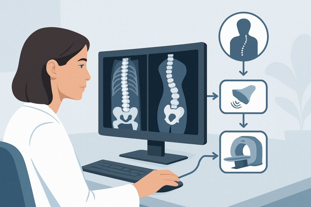
A 14-year-old comes in for a routine annual physical. They are healthy, active, and have no complaints of back pain or neurological symptoms. On examination, the Adams forward bend test reveals a right-sided thoracic prominence, raising suspicion for scoliosis. As the primary care physician, you need to confirm the diagnosis, quantify the curve, and determine the next steps in management. This requires ordering the correct initial imaging study, balancing diagnostic yield with the principle of minimizing radiation exposure in a young patient. This article details the American College of Radiology (ACR) Appropriateness Criteria workflow for this specific scenario: an adolescent with suspected idiopathic scoliosis and no risk factors. For the recommended first study, Radiography complete spine, the ACR rating is Usually Appropriate.
Who Fits This Clinical Scenario for Adolescent Idiopathic Scoliosis?
This guidance applies to a well-defined patient population: adolescents between 10 and 17 years of age with a clinical suspicion of idiopathic scoliosis who are undergoing their initial imaging workup. The key qualifier is the absence of risk factors or “red flags.” In this context, “no risk factors” means the patient does not present with any of the following:
- Significant or atypical back pain (e.g., night pain, pain that disrupts activities)
- Neurological signs or symptoms (e.g., weakness, numbness, gait disturbance, bowel/bladder changes)
- An atypical curve pattern on physical exam (e.g., a left-sided thoracic curve)
- Abnormal physical findings suggesting an underlying syndrome (e.g., cutaneous stigmata, significant joint laxity)
- A known history of congenital vertebral anomalies or neuromuscular disease
This workflow is distinct from other similar presentations. If your patient is younger than 10, they fall under the early-onset idiopathic scoliosis scenario, which has different considerations for progression. If the patient is an adolescent but does have one of the risk factors listed above, they fit the adolescent idiopathic scoliosis with risk factors scenario, where advanced imaging like MRI may be warranted upfront. Finally, if there is a known or highly suspected vertebral malformation, the workup follows the congenital scoliosis pathway.
What Diagnoses Are You Working Up in This Scenario?
When ordering initial imaging for a teenager with a spinal curve and no red flags, the primary goal is to confirm and characterize the suspected diagnosis while ruling out less common structural causes.
The most prevalent diagnosis by far is Adolescent Idiopathic Scoliosis (AIS). This is a three-dimensional deformity of the spine that occurs in otherwise healthy children around the time of puberty. It is a diagnosis of exclusion, meaning other causes must be ruled out. Imaging is essential to confirm the presence of a curve (defined as a Cobb angle of 10 degrees or more), measure its magnitude, identify the curve pattern, and assess the patient’s skeletal maturity, which is a critical predictor of future progression.
Though less common in this “no risk factors” presentation, imaging helps exclude Congenital Scoliosis. This condition results from malformed vertebrae present at birth, such as a hemivertebra (a wedge-shaped vertebra) or a failure of vertebral separation (a block vertebra). While often detected earlier in life, milder forms may not become apparent until the adolescent growth spurt. Radiographs are highly effective at identifying these structural bony abnormalities, which fundamentally change the diagnosis and management plan from idiopathic scoliosis.
Another consideration is Scheuermann’s Kyphosis, a structural rigidity of the thoracic spine characterized by vertebral wedging. While primarily a sagittal plane deformity (causing a “hunchback” appearance), it can be associated with a secondary coronal curve (scoliosis). A complete spine radiograph allows for evaluation of both planes to make an accurate diagnosis.
Why Is Complete Spine Radiography the Recommended First Study for Suspected AIS?
For the initial evaluation of an adolescent with suspected idiopathic scoliosis and no risk factors, the ACR designates Radiography complete spine as Usually Appropriate. This recommendation is based on the study’s ability to comprehensively answer the key clinical questions with the lowest necessary radiation dose. A properly performed scoliosis series provides the essential information to establish a diagnosis, create a prognosis, and guide initial treatment decisions. It allows for precise measurement of the Cobb angle, assessment of spinal balance, and determination of skeletal maturity via the Risser sign on the iliac crests.
Alternative imaging modalities are deemed less suitable for this specific initial workup.
- MRI complete spine without IV contrast is rated Usually not appropriate. While it involves no ionizing radiation (RRL: O 0 mSv), its utility in this scenario is very low. The prevalence of underlying intraspinal anomalies (like a syrinx or tethered cord) in patients with typical AIS and no neurological symptoms is minimal. Reserving MRI for patients with red flags avoids unnecessary costs, potential need for sedation, and patient anxiety without compromising the initial diagnosis.
- CT spine is also rated Usually not appropriate. CT provides exquisite bony detail but at the cost of a significantly higher radiation dose (RRL: Varies) compared to radiography. Given that these patients are young and may require multiple follow-up images over several years, minimizing cumulative radiation exposure is a primary safety concern. The additional detail from CT is not required for the initial diagnosis and classification of AIS.
The radiation dose for a complete spine radiograph (Pediatric RRL: ☢☢☢ 0.3-3 mSv) is considered a justifiable trade-off for the critical diagnostic and prognostic information it provides. Adherence to the ALARA (As Low As Reasonably Achievable) principle is paramount, and modern techniques like low-dose protocols and posteroanterior (PA) projections help protect sensitive tissues like the breast and thyroid.
What’s Next After Radiography? Downstream Workflow for AIS
The results of the complete spine radiograph directly guide the subsequent clinical pathway. The two most important data points from the report are the Cobb angle measurement and the assessment of skeletal maturity (e.g., Risser sign).
- If the study is positive for AIS (Cobb angle ≥ 10°): The patient should be referred to a pediatric orthopedic or spine specialist. The specialist’s management plan will be stratified by curve severity and skeletal maturity.
- Mild curves (<25°): Typically managed with observation and serial radiographic follow-up to monitor for progression, especially in skeletally immature patients.
- Moderate curves (25°-45°): In a growing adolescent, bracing is often the primary treatment to halt curve progression.
- Severe curves (>45°-50°): Surgical intervention is often considered to correct the deformity and prevent further progression and long-term complications.
- If the study is negative (Cobb angle < 10°): This is considered spinal asymmetry, not scoliosis. The patient and family can be reassured, and no further imaging or orthopedic referral is typically needed unless the clinical deformity worsens.
- If the study reveals atypical findings: Should the radiographs show an unusual curve pattern (e.g., a sharp, angular curve; a left thoracic curve), evidence of congenital vertebral anomalies, or other abnormalities, the patient’s presentation no longer fits the “idiopathic” category. This shifts the workflow toward the adolescent with risk factors scenario, and the consulting specialist will likely proceed with advanced imaging, such as an MRI, to evaluate for underlying pathology.
Common Pitfalls to Avoid in Initial Scoliosis Imaging
Navigating the initial workup for AIS is generally straightforward, but several common pitfalls can lead to incomplete data or unnecessary radiation exposure.
1. Ordering Incomplete Views: Requesting only a “thoracic spine” or “lumbar spine” X-ray is a frequent error. A “scoliosis series” or “radiography complete spine” is essential to visualize the entire curve from the cervical spine to the sacrum, allowing for accurate Cobb angle measurement and assessment of overall spinal balance.
2. Failing to Specify Standing Views: Scoliosis must be evaluated in a functional, weight-bearing position. Ordering supine films will underestimate the true magnitude of the curve. The order should always specify “standing” or “erect” PA and lateral views.
3. Omitting the Iliac Crests: The radiograph must include the iliac crests to allow for grading of the Risser sign, which is the primary method for assessing remaining skeletal growth. This information is crucial for predicting the risk of curve progression.
4. Prematurely Ordering Advanced Imaging: In a patient who perfectly fits this scenario (no pain, no neurologic deficits, typical curve pattern), ordering an MRI as the first step is a low-value action that deviates from established guidelines and increases healthcare costs.
If the clinical picture changes—for instance, if the patient develops new neurological symptoms or pain disproportionate to the curve—escalate care immediately to a spine specialist for further evaluation, which will likely include an MRI.
Related ACR Topics and Tools
For a comprehensive overview of all clinical variants related to pediatric scoliosis imaging, further reading and decision-support tools are available.
- For breadth across all scenarios in Scoliosis-Child, see our parent guide: Scoliosis-Child: ACR Appropriateness Decoded.
- To explore other clinical presentations, use the ACR Appropriateness Criteria Lookup.
- For details on imaging techniques, consult the Imaging Protocol Library.
- To help discuss radiation exposure with patients and families, refer to the Radiation Dose Calculator.
Frequently Asked Questions
What specific views are included in a ‘Radiography complete spine’ for scoliosis?
A standard scoliosis series typically includes two views: a standing posteroanterior (PA) and a standing lateral view. Both images are taken on a long 36-inch cassette to capture the entire spine from the cervical spine down to the sacrum and iliac crests in a single exposure.
Why is a PA (posteroanterior) view preferred over an AP (anteroposterior) view for scoliosis imaging?
A PA view, where the X-ray beam enters from the back and exits through the front, is preferred to reduce radiation dose to the breast and thyroid glands, which are more radiosensitive tissues. Given that patients with scoliosis are often young and may require multiple follow-up radiographs, this dose-saving technique is an important part of the ALARA (As Low As Reasonably Achievable) principle.
Is a screening Adam’s forward bend test sufficient for diagnosis without imaging?
No. The Adam’s forward bend test is a screening tool used to detect trunk asymmetry, which suggests the presence of scoliosis. However, it cannot confirm the diagnosis, measure the curve’s severity (Cobb angle), or assess skeletal maturity. Radiography is required to obtain this essential information for diagnosis and treatment planning.
If the initial radiograph is normal but I still have clinical suspicion, when should I re-image?
If the initial radiograph shows no significant curve (Cobb angle < 10 degrees) in an adolescent, routine follow-up imaging is generally not necessary. Re-imaging should only be considered if there is a clear clinical worsening of the trunk asymmetry on physical exam during a subsequent visit, especially if the child has significant remaining growth potential.
Does my patient need to see an orthopedist before I order the initial X-ray?
In many healthcare systems, the primary care physician (e.g., pediatrician, family medicine physician) orders the initial radiographs to confirm the diagnosis. This allows the referral to the orthopedic specialist to be more efficient, as the specialist will already have the necessary imaging to make an initial assessment and management plan during the first consultation.
Reviewed by Pouyan Golshani, MD, Interventional Radiologist — May 29, 2026