What Is the Right First Imaging Test for Lung Cancer Screening in High-Risk Patients?

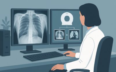
A 62-year-old patient with a 30-pack-year smoking history, who quit five years ago, is in your clinic for an annual wellness visit. They are asymptomatic and feel well, but you recognize they meet the high-risk criteria for lung cancer. The conversation turns to preventive care, and you decide it’s time to initiate annual screening. The central question is which imaging study to order first. This is not a diagnostic workup for symptoms, but a screening evaluation in an asymptomatic, high-risk individual. For this specific clinical scenario, the American College of Radiology (ACR) Appropriateness Criteria state that a `CT chest without IV contrast screening` is `Usually appropriate`, providing a clear, evidence-based starting point for your workflow.
Who Qualifies for This Lung Cancer Screening Scenario?
This guidance applies to a well-defined population for whom screening has a proven mortality benefit. The inclusion criteria are strict and based on the landmark National Lung Screening Trial (NLST) and subsequent recommendations from the U.S. Preventive Services Task Force (USPSTF).
To fit this scenario, the patient must meet all of the following criteria:
- Age: Between 50 and 80 years old.
- Smoking History: At least a 20-pack-year history of smoking. (Pack-years are calculated by multiplying the number of packs smoked per day by the number of years the person has smoked).
- Smoking Status: Is either a current smoker or has quit within the past 15 years.
It is critical to distinguish this screening scenario from others that may appear similar. This workflow does not apply if:
- The patient is symptomatic: A patient presenting with hemoptysis, new or changing cough, unexplained weight loss, or dyspnea requires a diagnostic workup, not a screening study.
- The patient is outside the age or smoking history criteria: A 48-year-old with a 30-pack-year history or a 60-year-old with a 15-pack-year history falls into a different risk category, where the benefit of screening is not as clearly established.
- The patient quit smoking more than 15 years ago: The risk of lung cancer decreases significantly after this period, and these individuals are no longer in the highest-risk cohort for whom annual screening is recommended.
What Diagnoses Are You Working Up in This Scenario?
Unlike a diagnostic workup, a screening study is designed to detect a specific condition before symptoms arise. The primary goal is to find disease at an earlier, more treatable stage.
The main target of this screening is early-stage, asymptomatic lung cancer. The rationale for screening this high-risk population is to identify non-small cell lung cancer (NSCLC) or small cell lung cancer (SCLC) when it is a small, localized nodule or mass. At this stage, surgical resection or definitive radiation therapy can be curative. By the time lung cancer causes symptoms like coughing up blood or chest pain, it has often spread, and the prognosis is significantly worse.
However, the CT scan will visualize the entire chest, and a range of incidental findings are common, effectively becoming part of the post-scan differential. These include:
- Benign Pulmonary Nodules: The most common finding. These are often granulomas from prior infections (like histoplasmosis or tuberculosis) or other benign growths like hamartomas. Managing these is the core challenge of a screening program.
- Emphysema or Chronic Obstructive Pulmonary Disease (COPD): Changes like parenchymal destruction and air trapping are frequently identified in this patient population.
- Coronary Artery Calcification (CAC): The scan provides a clear view of calcified plaque in the coronary arteries. While not the primary purpose, reporting CAC provides a powerful, actionable data point for cardiovascular risk stratification.
- Other Findings: The scan may also reveal aortic aneurysms, thyroid nodules, hiatal hernias, or vertebral body compression fractures.
Why Is CT Chest Without IV Contrast Screening the Recommended Study for This Presentation?
The ACR designates `CT chest without IV contrast screening` as `Usually appropriate` because it offers the best balance of sensitivity, safety, and practicality for detecting early-stage lung cancer in an asymptomatic population. This specific protocol is often referred to as a low-dose chest CT (LDCT).
The rationale is built on several key points:
- High Sensitivity: Low-dose CT is highly sensitive for detecting small, non-calcified pulmonary nodules, which are the earliest signs of lung cancer. Major clinical trials have demonstrated that annual screening with LDCT can reduce lung cancer-specific mortality in high-risk individuals compared to screening with chest radiography.
- Low Radiation Dose: The “low-dose” protocol is crucial. It uses significantly less radiation (`☢☢☢` 1-10 mSv) than a standard diagnostic chest CT while maintaining sufficient image quality to detect nodules. This minimizes the cumulative radiation exposure from annual scans over many years.
- No IV Contrast Needed: Intravenous contrast is not required to identify or initially characterize pulmonary nodules for screening. Omitting contrast avoids the associated risks of allergic reaction and contrast-induced nephropathy, which is an important consideration in a large, asymptomatic screening population that may have age-related renal function decline.
Alternative imaging studies are rated `Usually not appropriate` for this initial screening scenario for clear reasons:
- Radiography chest: This study has a low radiation dose (`☢` <0.1 mSv) but is `Usually not appropriate` due to its poor sensitivity. It can easily miss small nodules, especially those obscured by the heart, ribs, or diaphragm, defeating the purpose of early detection.
- FDG-PET/CT: This is `Usually not appropriate` as a screening tool. It is a functional imaging study used for staging confirmed malignancies, not for initial detection. It has a high rate of false positives from benign inflammatory processes, is expensive, and delivers a very high radiation dose (`☢☢☢☢` 10-30 mSv).
Once you’ve decided on the recommended study, our protocol guide covers the technique, contrast, and reading principles: CT Chest Without Contrast.
What’s Next After CT Chest Without IV Contrast Screening? Downstream Workflow
The result of the screening CT dictates the next steps, which are standardized by the Lung Cancer Screening Reporting and Data System (Lung-RADS). This system categorizes findings based on their risk of malignancy and provides clear management recommendations.
- Negative Result (Lung-RADS 1 or 2): If the scan is negative (no nodules or clearly benign nodules), the patient should continue with annual screening. This is the most common outcome. The recommendation is to repeat the low-dose CT in 12 months.
- Indeterminate/Potentially Suspicious Result (Lung-RADS 3 or 4A): If the scan shows a small nodule that is not clearly benign, follow-up is required. For a Lung-RADS 3 finding (probably benign), a short-term follow-up low-dose CT in 6 months is typically recommended to assess for stability or growth. For a Lung-RADS 4A finding (suspicious), a 3-month follow-up CT or PET/CT may be considered.
- Positive/Very Suspicious Result (Lung-RADS 4B or 4X): If the scan shows a larger, solid, or growing nodule, the workflow shifts from screening to diagnosis. The next step is typically a diagnostic chest CT with IV contrast, a PET/CT for staging, and/or referral to a pulmonologist or thoracic surgeon for consideration of a biopsy.
It is essential to have a system in place to track these results and ensure patients complete the recommended follow-up, as this is critical to realizing the mortality benefit of screening.
Pitfalls to Avoid (and When to Get Help)
Executing a successful lung cancer screening program requires attention to detail. Here are common pitfalls to avoid in this specific scenario:
- Ordering a “Diagnostic” CT Instead of a “Screening” CT: Be explicit in your order: “Low-Dose CT for Lung Cancer Screening.” A standard diagnostic CT uses a higher radiation dose and is not appropriate for routine annual screening.
- Screening Symptomatic Patients: Screening is for the asymptomatic. If a high-risk patient develops a new cough or hemoptysis, they need a diagnostic workup, not a screening study.
- Failing to Counsel on Smoking Cessation: The single most important intervention for this patient population is smoking cessation. An imaging order should always be paired with a robust conversation and offer of resources for quitting.
- Losing Patients to Follow-up: A positive or indeterminate finding is only useful if acted upon. Ensure a reliable tracking system is in place for Lung-RADS 3 and 4 follow-ups.
If a scan returns a Lung-RADS 4B or 4X result, or if the patient becomes symptomatic, it is time to escalate care by referring to a pulmonologist or a multidisciplinary thoracic oncology team.
Related ACR Topics and Tools
This article covers a single, common scenario in depth. For a broader view of all clinical variants and imaging options in this domain, please consult our parent guide. You can also use the tools below to explore adjacent scenarios or technical details of the recommended study.
- For breadth across all scenarios in Lung Cancer Screening, see our parent guide: Lung Cancer Screening: ACR Appropriateness Decoded.
- To explore other clinical presentations, use the ACR Appropriateness Criteria Lookup.
- For technical details on other studies, browse the Imaging Protocol Library.
- To discuss cumulative radiation exposure with your patients, consult the Radiation Dose Calculator.
Frequently Asked Questions
Does this patient need to stop smoking before starting screening?
No, the patient does not need to quit smoking to be eligible for screening. The criteria include both current smokers and those who have quit within the past 15 years. However, every screening visit should be used as an opportunity to provide smoking cessation counseling and resources, as quitting is the most effective way to reduce lung cancer risk.
What if the patient is 81 years old but otherwise meets all criteria?
The current USPSTF guidelines recommend screening for individuals up to age 80. For a patient older than 80, the decision to screen should be individualized, considering their overall health, life expectancy, and ability to undergo potential treatment if cancer is found. This falls outside the standard recommended scenario.
Why is a chest X-ray not sufficient for screening?
While a chest X-ray has a very low radiation dose, it is rated ‘Usually not appropriate’ because it lacks the sensitivity to detect the small, early-stage lung nodules that are the target of screening. The National Lung Screening Trial (NLST) demonstrated that low-dose CT is superior to chest radiography in reducing lung cancer mortality.
What is the role of Lung-RADS in this workflow?
Lung-RADS is a critical component of the screening process. It is a standardized reporting system used by radiologists to classify the findings on a screening CT, from ‘Negative’ (Lung-RADS 1) to ‘Very Suspicious’ (Lung-RADS 4X). Each category has a specific, evidence-based recommendation for the next step, such as ‘continue annual screening’ or ‘short-term follow-up CT,’ which guides your downstream management.
If the CT scan shows significant coronary artery calcification, what is the next step?
This is a key incidental finding. While the primary purpose of the scan is lung cancer screening, the presence of moderate to severe coronary artery calcification indicates a high risk for cardiovascular events. The next step should be a formal cardiovascular risk assessment, which may include initiating or adjusting statin therapy, optimizing blood pressure control, and reinforcing lifestyle modifications, independent of the lung nodule follow-up plan.
Reviewed by Pouyan Golshani, MD, Interventional Radiologist — May 30, 2026