What Is the Right Initial Imaging for Occupational Lung Disease Screening?

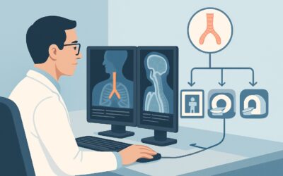
A 58-year-old male, a former shipyard worker with a 30-pack-year smoking history, presents for a routine physical. He reports a mild, chronic cough but denies acute dyspnea or hemoptysis. His occupational history is significant for asbestos exposure, and you are considering baseline imaging for screening and long-term surveillance. What is the most appropriate first step? This article addresses the initial imaging choice for patients with known or suspected occupational exposure who require screening or surveillance. According to the American College of Radiology (ACR) Appropriateness Criteria, a standard chest radiograph is the Usually Appropriate initial study.
Who Fits This Clinical Scenario for Occupational Lung Screening?
This guidance applies to a specific patient population: asymptomatic or minimally symptomatic individuals with a known or suspected history of significant occupational exposure to agents known to cause lung disease. This includes dusts, fibers, or fumes such as asbestos, silica, coal dust, or beryllium. The clinical context is either baseline screening (e.g., for a new employee in a high-risk occupation) or periodic surveillance for established workers or retirees with a history of exposure.
The key is the absence of significant, new, or progressive symptoms. This workflow is for proactive screening, not for a diagnostic workup of an acute problem.
This scenario should be distinguished from several related presentations that require a different imaging approach:
- Symptomatic patients: If the patient presents with new or worsening dyspnea, significant cough, or crackles on auscultation, they may fit the Occupational exposure, suspected interstitial lung disease scenario, which has a distinct workup.
- Airway-predominant symptoms: For patients whose primary symptoms are wheezing or a productive cough suggestive of bronchitis or asthma, the workup would follow the Occupational exposure, suspected airway disease scenario.
- Suspicion of malignancy: If a patient with known occupational lung disease develops concerning symptoms like hemoptysis, unexplained weight loss, or has a new suspicious finding on a prior study, the workup shifts to the Confirmed occupational lung disease, suspected thoracic neoplasm scenario.
What Diagnoses Are You Working Up in Occupational Lung Screening?
The goal of screening and surveillance is to detect early signs of disease, often before they become clinically apparent, allowing for intervention, exposure cessation, and monitoring. The differential diagnosis in this setting is focused on the long-term sequelae of inhaled agents.
Pneumoconioses
This is the primary diagnostic category. These are non-neoplastic lung reactions to inhaled mineral or organic dusts. The imaging goal is to identify characteristic patterns before significant pulmonary function decline. This includes asbestosis (basilar reticular opacities, pleural plaques), silicosis (upper-lobe predominant nodules, “egg-shell” calcification of lymph nodes), and coal workers’ pneumoconiosis (small, rounded opacities, potential for progressive massive fibrosis).
Malignancy (Lung Cancer and Mesothelioma)
A critical consideration, as many occupational exposures are carcinogenic. Asbestos exposure dramatically increases the risk of both lung cancer (especially in smokers) and malignant mesothelioma. While chest radiography has lower sensitivity for early-stage lung cancer than low-dose CT, it can detect larger nodules or masses. For mesothelioma, it may reveal a unilateral pleural effusion, diffuse pleural thickening, or a discrete pleural mass.
Chronic Obstructive Pulmonary Disease (COPD)
While strongly linked to smoking, many occupational inhalants (e.g., coal dust, cadmium, welding fumes) are independent risk factors for COPD or can exacerbate it. A screening radiograph can reveal signs of hyperinflation, flattened diaphragms, or bullous disease that suggest underlying emphysema.
Tuberculosis (TB)
Certain occupations, such as healthcare and corrections, carry a higher risk of TB exposure. Furthermore, silicosis significantly impairs macrophage function, increasing the risk of TB infection and reactivation. Radiography is the standard screening tool for both latent and active TB, looking for classic findings like apical opacities, cavitation, or hilar adenopathy.
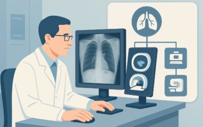
Why Is a Chest Radiograph the Recommended Initial Study for Screening?
For the initial screening and surveillance of an asymptomatic or minimally symptomatic patient with occupational exposure, the ACR designates Radiography chest as Usually Appropriate. This recommendation is based on a careful balance of diagnostic utility, safety, and practicality.
A standard chest radiograph is an excellent first-line tool due to its wide availability, low cost, and very low radiation dose (adult relative radiation level ☢ <0.1 mSv). In a screening context where imaging may be repeated over many years, minimizing cumulative radiation exposure is a primary concern. The radiograph provides a comprehensive initial overview of the lung parenchyma, pleura, mediastinum, and cardiac silhouette. It is sufficiently sensitive for detecting many of the hallmark findings of established pneumoconioses, such as the pleural plaques of asbestosis or the diffuse small nodules of silicosis.
Alternative imaging modalities are rated lower for this specific initial screening scenario:
- CT chest without IV contrast is rated May be appropriate. While CT is undoubtedly more sensitive for detecting subtle interstitial changes, early emphysema, and small nodules, its significantly higher radiation dose (☢☢☢ 1-10 mSv) and cost make it less suitable for routine, widespread screening of asymptomatic individuals. Its role is typically reserved for clarifying equivocal findings on a chest radiograph or for specific, structured screening programs (e.g., low-dose CT for lung cancer screening in high-risk smokers, which is a separate clinical indication).
- CT chest with IV contrast is rated Usually not appropriate. For the primary goals of screening—detecting interstitial patterns, nodules, or pleural disease—intravenous contrast provides no additional diagnostic value. It unnecessarily increases cost, carries a risk of contrast-related adverse events, and can increase radiation dose without benefit for this indication.
When ordering, specifying both posteroanterior (PA) and lateral views is standard practice for a complete evaluation. For pneumoconiosis surveillance, using the International Labour Organization (ILO) classification system for reading and reporting radiographs provides a standardized method for quantifying and describing abnormalities over time.
What’s Next After Radiography chest? Downstream Workflow
The results of the initial chest radiograph will guide the subsequent clinical and imaging pathway. The downstream workflow depends on whether the findings are negative, positive, or indeterminate.
If the radiograph is negative:
For an asymptomatic patient with a normal chest radiograph, the next step is typically continued surveillance. The frequency of follow-up imaging depends on the nature and intensity of the exposure, latency period, and specific regulatory requirements (e.g., OSHA standards). No immediate further imaging is usually required. The patient should be counseled on continued risk reduction and symptom monitoring.
If the radiograph is positive for suspected occupational lung disease:
If the radiograph shows findings suggestive of pneumoconiosis (e.g., pleural plaques, diffuse nodules, reticular opacities), the next step is often to obtain a CT chest without IV contrast. This study, rated May be appropriate for initial screening, becomes the primary tool for characterizing the extent and pattern of disease seen on the radiograph. This moves the patient from a screening scenario to a diagnostic one, specifically the “Occupational exposure, suspected interstitial lung disease based on radiography” ACR variant. Pulmonary function testing (PFTs) should also be performed to establish a functional baseline.
If the radiograph shows an indeterminate or suspicious nodule:
If a non-calcified pulmonary nodule is identified, the workup should follow established guidelines for nodule management, such as the Fleischner Society guidelines. This typically involves risk stratification (based on patient age, smoking history, nodule size, and morphology) and may lead to short-term follow-up with low-dose CT or further diagnostic procedures if the suspicion for malignancy is high.
Pitfalls to Avoid (and When to Get Help)
Navigating occupational lung disease screening requires attention to several potential pitfalls to ensure appropriate and effective patient care.
First, avoid using CT as a first-line screening tool in an unselected, asymptomatic population outside of established guidelines. The default initial study should be a chest radiograph to minimize radiation dose and cost. Second, do not overlook the importance of a high-quality occupational history; the radiologic findings of different pneumoconioses can overlap, and the exposure history is critical for narrowing the differential. Third, remember that a normal chest radiograph does not completely exclude early or microscopic disease. Clinical suspicion should remain if risk factors are high.
If a radiograph shows complex or advanced findings, such as progressive massive fibrosis, signs of active infection, or features suspicious for malignancy, it is crucial to escalate care. This typically involves referral to a pulmonologist for further evaluation, which may include bronchoscopy, advanced imaging, and comprehensive pulmonary function testing.
Related ACR Topics and Tools
For a comprehensive overview of imaging across all clinical variants related to occupational lung diseases, please see our parent topic hub article. For additional resources to help guide your imaging decisions, the following tools are available.
- Parent Topic Hub: For breadth across all scenarios in Occupational Lung Diseases, see our parent guide: Occupational Lung Diseases: ACR Appropriateness Decoded.
- ACR Criteria Lookup: To explore adjacent scenarios or other clinical questions, use the ACR Appropriateness Criteria Lookup.
- Imaging Protocols: For detailed technical specifications on how imaging studies are performed, consult the Imaging Protocol Library.
- Dose Calculation: To discuss cumulative radiation exposure with your patients, the Radiation Dose Calculator can be a useful aid.
Frequently Asked Questions
Why not start with a low-dose CT (LDCT) for everyone with occupational exposure?
While LDCT is more sensitive than a chest radiograph for subtle lung disease and early cancer, it is not recommended as a universal first-line screening tool for all occupational exposures. The primary reasons are the higher cumulative radiation dose over a lifetime of surveillance and higher costs. The ACR rates CT as ‘May be appropriate,’ reserving it for specific high-risk situations or as a follow-up to an abnormal radiograph. The exception is structured lung cancer screening programs for high-risk smokers, which is a distinct clinical indication.
How often should screening chest radiographs be repeated?
The frequency of surveillance imaging is not one-size-fits-all. It depends on the specific substance of exposure (e.g., asbestos vs. silica), the duration and intensity of the exposure, the time since last exposure, and regulatory guidelines from bodies like OSHA or NIOSH. For example, asbestos-exposed workers may require periodic screening every 3-5 years, while silica-exposed workers might have a different schedule. The decision should be individualized based on these factors.
What is an ILO B-read and is it necessary for screening?
An ILO B-read is a specialized interpretation of a chest radiograph by a NIOSH-certified B-reader, who uses the International Labour Organization (ILO) classification system to standardize the reporting of pneumoconiosis. While not required for every screening radiograph, it is the standard for medico-legal contexts, epidemiologic studies, and in regulated surveillance programs to ensure consistent and quantifiable assessment of dust-related lung disease over time.
If a patient with asbestos exposure has a normal chest radiograph, are they ‘clear’?
Not necessarily. A normal chest radiograph is reassuring, but it does not eliminate all future risk. Asbestosis and mesothelioma have long latency periods, often 20-40 years or more after exposure begins. A normal baseline radiograph is a starting point for future comparison. The patient still requires long-term surveillance and counseling on recognizing symptoms (like worsening shortness of breath or chest pain) and the synergistic risk of smoking.
Should IV contrast ever be used in the workup of occupational lung disease?
For initial screening and surveillance, IV contrast is ‘Usually not appropriate.’ However, it becomes essential in specific downstream scenarios. For instance, if a lung mass is found and there is concern for malignancy, a contrast-enhanced CT is necessary to evaluate the mass, check for mediastinal or hilar lymph node involvement, and assess for metastatic disease. It is also used to evaluate suspected pulmonary embolism or other vascular complications, but not for the primary workup of parenchymal or pleural changes from pneumoconiosis.
Reviewed by Pouyan Golshani, MD, Interventional Radiologist — May 29, 2026