What Is the Right Initial Imaging for Suspected COPD and Chronic Dyspnea?
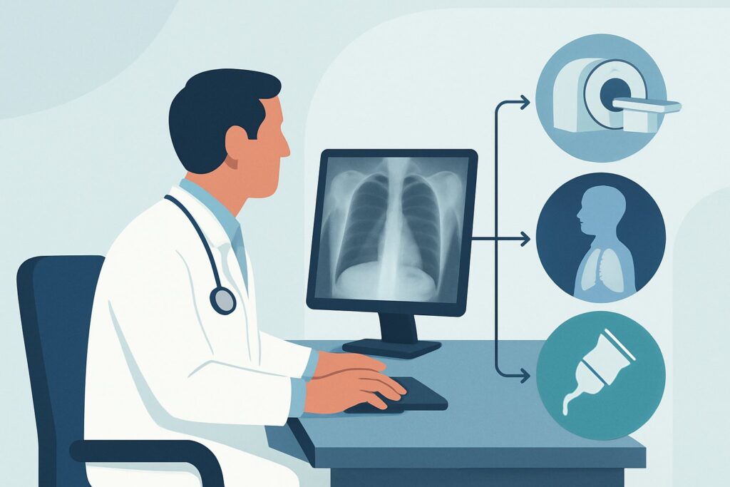
A 68-year-old patient with a significant smoking history sits in your exam room, describing a shortness of breath that has slowly worsened over the past year. It’s most noticeable when he climbs a flight of stairs. His spirometry results are on your screen, showing an FEV1/FVC ratio consistent with obstructive lung disease. Your leading diagnosis is chronic obstructive pulmonary disease (COPD), but you need to confirm parenchymal changes and rule out other contributing factors. The clinical question is clear: what is the most appropriate initial imaging study to order? This article provides a detailed workflow for this specific scenario, grounded in the American College of Radiology (ACR) Appropriateness Criteria, which rates a chest radiograph as Usually Appropriate for this presentation.
Who Fits This Clinical Scenario?
This guidance applies to a specific patient population: adults presenting with chronic dyspnea (persisting for weeks to months) where the clinical suspicion points strongly toward chronic obstructive pulmonary disease (COPD). The typical patient has significant risk factors, most commonly a long-term history of tobacco smoking, but may also have occupational exposures (e.g., dust, fumes) or a known alpha-1 antitrypsin deficiency. The “initial imaging” component is key; this workflow is for the first imaging study ordered for this indication, not for follow-up or acute exacerbations.
This workflow is NOT intended for:
- Patients with acute or subacute dyspnea: A sudden onset of shortness of breath requires a different workup to evaluate for urgent conditions like pulmonary embolism, pneumonia, or pneumothorax.
- Patients with dyspnea of unclear etiology: If there is no clear risk factor profile for COPD and the cause of dyspnea is broad, the imaging strategy may differ. This falls under a separate ACR variant, Adult. Chronic dyspnea. Unclear etiology.
- Patients with known interstitial lung disease (ILD): If ILD is already diagnosed, imaging is typically for monitoring disease progression, not initial diagnosis.
- Patients where small airways disease is the primary suspicion: While related to COPD, if obliterative bronchiolitis or a similar condition is the main concern, the imaging approach is more specific and covered in its own variant.
What Diagnoses Are You Working Up in This Scenario?
When ordering initial imaging for suspected COPD, the goal is twofold: to find evidence supporting the diagnosis and to exclude common or consequential mimics. The differential diagnosis guides the interpretation of the study.
Chronic Obstructive Pulmonary Disease (COPD): This is the primary diagnosis under consideration. Imaging seeks to identify characteristic features of emphysema, such as lung hyperinflation (seen as flattened hemidiaphragms or an increased retrosternal clear space), parenchymal destruction, and the formation of bullae. Signs of chronic bronchitis, like bronchial wall thickening, may also be visible.
Interstitial Lung Disease (ILD): Many forms of ILD, particularly idiopathic pulmonary fibrosis, can present with chronic dyspnea and are more common in smokers. A chest radiograph can reveal early signs like reticular or linear opacities, especially at the lung bases, prompting a different diagnostic pathway. Combined pulmonary fibrosis and emphysema (CPFE) is a specific entity seen in smokers.
Lung Cancer: Given that the primary risk factor for COPD (smoking) is also the primary risk factor for lung cancer, every initial evaluation must consider this possibility. Chronic dyspnea can be a presenting symptom of a central or advanced malignancy. The imaging study serves as a crucial screen for nodules, masses, or hilar adenopathy.
Bronchiectasis: This condition, characterized by permanent dilation of the airways, can cause chronic cough and dyspnea and may coexist with or mimic COPD. Radiographs may show “tram tracking” (thickened bronchial walls) or cystic changes, though CT is more sensitive.
Why Is a Chest Radiograph the Recommended Initial Study?
The ACR rates both chest radiography and non-contrast chest CT as “Usually Appropriate” for this scenario. However, the chest radiograph is the recommended initial study due to its excellent balance of diagnostic utility, low radiation dose, cost-effectiveness, and wide availability.
A standard posteroanterior (PA) and lateral chest radiograph is a powerful first-line tool. It is highly effective for identifying the classic signs of moderate to severe COPD, such as hyperinflated lungs, flattened hemidiaphragms, and bullous changes. More importantly, it is an excellent examination for evaluating for the key alternative diagnoses on the differential. It can readily detect a suspicious lung mass, overt signs of interstitial lung disease, a large pleural effusion, or cardiomegaly that might suggest a cardiac component to the patient’s dyspnea.
The radiation dose is minimal (Relative Radiation Level ☢ <0.1 mSv), which is a significant advantage over CT for an initial diagnostic test.
Why are other studies rated lower for this initial workup?
- CT chest without IV contrast: While also rated Usually Appropriate, its higher radiation dose (☢☢☢ 1-10 mSv) makes it better suited as a second-line test. It is typically reserved for cases where the chest radiograph is normal or equivocal despite high clinical suspicion, or to better characterize abnormalities seen on the radiograph (e.g., a lung nodule, suspected ILD).
- CT chest with IV contrast: This is rated Usually Not Appropriate. Intravenous contrast is unnecessary for evaluating the lung parenchyma for emphysema or ILD. It adds risks (e.g., contrast reaction, post-contrast acute kidney injury) and should be reserved for when there is a specific concern for a vascular issue, such as pulmonary embolism or a suspected vascular malformation, which represents a different clinical question.
- US chest: Ultrasound is rated Usually Not Appropriate because it cannot visualize the lung parenchyma. Its utility in the chest is limited to evaluating the pleura for effusions or pneumothorax, not for diagnosing COPD.
What’s Next After a Chest Radiograph? Downstream Workflow
The results of the initial chest radiograph will guide your next steps in a branching clinical pathway.
If the radiograph is positive for COPD: Findings like hyperinflation and bullae, in the context of supportive clinical history and spirometry, confirm the diagnosis. The primary next step is clinical management, including smoking cessation, initiation of bronchodilator therapy, and referral to pulmonology or pulmonary rehabilitation. Further imaging, such as a non-contrast CT, may be considered later for lung cancer screening in eligible individuals or for COPD phenotyping (e.g., to assess the extent of emphysema before considering procedures like lung volume reduction surgery).
If the radiograph is negative or normal: A normal chest X-ray does not rule out COPD, especially in its early stages. If spirometry is clearly obstructive and clinical suspicion remains high, the diagnosis of COPD can still be made. A non-contrast high-resolution CT (HRCT) may be the appropriate next step if you need to confirm subtle emphysema or evaluate for an alternative diagnosis like small airways disease or early ILD not visible on the radiograph.
If the radiograph shows an unexpected finding: The workflow pivots to address the new finding. A lung nodule will trigger a follow-up plan based on its size, morphology, and patient risk factors, often guided by Fleischner Society criteria, which typically involves a follow-up CT scan. If the radiograph suggests ILD (e.g., reticulation, honeycombing), the next step is usually an HRCT to characterize the pattern of fibrosis and a referral to a pulmonologist specializing in ILD.
Pitfalls to Avoid (and When to Get Help)
Navigating the initial workup for suspected COPD requires careful interpretation and an awareness of common diagnostic traps.
- The “Normal X-ray” Fallacy: Do not dismiss a diagnosis of COPD based on a normal chest radiograph. The diagnosis is ultimately established by spirometry in a patient with appropriate symptoms and risk factors.
- Satisfaction of Search: When clear signs of COPD are present, avoid the pitfall of missing a second, more sinister diagnosis, such as a small, superimposed lung cancer. Scrutinize the entire film methodically.
- Attribution Error: In a patient with established COPD, resist the urge to attribute new or worsening dyspnea solely to their known disease. Consider and evaluate for superimposed conditions like heart failure, pneumonia, or pulmonary embolism.
If a patient presents with acute worsening of their chronic dyspnea, especially with signs of infection or hemodynamic instability, escalate immediately to an acute care setting and consider imaging protocols designed for acute pathology, such as CT angiography to rule out pulmonary embolism.
Related ACR Topics and Tools
This article focuses on a single, common clinical scenario. For a comprehensive overview of all variants related to noncardiovascular chronic dyspnea, please consult the parent topic hub article. Additionally, GigHz provides several tools to assist with evidence-based imaging decisions.
- For breadth across all scenarios in Chronic Dyspnea-Noncardiovascular Origin, see our parent guide: Chronic Dyspnea-Noncardiovascular Origin: ACR Appropriateness Decoded.
- To explore other clinical presentations, use the ACR Appropriateness Criteria Lookup.
- For detailed imaging techniques, refer to the Imaging Protocol Library.
- To discuss radiation exposure with patients, the Radiation Dose Calculator can help frame the conversation.
Frequently Asked Questions
Why is a chest X-ray recommended over a CT scan if both are rated ‘Usually Appropriate’ for suspected COPD?
For an initial diagnostic evaluation, the principle of using the lowest necessary radiation dose (ALARA) applies. A chest radiograph provides sufficient information to support a COPD diagnosis and rule out major alternative causes with a fraction of the radiation of a CT scan. It is also more widely available and less costly. A CT scan is reserved for situations where the X-ray is inconclusive or when more detailed anatomical information is needed to answer a specific secondary question.
Can a normal chest X-ray definitively rule out COPD?
No. COPD is a physiologic diagnosis confirmed by spirometry showing airflow obstruction. A chest radiograph can be completely normal in early or mild COPD. The role of imaging is to look for characteristic structural changes and, importantly, to exclude other diseases that can cause similar symptoms.
If I order a chest X-ray for suspected COPD, should I order it as a ‘low-dose CT for lung cancer screening’ instead?
No, these are different tests for different purposes. A diagnostic chest radiograph is for evaluating a symptomatic patient. A low-dose CT (LDCT) for lung cancer screening is a specific protocol for asymptomatic, high-risk individuals based on age and smoking history, as defined by USPSTF guidelines. Ordering the correct test for the correct indication is crucial for appropriate patient care and reimbursement.
What specific findings on a chest radiograph support a diagnosis of COPD?
Key findings include signs of lung hyperinflation, such as flattening of the hemidiaphragms on the lateral view, an increased retrosternal (behind the sternum) clear space, and an overall increase in lung volume. Other signs can include a narrow cardiac silhouette, bullae (large air sacs from destroyed lung tissue), and prominent bronchial wall markings (‘dirty lungs’) suggestive of chronic bronchitis.
When should I skip the chest X-ray and go directly to a CT scan in a patient with chronic dyspnea?
While a chest radiograph is the standard initial step, you might proceed directly to a non-contrast CT if the clinical suspicion for an alternative diagnosis that is better seen on CT is very high. For example, if you strongly suspect interstitial lung disease based on fine ‘crackles’ on auscultation, or if the patient has a known prior malignancy and you are concerned about metastatic disease. However, for the typical presentation of suspected COPD, the radiograph-first approach remains the standard.
Reviewed by Pouyan Golshani, MD, Interventional Radiologist — May 29, 2026