What Is the Best Initial Imaging for a Clinically Suspected Mediastinal Mass?
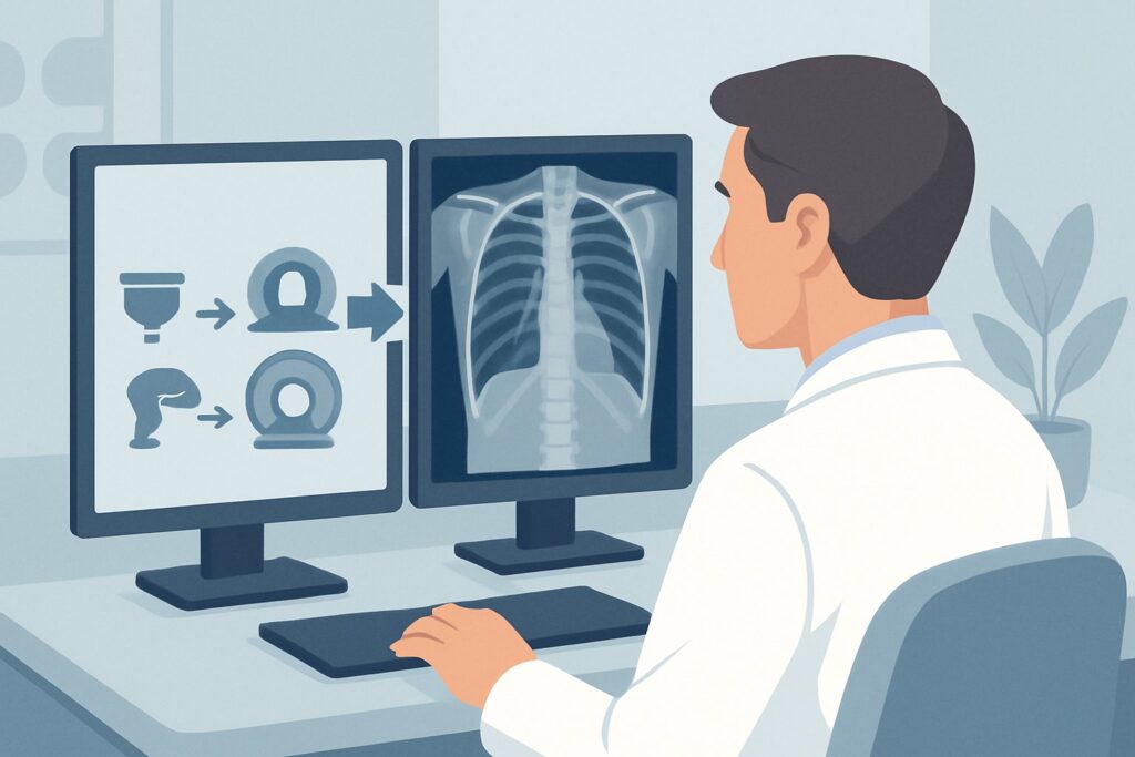
A 45-year-old patient presents to your clinic with a persistent, non-productive cough and a vague sensation of chest fullness that has progressed over three months. They also report mild dysphagia. Physical exam is largely unremarkable, but you have a clinical suspicion for a mediastinal mass compressing adjacent structures. The immediate question is which imaging study to order first to confirm or refute this suspicion without exposing the patient to unnecessary radiation or cost. This article details the American College of Radiology (ACR) Appropriateness Criteria workflow for this exact scenario: the initial imaging workup of a clinically suspected mediastinal mass. For this presentation, the ACR rates a standard chest radiograph as Usually Appropriate.
Who Fits This Clinical Scenario?
This guidance applies to adult and pediatric patients who present with signs or symptoms suggestive of a mediastinal mass but have not yet had any imaging. These symptoms are often non-specific and can include persistent cough, dyspnea, chest pain, dysphagia, hoarseness (due to recurrent laryngeal nerve involvement), or constitutional symptoms like fever and weight loss. More specific findings, such as superior vena cava (SVC) syndrome (facial swelling, plethora, distended neck veins), are also strong indicators.
This workflow is intended for the initial diagnostic step. It is crucial to distinguish this scenario from related but distinct clinical situations:
- Exclusion 1: Indeterminate mass already found on imaging. If a patient has already had a chest radiograph or CT for another reason that revealed an indeterminate mediastinal mass, this scenario does not apply. The appropriate next step is covered in our guide for an indeterminate mediastinal mass on radiography.
- Exclusion 2: Staging of a known malignancy. This guidance is for initial detection and localization, not for staging a patient with a known primary cancer elsewhere that may have metastasized to the mediastinum. Staging protocols are different and often involve PET/CT.
- Exclusion 3: Suspected primary lung cancer. While a large lung cancer can extend into the mediastinum, the workup for a suspected primary bronchogenic carcinoma follows a different diagnostic pathway.
What Diagnoses Are You Working Up in This Scenario?
The mediastinum is anatomically complex, and the differential diagnosis for a mass is broad, often categorized by its location within the anterior, middle, or posterior compartments. The initial imaging aims to localize the mass to one of these compartments, which dramatically narrows the differential.
In the anterior mediastinum, the classic differential is often remembered by the “4 T’s.” The most common etiologies include Thymoma (or other thymic neoplasms like cysts or carcinomas), Teratoma and other germ cell tumors (which can be benign or malignant), Thyroid masses (typically a substernal goiter), and “Terrible” Lymphoma (both Hodgkin and non-Hodgkin types).
In the middle mediastinum, the differential is most commonly dominated by lymphadenopathy, which can be from metastatic disease, lymphoma, or granulomatous diseases like sarcoidosis or tuberculosis. Congenital foregut duplication cysts, such as bronchogenic cysts, are also found here.
The posterior mediastinum is the primary location for neurogenic tumors, arising from nerve sheaths (schwannomas, neurofibromas) or sympathetic ganglia (ganglioneuromas). Other less common possibilities include esophageal pathology or extramedullary hematopoiesis. The initial imaging study is the first step in differentiating these possibilities.
Why Is a Chest Radiograph the Recommended Initial Study?
For the initial evaluation of a suspected mediastinal mass, a simple posteroanterior (PA) and lateral chest radiograph is rated as Usually Appropriate and serves as the ideal first-line test. The rationale is based on its high utility as a screening and localization tool, combined with its low cost, wide availability, and minimal radiation dose.
A two-view chest radiograph can effectively:
- Confirm the presence of a mediastinal abnormality.
- Localize the mass to the anterior, middle, or posterior compartment, which is the most critical first step in narrowing the differential diagnosis.
- Provide initial characterization, such as identifying calcification (common in goiters or teratomas), fat (suggesting a teratoma), or assessing the contour of the mass (e.g., the “sail sign” of a normal thymus in an infant).
While CT and MRI are also rated Usually Appropriate, they are not the recommended initial study. Starting with these advanced modalities is often inefficient. A CT chest (with or without contrast) delivers a significantly higher radiation dose (☢☢☢ 1-10 mSv) compared to a radiograph (☢ <0.1 mSv). An MRI chest (O 0 mSv) involves no ionizing radiation but is more expensive, less accessible, and takes longer to perform, making it unsuitable as a first-line screening tool for this indication. The radiograph provides the necessary information to determine if cross-sectional imaging is needed and helps tailor the protocol for that subsequent CT or MRI.
Studies rated Usually Not Appropriate for this initial workup include FDG-PET/CT and ultrasound. PET/CT is a high-radiation (☢☢☢☢ 10-30 mSv) functional study best used for staging known malignancies, not for initial detection. Ultrasound is severely limited by the overlying sternum, ribs, and air-filled lungs, and it cannot visualize the majority of the mediastinum.
What’s Next After a Chest Radiograph? Downstream Workflow
The results of the initial chest radiograph will dictate the subsequent diagnostic pathway. The goal of the radiograph is not to make a final diagnosis but to guide the next, more definitive imaging step.
- If the radiograph is positive: A discrete mass or abnormal mediastinal contour confirms the clinical suspicion. The patient’s workflow now moves to the next clinical scenario: Indeterminate mediastinal mass on radiography. The next step is almost always cross-sectional imaging. A CT scan with intravenous contrast is typically the workhorse for further characterization, providing detailed anatomic information about the mass, its density, enhancement pattern, and relationship to adjacent vascular and airway structures. MRI may be preferred if the mass is in the posterior mediastinum (for better evaluation of neural foramina) or if a cystic nature is suspected.
- If the radiograph is negative: A normal chest radiograph significantly lowers the likelihood of a large mediastinal mass. However, it cannot definitively exclude a small or subtle lesion. If clinical suspicion remains high (e.g., in a patient with classic SVC syndrome symptoms), it is appropriate to proceed directly to a CT chest with IV contrast.
- If the radiograph is indeterminate: Findings like subtle mediastinal widening or an unusual contour may be inconclusive. In these cases, a CT chest is the logical next step to clarify the anatomy and determine if a true mass is present.
Pitfalls to Avoid (and When to Get Help)
Several common pitfalls can occur during the initial workup of a suspected mediastinal mass. Awareness of these can help ensure a timely and accurate diagnosis.
- Stopping with a negative radiograph: Do not dismiss strong clinical signs (like those of SVC syndrome) based on a normal chest X-ray. A CT scan is warranted if suspicion remains high.
- Omitting the lateral view: A PA-only chest radiograph is insufficient. The lateral view is essential for localizing a mass to the correct mediastinal compartment, which is the key to narrowing the differential.
- Prematurely ordering PET/CT: Ordering a PET/CT as the initial imaging test is inappropriate. It leads to unnecessary radiation exposure and cost and is best reserved for staging or assessing metabolic activity after a mass has been identified and characterized on CT or MRI.
- Misinterpreting normal variants: Be aware of normal anatomic structures that can mimic a mass, such as a prominent thymus in a young patient, a tortuous aorta, or a pericardial fat pad.
If you identify a mass that appears to be actively invading critical structures like the great vessels or trachea, or if the patient is in clinical distress, an urgent consultation with thoracic surgery or interventional radiology is warranted.
Related ACR Topics and Tools
This article covers one specific scenario within the broader topic of mediastinal masses. For a comprehensive overview of all related scenarios, from initial workup to post-treatment surveillance, please consult our parent guide. The following GigHz resources can also support your clinical decision-making:
- For breadth across all scenarios in Imaging of Mediastinal Masses, see our parent guide: Imaging of Mediastinal Masses: ACR Appropriateness Decoded.
- To look up other clinical presentations, use the ACR Appropriateness Criteria Lookup.
- For detailed imaging techniques, browse the Imaging Protocol Library.
- To discuss radiation exposure with patients, use the Radiation Dose Calculator.
Frequently Asked Questions
Why not just order a CT scan first for every patient with a suspected mediastinal mass?
While a CT scan provides more detail, a chest radiograph is the recommended first step due to its role as an effective, low-cost, and low-radiation screening tool. It can confirm the presence of a mass and localize it, which is often sufficient to guide the next steps. This staged approach avoids unnecessary radiation and healthcare costs for patients whose symptoms may be due to other causes.
What if the patient has severe symptoms, like those of superior vena cava (SVC) syndrome?
In cases of high clinical urgency, such as suspected SVC syndrome, it is appropriate to proceed directly to a CT chest with intravenous contrast. This allows for rapid evaluation of the great vessels and identification of the cause of compression. While a chest radiograph is still useful, the acuity of the presentation justifies expediting the workup with cross-sectional imaging.
Is a chest radiograph sensitive enough to rule out a mediastinal mass?
A high-quality two-view chest radiograph is sensitive for detecting most clinically significant mediastinal masses. However, small masses or those obscured by other structures can be missed. Therefore, a negative radiograph does not completely rule out a mass, especially if clinical suspicion is high. In such cases, further imaging with CT is warranted.
Does the initial imaging recommendation change for pediatric patients?
No, the recommendation to start with a chest radiograph remains the same for pediatric patients. The principle of using the lowest possible radiation dose (ALARA – As Low As Reasonably Achievable) is especially important in children. A radiograph is an excellent first step and helps avoid the higher radiation dose of a CT scan unless it is clearly indicated by the radiograph’s findings or persistent, high-risk symptoms.
If the chest X-ray shows a mass, should I order a CT or an MRI next?
After a mass is identified on a radiograph, a CT with intravenous contrast is the most common next step. It excels at showing calcification, vascular involvement, and lymphadenopathy. MRI is typically reserved for specific situations, such as characterizing cystic lesions, evaluating for spinal canal extension of posterior mediastinal masses (neurogenic tumors), or for patients who cannot receive iodinated contrast.
Reviewed by Pouyan Golshani, MD, Interventional Radiologist — May 29, 2026