What Imaging Should You Order for Chest Wall Pain After a Prior Intervention?
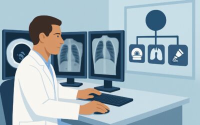
A 68-year-old patient presents to your clinic with three weeks of persistent, focal pain over the right lateral chest wall. Six months ago, they underwent a thoracotomy for a wedge resection of a pulmonary nodule. The pain is sharp, non-pleuritic, and tender to palpation directly over the surgical scar. An initial chest radiograph is unremarkable, showing expected post-surgical changes but no acute process. You suspect a localized complication but need to rule out more serious pathology. This article details the American College of Radiology (ACR) recommended imaging workflow for this specific scenario: nontraumatic chest wall pain in a patient with a history of prior chest intervention, following a normal chest radiograph. For this presentation, the ACR rates CT chest with IV contrast as Usually Appropriate.
Who Fits This Clinical Scenario?
This guidance is for a specific patient population: individuals presenting with new or worsening nontraumatic chest wall pain who have a history of a prior intervention involving the chest. This history is the critical anchor. “Intervention” is a broad category that includes:
- Thoracic Surgery: Such as thoracotomy, sternotomy, video-assisted thoracoscopic surgery (VATS), or rib resection.
- Radiation Therapy: To the chest wall, mediastinum, or breast.
- Device Placement: Including pacemakers, defibrillators, vascular access ports, or pain pumps.
- Prior Procedures: Such as chest tube placement or percutaneous biopsy.
The workflow applies when this history is coupled with a recent, unrevealing chest radiograph. The key decision is selecting the next, more advanced imaging study.
This article does not apply to patients with acute trauma, those with no history of prior intervention, or those with a known active malignancy being evaluated for new metastatic disease. Those presentations follow different diagnostic pathways. For example, a patient with suspected malignancy but no prior intervention would fall under a different ACR variant, which may alter the choice or urgency of imaging.
What Diagnoses Are You Working Up in This Scenario?
In a patient with a history of chest intervention, the differential diagnosis for new chest wall pain is distinct from that in a patient with no such history. The imaging strategy is designed to differentiate between benign post-procedural changes and clinically significant pathology.
Post-procedural Fluid Collections (Abscess, Hematoma, Seroma)
The most common etiologies relate directly to the prior intervention. A localized infection can manifest as an abscess, often appearing as a rim-enhancing fluid collection. A hematoma or seroma can also develop, and while often self-limiting, they can become secondarily infected or cause significant mass effect and pain. Differentiating these is a primary goal of cross-sectional imaging.
Tumor Recurrence or Metastasis
If the initial intervention was for a malignancy, local recurrence in the chest wall is a primary concern. Tumor implants can occur along surgical tracts, chest tube sites, or in the soft tissues. Imaging must be sensitive enough to detect enhancing soft tissue nodules or masses that are distinct from normal scar tissue or granulation tissue.
Suture Granuloma or Foreign Body Reaction
Non-absorbable sutures or other surgical materials can incite a chronic inflammatory reaction, forming a painful mass known as a suture granuloma. While benign, these can mimic tumor recurrence on imaging and may require biopsy for definitive diagnosis. CT can help characterize the mass and its relationship to prior surgical clips or sutures.
Musculoskeletal and Osseous Pathology
Less common but consequential possibilities include osteomyelitis of a rib or the sternum, particularly after sternotomy. Radiation therapy can also lead to osteoradionecrosis or stress fractures. These conditions are poorly visualized on a plain radiograph but are well-delineated on CT.
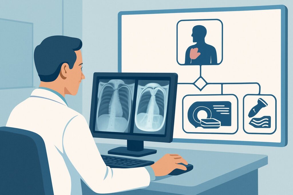
Why Is CT Chest with IV Contrast the Recommended Study for This Presentation?
The ACR designates CT chest with IV contrast as Usually Appropriate because it provides the comprehensive anatomic detail needed to assess the diverse differential diagnoses in this scenario. It excels at evaluating the bones, pleura, and soft tissues of the chest wall simultaneously.
The addition of intravenous contrast is critical. Contrast enhancement helps distinguish between a simple fluid collection (non-enhancing), an abscess (rim-enhancing), and a solid tumor (variably enhancing). This distinction is fundamental to the downstream clinical workflow, guiding decisions between antibiotic therapy, percutaneous drainage, or biopsy. Without contrast, these entities can appear frustratingly similar.
CT chest without IV contrast is also rated Usually Appropriate. It is an excellent choice for evaluating osseous structures for fracture, hardware complication, or osteomyelitis. However, it is less sensitive for detecting abscesses or characterizing soft tissue masses, which are key considerations in this scenario. The choice between a contrasted and non-contrasted study often hinges on the specific concern; if infection or tumor recurrence is high on the differential, the contrasted study is superior.
How do alternative studies compare?
- Ultrasound (US) chest: Rated as May be appropriate, ultrasound is a valuable tool for targeted evaluation of a palpable abnormality or suspected superficial fluid collection. It is non-invasive and uses no ionizing radiation. However, its utility is limited by the overlying ribs and its inability to visualize deep structures or intrathoracic extension. It is often best used as an adjunct to CT or to guide a bedside drainage procedure.
- Rib Views: Rated as Usually not appropriate. A standard chest radiograph includes the ribs. Dedicated rib views offer little additional diagnostic information for the soft tissue and post-procedural complications being considered here and result in unnecessary radiation exposure.
The radiation dose for a CT chest is moderate (ACR RRL ☢☢☢, 1-10 mSv), a consideration that should be weighed against the significant diagnostic value it provides in clarifying the cause of pain and guiding subsequent management. Once you’ve decided on CT chest with IV contrast, our protocol guide covers key technical considerations for contrast-enhanced torso imaging, including technique, contrast timing, and reading principles: CT Chest/Abdomen/Pelvis with IV Contrast.
What’s Next After CT Chest with IV Contrast? Downstream Workflow
The results of the CT scan will direct the subsequent clinical pathway. The goal is to move from a broad differential to a specific diagnosis and treatment plan.
If the CT is positive for a drainable collection (abscess or symptomatic hematoma/seroma):
The next step is typically a consultation with interventional radiology for image-guided percutaneous drainage. This can be both diagnostic (by providing fluid for culture) and therapeutic. A surgical consultation may be required for complex, loculated collections or those not amenable to a percutaneous approach.
If the CT shows a solid, enhancing mass suspicious for tumor recurrence:
This finding necessitates a tissue diagnosis. An image-guided (CT or ultrasound) core needle biopsy is the standard next step. The results will guide consultation with oncology to determine treatment options, which may include further surgery, radiation, or systemic therapy.
If the CT is negative or shows only nonspecific post-surgical changes:
If a high clinical suspicion for pathology remains despite a negative CT, the workup may proceed to a more sensitive but less specific modality. An MRI of the chest wall (May be appropriate) can be a powerful problem-solving tool, offering superior soft tissue contrast to differentiate scar from subtle recurrent tumor or to evaluate for marrow abnormalities. An FDG-PET/CT scan (May be appropriate) may also be considered to look for metabolically active processes like occult infection or malignancy.
Pitfalls to Avoid (and When to Get Help)
Navigating this clinical scenario requires careful correlation of imaging with the patient’s history and physical exam. Here are a few common pitfalls to avoid:
- Omitting IV Contrast: Ordering a non-contrast CT when infection or tumor is a primary concern can lead to a non-diagnostic study, delaying diagnosis and requiring a second, contrasted scan.
- Ignoring the Operative Report: Providing the radiologist with details from the prior intervention (e.g., type of surgery, materials used like mesh or plates) can be invaluable for interpreting subtle post-surgical findings.
- Dismissing Pain as “Just Scar Tissue”: While scar pain is common, new or changing pain warrants a thorough investigation to rule out an actionable cause.
- Underestimating Superficial Pathology: Do not forget the utility of a focused physical exam. A palpable fluid wave or focal erythema may point directly to a superficial collection best evaluated first with ultrasound.
If the CT findings are equivocal or conflict with the clinical picture, a discussion with the reading radiologist is the most effective next step to determine if alternative sequences, different modalities, or short-term follow-up imaging is warranted.
Related ACR Topics and Tools
This article covers one specific variant within the broader topic of nontraumatic chest wall pain. For a comprehensive overview of all clinical scenarios and their corresponding imaging recommendations, please refer to our parent guide. Additionally, GigHz offers several tools to assist in evidence-based imaging decisions.
- For breadth across all scenarios in Nontraumatic Chest Wall Pain, see our parent guide: Nontraumatic Chest Wall Pain: ACR Appropriateness Decoded.
- ACR Appropriateness Criteria Lookup — for adjacent scenarios
- Imaging Protocol Library — for technique on the recommended study
- Radiation Dose Calculator — for cumulative dose conversations
Frequently Asked Questions
Why not just order an MRI first to avoid radiation?
While MRI has excellent soft tissue contrast and no ionizing radiation, CT is generally faster, more widely available, and provides superior evaluation of the osseous structures of the chest wall. For the initial workup of a post-procedural complication, CT provides a comprehensive and rapid assessment. The ACR rates MRI as ‘May be appropriate’, and it is often best reserved as a problem-solving tool if the CT is negative or equivocal.
The ACR lists CT chest *without* contrast as also ‘Usually Appropriate’. When should I choose that?
A non-contrast CT is a reasonable choice if your primary clinical suspicion is for a purely osseous issue, such as a sternal wire complication, non-displaced fracture, or bony destruction. However, if you are concerned about an abscess, fluid collection, or tumor recurrence—all of which are common in this scenario—the IV contrast is critical for characterization. In most cases, the contrasted study provides more diagnostic information.
My patient has severe chronic kidney disease. What are the options if I can’t give iodinated IV contrast?
In patients with contraindications to iodinated contrast, a non-contrast CT is the first step. If this study is unrevealing, the next best modality is often an MRI of the chest wall, which can be performed without and with a gadolinium-based contrast agent (after assessing the risk based on the patient’s specific GFR and the type of agent used). Ultrasound can also be used to evaluate superficial abnormalities without any risk to the kidneys.
Does the imaging choice change if the pain is specifically over the sternum after a median sternotomy?
The primary imaging choice remains CT, but the specific concerns are slightly different. In a post-sternotomy patient, the radiologist will pay special attention to sternal dehiscence, nonunion, and osteomyelitis. CT is the best modality for evaluating the sternal wires and bone integrity. IV contrast remains important for assessing for associated collections like a mediastinal abscess.
The initial chest radiograph was normal. Why is another imaging study needed at all?
A chest radiograph is excellent for evaluating the lungs, large pleural effusions, and significant bony abnormalities. However, it is very insensitive for subtle chest wall pathology. Soft tissue abscesses, small fluid collections, early osteomyelitis, and most tumor recurrences confined to the chest wall will be completely invisible on a plain radiograph. Cross-sectional imaging like CT is required to visualize these structures.
Reviewed by Pouyan Golshani, MD, Interventional Radiologist — May 29, 2026