What Surveillance Imaging Is Best for Stable Chronic Cardiopulmonary Disease?
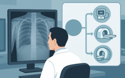
You are seeing a 72-year-old patient with a long history of chronic obstructive pulmonary disease (COPD) for a routine six-month follow-up. His breathing is at his baseline, with no new cough, fever, or increased sputum production. He feels well. As you review his chart, you note his last chest x-ray was over a year ago. You pause, considering whether routine surveillance imaging is indicated today. Is it time for another chest radiograph, or is that unnecessary medical radiation? This article details the American College of Radiology (ACR) Appropriateness Criteria workflow for this exact scenario: surveillance imaging in a patient with stable, chronic cardiopulmonary disease. For this presentation, a chest radiograph is rated as May be appropriate.
Who Fits This Clinical Scenario?
This guidance applies specifically to patients with a known, established diagnosis of chronic cardiopulmonary disease who are clinically stable. This includes conditions like Chronic Obstructive Pulmonary Disease (COPD), stable interstitial lung disease (ILD), or chronic congestive heart failure (CHF) without acute decompensation. The key qualifier is “stable”—meaning the patient’s symptoms are at their usual baseline, with no new or accelerating complaints like increased dyspnea, chest pain, or fever.
This workflow is intended for routine, scheduled surveillance, not for the evaluation of new problems. It is crucial to distinguish this scenario from several similar but distinct clinical situations that require a different imaging approach:
- Patients with acute exacerbation: If the patient presents with worsening symptoms (e.g., increased shortness of breath, new cough, fever), they are no longer “stable.” Their workup falls under a different clinical pathway for acute respiratory illness.
- Preoperative evaluation: Patients undergoing preoperative assessment for noncardiothoracic surgery, even with a history of cardiopulmonary disease, are covered by a separate ACR variant focused on assessing surgical risk.
- Hospital admission: A patient being admitted to the hospital, even if asymptomatic from a pulmonary perspective, may undergo imaging based on admission protocols, which is a distinct scenario.
Applying this surveillance guidance to a patient with changing symptoms is a common pitfall that can delay the diagnosis of an acute process.
What Diagnoses Are You Working Up in This Scenario?
In surveillance imaging for a stable patient, the goal is not to diagnose a new, symptomatic condition but rather to monitor for subtle, clinically silent changes. The “differential” in this context is a list of potential findings you are monitoring for over time.
A primary goal is to detect the slow progression of the underlying chronic disease. For a patient with interstitial lung disease, this might mean identifying subtle increases in reticulation or honeycombing. In a patient with congestive heart failure, it could involve tracking changes in cardiomegaly or early signs of pulmonary venous congestion that predate a clinical decompensation. For emphysema, it might be assessing the progression of hyperinflation or bullous changes.
Another key objective is to screen for superimposed processes that may be asymptomatic in their early stages. This is particularly relevant for identifying an early lung malignancy in high-risk patients, such as a long-term smoker with COPD. While a routine chest radiograph is not a substitute for dedicated low-dose CT lung cancer screening, it can occasionally reveal an incidental nodule that warrants further investigation.
Finally, surveillance can identify complications related to the chronic condition. This could include the development of significant pleural effusions in a patient with chronic heart failure or liver disease, or detecting a spontaneous pneumothorax in a patient with severe bullous emphysema, which can sometimes present with minimal symptoms initially.
Why Is a Chest Radiograph the Recommended Study for This Presentation?
The ACR designates a chest radiograph as May be appropriate for surveillance in stable chronic cardiopulmonary disease, reflecting a nuanced balance of benefit versus risk. The recommendation is not a strong “usually appropriate” because the value of routine imaging in a truly asymptomatic and stable patient can be limited. However, when surveillance is deemed clinically useful, the chest radiograph is the clear first-line choice.
The primary rationale is its favorable risk-benefit profile. A chest radiograph provides a global assessment of the heart and lungs with minimal radiation exposure (relative radiation level ☢, <0.1 mSv). It is highly effective for detecting significant interval changes, such as the development of a large pleural effusion, a new consolidation concerning for pneumonia, a significant change in heart size, or a large, obvious mass. It is inexpensive, widely accessible, and fast.
Alternative imaging modalities are rated lower for this specific scenario due to higher radiation, cost, or lack of added clinical value in an asymptomatic patient:
- CT chest without IV contrast: While also rated May be appropriate, it delivers a substantially higher radiation dose (☢☢☢, 1-10 mSv). It is not justified for routine annual surveillance in every stable patient but becomes the logical next step if a chest radiograph shows an indeterminate or concerning finding. It offers superior detail for evaluating subtle interstitial disease progression or characterizing a small nodule.
- CT chest with IV contrast or CTA chest: These are rated Usually not appropriate. In a stable patient with no signs of infection, pulmonary embolism, or suspected vascular pathology, intravenous contrast adds no diagnostic value and introduces risks of allergic reaction and contrast-induced nephropathy.
- MRI chest: This is also Usually not appropriate. While it avoids ionizing radiation, MRI is more expensive, less available, and prone to motion artifact from breathing and cardiac motion. It offers little advantage over radiography or CT for evaluating the lung parenchyma in most chronic diseases.
The choice to perform surveillance imaging at all should be deliberate. For many stable patients, annual imaging may not change management. However, for specific conditions like ILD or in patients at high risk for complications, periodic radiographic assessment can be a valuable component of long-term care.
What’s Next After a Chest Radiograph? Downstream Workflow
The results of the surveillance chest radiograph will guide the subsequent clinical workflow. The decision tree branches based on whether the findings are stable, show progression, or are indeterminate.
If the study is negative or stable compared to prior exams: This is the most common outcome. The finding provides reassurance that no major interval changes have occurred. The next step is to continue routine clinical management and schedule the next surveillance imaging study according to the patient’s specific condition and established guidelines (e.g., in 12-24 months), assuming they remain clinically stable.
If the study shows a new or worsening finding: This result effectively transitions the patient from a “surveillance” to a “diagnostic” pathway.
- A new focal opacity or lung nodule would typically prompt a CT chest without IV contrast to better characterize the lesion.
- Worsening cardiomegaly or signs of pulmonary edema would lead to clinical re-evaluation of volume status, medication adjustment, and potentially an echocardiogram.
- A new or significantly larger pleural effusion may require diagnostic thoracentesis.
If the study is indeterminate: Sometimes, a finding is ambiguous—for example, a vague opacity that could represent scarring, atelectasis, or an early infiltrate. The first and most critical step is a careful comparison with all available prior imaging studies. If the finding is old and stable, no further action is needed. If it is new or priors are unavailable, a CT chest without IV contrast is often the most appropriate next step to resolve the ambiguity.
Pitfalls to Avoid (and When to Get Help)
Navigating surveillance imaging requires careful clinical judgment to maximize benefit and minimize harm. Here are a few common pitfalls to avoid in this specific scenario:
- Over-imaging the Stable Patient: Performing chest radiographs too frequently (e.g., every few months) in a patient with no clinical changes can lead to unnecessary radiation exposure and costs without improving outcomes.
- Misclassifying the Patient as “Stable”: Do not apply this surveillance workflow to a patient with new or worsening symptoms. A subtle increase in dyspnea or a new cough warrants a diagnostic workup, not a routine check.
- Ignoring Prior Images: The value of a surveillance radiograph is magnified exponentially when compared to previous studies. Always ensure the interpreting radiologist has access to historical images. A “new” finding may turn out to be a stable, chronic one.
- Automatic CT Escalation: Avoid ordering a CT scan as the initial surveillance study in a stable patient. The chest radiograph should be the first step unless there is a highly specific question that only CT can answer (e.g., precise monitoring of fibrotic lung disease).
If a surveillance image reveals a complex or unexpected finding, such as multiple new nodules or diffuse parenchymal changes, consultation with a pulmonologist or thoracic radiologist is the appropriate next step.
Related ACR Topics and Tools
For a comprehensive overview of all clinical variants related to routine chest imaging, from preoperative clearance to hospital admission, please see our parent guide. For other tools to assist in imaging decisions, see the resources below.
- For breadth across all scenarios in Routine Chest Imaging, see our parent guide: Routine Chest Imaging: ACR Appropriateness Decoded.
- To explore other clinical scenarios, use the ACR Appropriateness Criteria Lookup.
- For details on imaging techniques, visit the Imaging Protocol Library.
- To discuss cumulative radiation exposure with patients, consult the Radiation Dose Calculator.
Frequently Asked Questions
How often should I order a surveillance chest x-ray for a stable COPD patient?
There is no universal consensus, and guidelines vary. The ACR rates chest radiography as ‘May be appropriate,’ indicating it should not be an automatic annual order. The decision should be individualized. For a truly stable patient with no new symptoms, ordering an x-ray less frequently (e.g., every 2-3 years) or only when clinically indicated is a reasonable approach to minimize radiation.
If my patient with stable interstitial lung disease (ILD) needs surveillance, is a chest x-ray enough?
A chest x-ray is the appropriate first-line surveillance tool. However, for ILD, subtle changes in fibrosis may be difficult to appreciate on a radiograph. For this reason, many pulmonologists alternate chest x-rays with periodic high-resolution CT scans (a non-contrast study) to more accurately assess for disease progression, though this falls into a more specialized surveillance category.
Why is CT chest without contrast also rated ‘May be appropriate’ for this scenario?
While chest radiography is the preferred initial test due to lower radiation and cost, a non-contrast CT is considered ‘May be appropriate’ because it can serve as a problem-solving tool or a more detailed surveillance method in select cases. For instance, if a prior radiograph showed an indeterminate nodule, follow-up might be with CT. It is not intended as the primary, routine screening tool for all stable patients.
Does a normal surveillance chest x-ray rule out lung cancer?
No. A chest radiograph can miss small or subtle lung nodules. For high-risk individuals (based on age and smoking history), the appropriate screening test for lung cancer is a low-dose CT (LDCT) scan, which is a separate clinical indication from routine surveillance of chronic cardiopulmonary disease.
My patient has stable CHF. What am I looking for on a surveillance chest x-ray?
In a stable CHF patient, you are primarily monitoring for signs of impending decompensation that may not yet be clinically obvious. Key findings include an increase in cardiac silhouette size, development of pulmonary venous congestion (e.g., cephalization of pulmonary vessels), Kerley B lines, or new or enlarging pleural effusions. These findings may prompt medication adjustments before the patient becomes overtly symptomatic.
Reviewed by Pouyan Golshani, MD, Interventional Radiologist — May 30, 2026