What Is the Next Imaging Step for Chest Wall Pain in an Oncology Patient with a Normal X-ray?
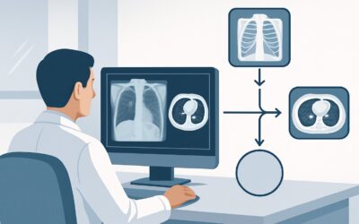
A 68-year-old man with a history of prostate cancer presents to your clinic with two weeks of new, deep, nontraumatic pain over his left lateral chest wall. It’s worse at night and not improving with simple analgesics. You obtain a chest radiograph, which shows no acute cardiopulmonary disease, definite fracture, or obvious lytic or blastic lesion. Your primary concern is osseous metastatic disease, but you need to confirm the diagnosis and assess for other sites of involvement. What is the most appropriate next imaging study to order? This clinical workflow article details the American College of Radiology (ACR) guidance for this specific scenario. For this presentation, a whole body bone scan is rated Usually Appropriate as the next step in evaluation.
Who Fits This Clinical Scenario for Chest Wall Pain?
This guidance applies to a specific patient population: individuals with a known or suspected malignancy who present with nontraumatic chest wall pain and have already had a normal or non-diagnostic chest radiograph. The key inclusion criteria are the oncologic history (which raises the pre-test probability of metastatic disease) and the negative initial radiograph (which necessitates more sensitive secondary imaging).
This workflow is distinct from other presentations of chest wall pain. It is crucial to ensure your patient fits this scenario and not a neighboring one, as the recommended imaging may differ significantly. This guidance does not apply if:
- There is no history or suspicion of malignancy. For patients without an oncologic history, the initial workup for nontraumatic chest wall pain follows a different pathway, often focusing on musculoskeletal or cardiopulmonary causes.
- An infectious or inflammatory cause is suspected. If the patient presents with fever, erythema, swelling, or elevated inflammatory markers (e.g., C-reactive protein, erythrocyte sedimentation rate), the differential diagnosis shifts toward conditions like osteomyelitis or costochondritis, which may require different imaging modalities.
- The pain is related to a prior chest intervention. Patients with a history of recent surgery, radiation therapy, or biopsy in the area of pain require a workup tailored to evaluating post-procedural complications, such as seroma, infection, or radiation-induced changes.
What Diagnoses Are You Working Up in This Malignancy Scenario?
After a normal chest radiograph, the imaging workup is driven by a differential diagnosis heavily weighted toward oncologic causes. While benign etiologies are still possible, the primary goal is to rule in or rule out diagnoses that would significantly alter patient management.
The most common and consequential diagnosis in this setting is osseous metastatic disease. Many primary cancers, particularly breast, prostate, lung, kidney, and thyroid, have a high propensity to spread to bone. The ribs and thoracic spine are common sites. The pain from bone metastases is often described as deep, dull, and progressive, and it may be worse at night. A normal radiograph does not exclude this diagnosis, as early lesions or those without significant cortical destruction can be radiographically occult.
A direct and urgent complication of a bone metastasis is a pathologic fracture. An underlying metastatic lesion can weaken the bone, leading to a fracture with minimal or no trauma. A plain film may miss a non-displaced fracture through an abnormal-appearing bone, necessitating cross-sectional or functional imaging for detection.
Less commonly, the pain could represent a primary bone or soft tissue tumor, such as a sarcoma. While rare compared to metastatic disease, it remains a critical consideration, especially if the pain is associated with a new, palpable soft tissue mass.
Finally, benign musculoskeletal causes like costochondritis or muscle strain can certainly occur in patients with cancer. However, in the setting of a new, persistent, and focal pain, they become diagnoses of exclusion after more serious pathology has been ruled out.
Why Is a Whole Body Bone Scan Usually Appropriate for This Presentation?
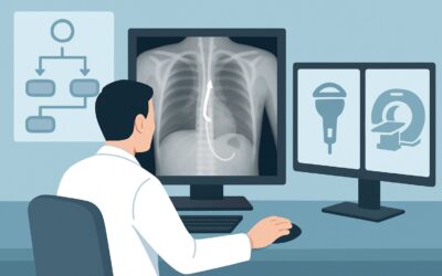
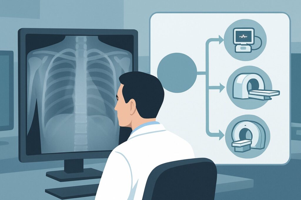
The ACR Appropriateness Criteria rate several studies as Usually Appropriate for this scenario, including a whole body bone scan, CT chest with IV contrast, and CT chest without IV contrast. The choice among them often depends on the specific clinical question, the type of primary malignancy, and local availability.
A Technetium-99m whole body bone scan is a cornerstone of this workup due to its high sensitivity for detecting osteoblastic activity. Many common metastases, particularly from prostate and breast cancer, are blastic or mixed blastic/lytic and will demonstrate intense radiotracer uptake. The primary advantage of a bone scan is its ability to survey the entire skeleton in a single examination, which is essential for staging. It can identify asymptomatic sites of disease in addition to evaluating the symptomatic area in the chest wall.
While highly sensitive, a bone scan has lower specificity. Arthritis, healing fractures, and other benign processes can also show increased uptake. Therefore, a positive finding often requires correlation with other imaging.
Computed Tomography (CT) of the chest (with or without contrast) is also Usually Appropriate. CT provides excellent anatomic detail of the bone cortex and is superior to bone scan for detecting purely lytic lesions, which may not be metabolically active enough to show significant uptake. It can also precisely characterize the extent of bone destruction, assess for an impending pathologic fracture, and evaluate adjacent soft tissues for extension of the tumor.
Other modalities are rated lower for this initial secondary evaluation:
- Magnetic Resonance Imaging (MRI) of the chest (May be appropriate) offers unparalleled evaluation of bone marrow and soft tissues. It is highly sensitive for early marrow replacement by tumor. However, its field of view is limited, making it a poor choice for a primary whole-body screening tool. It is better suited as a problem-solving modality to evaluate an indeterminate finding on a bone scan or CT, or if there are neurologic symptoms suggesting spinal cord compression.
- FDG-PET/CT (May be appropriate) is a powerful tool for whole-body staging of many metabolically active cancers. However, it carries a higher radiation dose (☢☢☢☢ 10-30 mSv) compared to a bone scan (☢☢☢ 1-10 mSv). Furthermore, it can be less sensitive for purely sclerotic (blastic) metastases, which often have low glucose metabolism and may not be FDG-avid.
Once you’ve decided on a whole body bone scan, our protocol guide covers the technique, radiotracer, and reading principles in detail: Nuclear Medicine Bone Scan (Whole Body).
What’s Next After a Bone Scan? Downstream Workflow
The results of the bone scan will guide the subsequent clinical and diagnostic pathway. The goal is to move from detection to definitive characterization and treatment planning.
- If the bone scan is positive for metastatic disease: A finding of multiple areas of abnormal uptake in a pattern typical for metastases (e.g., axial skeleton, ribs) in a patient with a known primary cancer is often sufficient for diagnosis. The next steps involve consultation with oncology to discuss systemic therapy, radiation oncology for palliative radiotherapy to symptomatic sites, and potentially orthopedic surgery if a pathologic fracture is impending. Sometimes, a correlative CT or MRI may be ordered to better define the anatomy of a specific lesion, especially if it is in a weight-bearing bone.
- If the bone scan is negative: A negative bone scan significantly lowers the likelihood of widespread osseous metastatic disease, especially of the blastic type. If clinical suspicion remains high (e.g., severe, persistent, localized pain), the next step may be a more focused, high-resolution study of the symptomatic area. An MRI of the chest wall or thoracic spine would be an excellent choice to evaluate for marrow-based pathology or soft tissue abnormalities missed by the bone scan.
- If the bone scan is indeterminate: A single, equivocal focus of uptake is a common clinical challenge. This could represent an early metastasis or a benign process like a healing rib fracture or degenerative change. The next step is typically correlative imaging with CT or MRI of the specific location to provide anatomic detail and characterize the lesion. In some cases, a PET/CT may be used to assess metabolic activity, or a biopsy may be required for a definitive tissue diagnosis.
Pitfalls to Avoid (and When to Get Help)
Navigating this clinical scenario requires careful consideration to avoid common diagnostic errors.
- Stopping the workup after a normal radiograph: A plain film has low sensitivity for early bone metastases. Persistent, focal bone pain in an oncology patient warrants further investigation even if the x-ray is clear.
- Misinterpreting benign uptake on a bone scan: Be aware of common mimics of metastatic disease, such as degenerative joint disease, healing fractures, and Paget’s disease. Clinical correlation and review with a radiologist are essential.
- Ignoring the primary cancer type: The likelihood of a lesion being lytic versus blastic varies by tumor histology. For primary cancers known for lytic lesions (e.g., multiple myeloma, renal cell carcinoma), a CT may be a better initial choice than a bone scan.
- Delaying imaging for neurologic symptoms: If chest wall pain is accompanied by radicular symptoms, weakness, or sensory changes, this is a red flag for spinal involvement. Escalate immediately to an MRI of the thoracic spine to evaluate for epidural extension and potential spinal cord compression.
Related ACR Topics and Tools
This article focuses on one specific clinical variant. For a comprehensive overview of imaging for all related patient presentations, see our parent guide. For tools to help select the right study and understand its technical details, see the resources below.
- For breadth across all scenarios in Nontraumatic Chest Wall Pain, see our parent guide: Nontraumatic Chest Wall Pain: ACR Appropriateness Decoded.
- ACR Appropriateness Criteria Lookup — For exploring adjacent clinical scenarios and alternative imaging options.
- Imaging Protocol Library — For detailed technical specifications on recommended studies.
- Radiation Dose Calculator — For discussing cumulative radiation exposure with patients.
Frequently Asked Questions
Why not just order a PET/CT for every patient with suspected metastases?
While FDG-PET/CT is a powerful staging tool, it is not always the best first choice for this specific scenario. It has a higher radiation dose than a bone scan and can be less sensitive for detecting purely sclerotic (blastic) metastases, which are common in prostate and breast cancer. The ACR rates it as ‘May be appropriate,’ reserving it for specific clinical situations or as a problem-solving tool.
If both bone scan and CT are ‘Usually Appropriate’, how do I choose between them?
The choice often depends on the primary tumor type and the specific clinical question. A bone scan is excellent for a sensitive, whole-body survey for blastic lesions (common in prostate/breast cancer). A CT provides superior anatomic detail, is better for lytic lesions (common in lung/renal cancer), and can assess the risk of pathologic fracture and soft tissue extension. Often, the two are complementary.
What if the patient has renal insufficiency and cannot receive IV contrast for a CT?
A non-contrast CT of the chest is also rated as ‘Usually Appropriate’ by the ACR. It is excellent for evaluating bone detail, including cortical destruction from lytic or blastic lesions, and for detecting pathologic fractures. The primary limitation is the inability to assess vascular structures or the enhancement patterns of soft tissue masses.
The patient’s pain is very localized to one rib. Should I order a dedicated rib series radiograph?
A dedicated rib series is rated as ‘May be appropriate.’ While it can provide more detailed views of the ribs than a standard chest radiograph, it has the same fundamental limitation: low sensitivity for early metastatic disease that has not yet caused significant bone destruction. In a patient with a known malignancy, a more sensitive systemic study like a bone scan or CT is generally preferred to avoid missing the diagnosis.
Is there a role for ultrasound in this scenario?
Ultrasound of the chest is rated as ‘Usually not appropriate’ for this indication. While ultrasound is excellent for evaluating superficial soft tissue abnormalities, fluid collections, or pleural effusions, it cannot penetrate bone and is not a suitable modality for assessing for osseous metastatic disease, which is the primary concern in this clinical setting.
Reviewed by Pouyan Golshani, MD, Interventional Radiologist — May 29, 2026