Suspected Occupational ILD on Radiograph: Why Is a Non-Contrast CT the Recommended Next Study?
A 68-year-old retired shipyard worker presents with a multi-year history of progressive dyspnea on exertion and a dry cough. His primary care physician ordered a chest radiograph, which reveals bibasilar reticular opacities and thickened pleura. Given his extensive occupational history involving potential asbestos exposure, you suspect asbestosis or another form of interstitial lung disease (ILD). The immediate clinical question is how to best characterize these radiographic findings to confirm a diagnosis and assess the extent of disease. This article provides a focused, step-by-step workflow for this exact scenario, guided by the American College of Radiology (ACR) Appropriateness Criteria, which rate a CT chest without IV contrast as Usually Appropriate.
## Who Fits This Clinical Scenario for Occupational Lung Disease Imaging?
This guidance is specifically for patients who meet two key criteria: a documented or highly suspected history of significant occupational exposure to fibrogenic dusts or agents, and an abnormal chest radiograph that is suspicious for interstitial lung disease. The initial radiograph serves as the trigger, having already identified findings like reticular patterns, nodules, ground-glass opacities, or pleural abnormalities.
This workflow is designed to characterize known radiographic abnormalities, not for initial detection. It is crucial to distinguish this situation from several related but distinct clinical presentations:
- Exclusion 1: Asymptomatic Screening: If the patient has a known exposure history but is asymptomatic with a normal or no recent chest radiograph, the imaging strategy falls under the ACR variant for screening and surveillance. That pathway involves different considerations about the timing and modality of initial imaging.
- Exclusion 2: Initial Workup Without Radiography: If a patient presents with symptoms suggestive of ILD (e.g., dyspnea, cough) and a relevant exposure history but has not yet had any imaging, the workup starts with the ACR variant for suspected interstitial lung disease, initial imaging, where chest radiography is often the first step.
- Exclusion 3: Suspected Airway Disease: If the patient’s symptoms and pulmonary function tests point toward an obstructive process like occupational asthma or chronic bronchitis (e.g., wheezing, airflow limitation), the imaging workup would follow the ACR variant for suspected airway disease, which may involve different CT protocols like expiratory imaging.
This article focuses solely on the patient who already has an abnormal radiograph, and the goal is to select the best next imaging study for definitive characterization.
## What Diagnoses Are You Working Up in This Scenario?
When an abnormal radiograph follows a history of occupational exposure, the differential diagnosis centers on the pneumoconioses—a group of interstitial lung diseases caused by the inhalation of mineral dusts. The next imaging study, a high-resolution CT, is intended to differentiate these conditions, which often have characteristic, though sometimes overlapping, features.
Asbestosis: Caused by inhaling asbestos fibers, common in construction, shipbuilding, and insulation work. The classic CT findings include parenchymal bands, subpleural curvilinear lines, and bibasilar, peripheral-predominant honeycombing. The presence of associated pleural plaques or diffuse pleural thickening is a strong clue pointing toward asbestos as the causative agent.
Silicosis: Resulting from the inhalation of crystalline silica dust in occupations like mining, quarrying, sandblasting, and stone cutting. CT typically reveals multiple small, well-defined nodules, often with a predilection for the upper lung zones. In advanced cases, these nodules can merge into large conglomerate masses, a condition known as progressive massive fibrosis (PMF). Hilar and mediastinal lymph node calcification, sometimes in an “egg-shell” pattern, is highly characteristic.
Coal Worker’s Pneumoconiosis (CWP): Seen in coal miners, CWP has imaging findings that can be very similar to silicosis. Simple CWP presents with small centrilobular and perilymphatic nodules, while complicated CWP involves the development of progressive massive fibrosis. Differentiating CWP from silicosis on imaging alone can be challenging without a clear occupational history.
Chronic Beryllium Disease (CBD): An immune-mediated response to beryllium exposure (aerospace, electronics manufacturing). CT findings can be variable but often include poorly defined centrilobular nodules, ground-glass opacities, and thickened interlobular septa, sometimes mimicking sarcoidosis.
Hypersensitivity Pneumonitis (HP): While not a classic pneumoconiosis, HP is a common occupational ILD resulting from an immunologic reaction to inhaled organic antigens (e.g., “farmer’s lung” from moldy hay). The fibrotic form can show centrilobular nodules, ground-glass opacities, and signs of air trapping on expiratory images, often with mid-lung zone predominance.
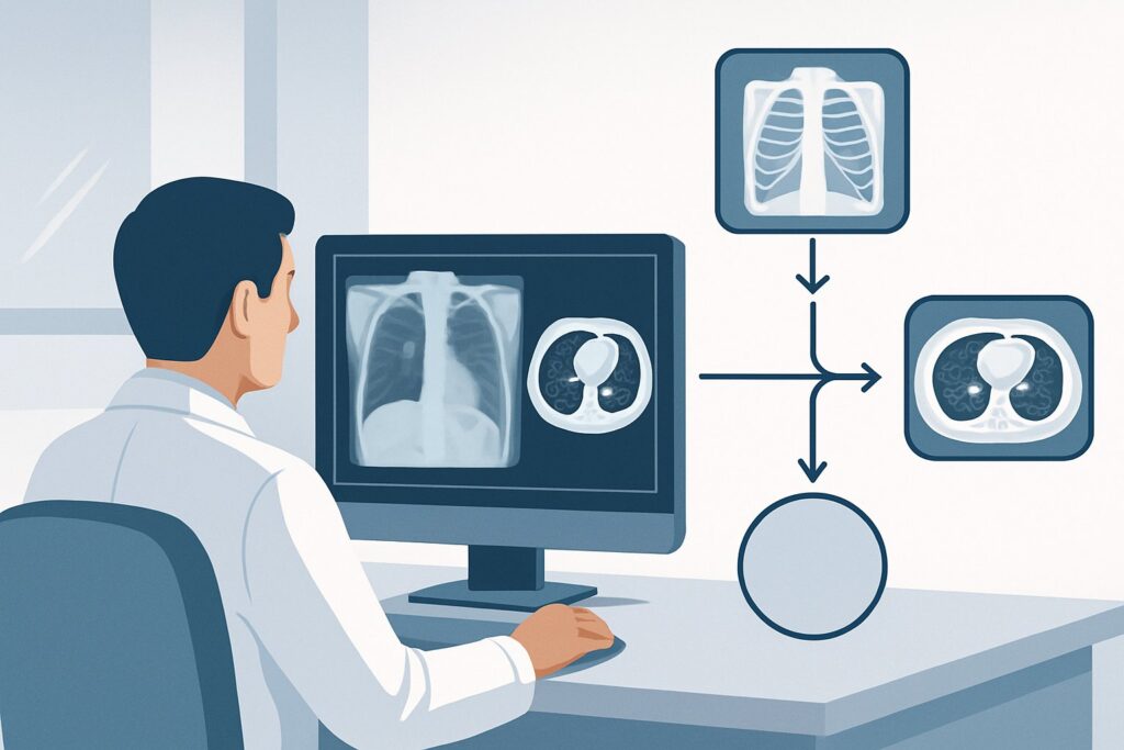
## Why Is CT Chest Without IV Contrast the Recommended Next Study for Suspected Occupational ILD?
For a patient with a suspected occupational ILD based on an abnormal radiograph, the ACR designates CT chest without IV contrast as Usually Appropriate. The rationale is rooted in the superior ability of this modality to visualize the fine architectural details of the lung parenchyma, pleura, and airways.
The key is ordering the study using a high-resolution CT (HRCT) protocol. This technique utilizes very thin slices (typically 1-1.5 mm) and a sharp reconstruction algorithm to provide exquisite detail of the secondary pulmonary lobule—the fundamental unit where the pathology of ILD unfolds. HRCT is exceptionally sensitive for detecting and characterizing the patterns of fibrosis, nodularity, ground-glass opacity, and honeycombing that distinguish different interstitial diseases. It can clearly delineate the distribution of disease (e.g., upper vs. lower lobe, peripheral vs. central), which is a critical clue in the differential diagnosis.
In contrast, several alternative studies are rated Usually Not Appropriate for this specific clinical question:
- CT Chest with IV Contrast: Intravenous contrast is designed to enhance vascular structures and highlight areas of inflammation or malignancy within solid organs. For the primary goal of evaluating the lung interstitium, it provides no additional diagnostic information. It also introduces the risks of allergic reaction and contrast-induced nephropathy and can sometimes create artifacts that obscure subtle parenchymal findings.
- MRI Chest (with or without contrast): While MRI avoids ionizing radiation, it lacks the spatial resolution and signal-to-noise ratio needed to visualize the delicate structures of the lung parenchyma. It is highly susceptible to motion artifacts from breathing and cardiac pulsation, rendering it unsuitable for the detailed assessment required for ILD.
- Image-guided Transthoracic Needle Biopsy: Biopsy is an invasive procedure with risks of pneumothorax and hemorrhage. It is generally reserved for cases where imaging is non-diagnostic or when a superimposed process, like a suspected malignancy, needs to be sampled. It is not an appropriate next step for the initial characterization of diffuse disease seen on a radiograph.
The radiation dose for a non-contrast chest CT is moderate (ACR Relative Radiation Level ☢☢☢, corresponding to 1-10 mSv), but this exposure is justified by the high diagnostic yield that directly guides patient management.
Once you’ve decided on this study, our protocol guide covers the technique and reading principles in detail. For more information, see our complete guide: CT Chest Without Contrast.
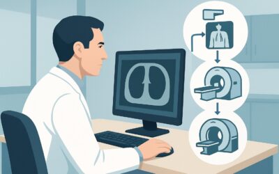
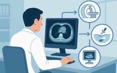
## What’s the Next Step After a Non-Contrast Chest CT?
The results of the high-resolution chest CT will guide the subsequent clinical workflow, which often involves a multidisciplinary discussion with pulmonology and sometimes rheumatology or occupational medicine specialists.
- If the CT shows a classic pattern: When the CT findings are highly characteristic of a specific pneumoconiosis in the context of a clear exposure history (e.g., bilateral pleural plaques and basilar fibrosis in a shipyard worker), the diagnosis of asbestosis can often be made with high confidence. Further invasive testing may be unnecessary, and management can focus on supportive care, smoking cessation, and surveillance for complications like lung cancer or mesothelioma.
- If the CT is suggestive but not definitive: Sometimes, the imaging pattern is consistent with an occupational ILD but has overlapping features (e.g., distinguishing CWP from silicosis, or fibrotic HP from other patterns). In these cases, the next steps may include a comprehensive occupational history review, serologic testing (e.g., for beryllium), and detailed pulmonary function testing. Bronchoalveolar lavage (BAL) may be performed to analyze cell counts and rule out infection.
- If the CT is indeterminate or atypical: If the findings do not fit a clear pattern or if an alternative diagnosis like idiopathic pulmonary fibrosis (IPF) or connective tissue disease-related ILD is suspected, the case should be discussed in a multidisciplinary setting. This may lead to a recommendation for a surgical lung biopsy to obtain a definitive histopathologic diagnosis.
- If the CT reveals a suspected malignancy: Occupational lung diseases, particularly asbestosis and silicosis, significantly increase the risk of lung cancer. If the CT identifies a suspicious nodule, mass, or other finding concerning for a thoracic neoplasm, the workup will pivot to follow the appropriate ACR variant for that scenario, which may involve PET/CT and biopsy.
## Pitfalls to Avoid (and When to Get Help)
Navigating the workup for occupational ILD requires careful attention to detail to avoid common missteps.
- Pitfall 1: Ordering the wrong CT protocol. Simply ordering a “CT Chest” may result in a routine study with thick slices and IV contrast, which is suboptimal for ILD evaluation. Be specific: order a “High-Resolution CT (HRCT) Chest without IV contrast for Interstitial Lung Disease.”
- Pitfall 2: Overlooking coexisting conditions. Patients with pneumoconiosis are often older and may have comorbidities like emphysema (especially in smokers) or heart failure, which can confound the clinical and imaging picture.
- Pitfall 3: Neglecting the exposure history. The imaging pattern must always be interpreted in the context of a detailed and accurate occupational and environmental exposure history. A pattern that is indeterminate on its own may become highly specific when linked to a known exposure.
If the CT findings are complex, atypical, or discordant with the clinical history, escalation to a multidisciplinary ILD conference including thoracic radiologists, pulmonologists, and pathologists is the standard of care.
## Related ACR Topics and Tools
This article is a deep dive into one specific clinical scenario. For a comprehensive overview of imaging for all related presentations, from screening to malignancy, please see our parent guide.
- For breadth across all scenarios in Occupational Lung Diseases, see our parent guide: Occupational Lung Diseases: ACR Appropriateness Decoded.
To explore other scenarios, look up specific study techniques, or discuss radiation dose with your patients, GigHz offers several focused resources:
- ACR Appropriateness Criteria Lookup — for adjacent scenarios
- Imaging Protocol Library — for technique on the recommended study
- Radiation Dose Calculator — for cumulative dose conversations
Frequently Asked Questions
Why is a non-contrast CT preferred over a CT with contrast for suspected occupational ILD?
A non-contrast, high-resolution CT (HRCT) is preferred because it provides the best possible detail of the lung parenchyma, airways, and pleura, which are the structures affected by interstitial lung disease. Intravenous contrast enhances blood vessels and solid organs but adds no diagnostic value for characterizing fibrosis or nodules in the lung itself. It also adds unnecessary risk and cost.
What if the patient has renal insufficiency? Can they still get the recommended CT scan?
Yes. Since the recommended study is a CT chest *without* IV contrast, there is no risk of contrast-induced nephropathy. The patient’s renal function is not a contraindication to this specific imaging test.
Should I order inspiratory and expiratory images for this CT scan?
Including expiratory images can be very helpful, particularly if hypersensitivity pneumonitis (HP) or a mixed obstructive/restrictive process is on the differential. Expiratory scans are excellent for detecting air trapping, a key feature of HP and small airways disease. It is reasonable to request both inspiratory and expiratory series when ordering the HRCT.
The radiograph mentioned a suspicious nodule in addition to interstitial changes. Does that change the imaging plan?
It reinforces the choice of CT but may influence the follow-up plan. A non-contrast HRCT is still the best initial step to characterize both the interstitial disease and the nodule. If the nodule has suspicious features on CT, the workup will then also proceed down a lung cancer screening or diagnostic nodule pathway, which could involve subsequent PET/CT or biopsy, depending on the nodule’s characteristics.
How does HRCT differentiate between asbestosis and silicosis?
While there can be overlap, HRCT often shows distinct patterns. Asbestosis typically causes fibrosis and honeycombing with a bibasilar and peripheral predominance, and is frequently associated with pleural plaques. Silicosis classically presents with small, well-defined nodules in the upper lobes and can cause ‘egg-shell’ calcification in the hilar lymph nodes. The distribution and specific ancillary findings are key differentiators.
Reviewed by Pouyan Golshani, MD, Interventional Radiologist — May 29, 2026