Which Imaging Study Is Best for Suspected Tracheomalacia in an Adult?
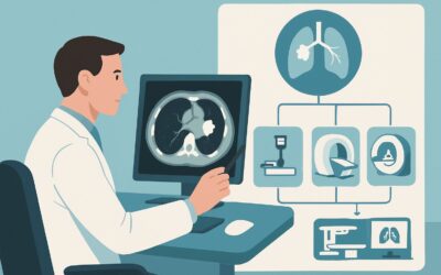
A 62-year-old man with a history of severe COPD presents with a persistent, barking cough and worsening dyspnea on exertion, particularly when he bends over or lies flat. His symptoms have been refractory to aggressive bronchodilator therapy. Pulmonary function tests show expiratory airflow limitation, but the pattern is suggestive of a dynamic central airway process. You suspect tracheomalacia, a condition characterized by excessive airway collapse during breathing. The critical question now is how to confirm this diagnosis non-invasively. This article details the ACR-guided imaging workflow for this specific scenario, explaining why a particular study is recommended for the initial workup. For this presentation, the American College of Radiology rates CT chest without IV contrast as Usually appropriate.
Who Fits This Clinical Scenario for Suspected Tracheomalacia?
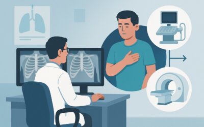
This guidance applies specifically to adult patients undergoing an initial imaging evaluation for clinically suspected tracheomalacia or bronchomalacia. The typical patient presents with symptoms of dynamic airway obstruction, which may include a harsh, barking “seal-like” cough, exertional dyspnea, expiratory wheezing, or a sensation of airway collapse. These symptoms are often exacerbated by coughing, laughing, or changes in position. The condition may be idiopathic or secondary to chronic inflammation (like in COPD or relapsing polychondritis), trauma from prior intubation, or external compression.
This workflow is distinct from several related but different clinical situations. It is crucial to ensure your patient fits this specific scenario:
- Exclusion 1: Suspected Fixed Stenosis. If the clinical suspicion is for a fixed, non-dynamic narrowing of the airway, such as from a tumor, scar tissue, or foreign body, the patient fits the ACR scenario for suspected tracheal or bronchial stenosis, which has a different set of imaging considerations.
- Exclusion 2: Post-Treatment Assessment. This article covers the initial diagnosis. If the patient has already been diagnosed with tracheomalacia and requires imaging to assess their response to stenting, surgery, or other therapies, they fall under the post-treatment assessment scenario.
- Exclusion 3: Suspected Bronchiectasis. If the primary clinical picture is one of chronic productive cough, recurrent infections, and potential hemoptysis, suggesting permanent bronchial dilation rather than dynamic collapse, the workup should follow the guidelines for suspected bronchiectasis.
What Diagnoses Are You Working Up in This Scenario?
When ordering imaging for suspected tracheomalacia, you are evaluating a differential diagnosis centered on large airway pathology and its mimics. The imaging study is chosen to confirm or exclude these possibilities.
Tracheobronchomalacia (TBM) is the primary diagnosis of concern. This condition involves excessive flaccidity of the tracheal and/or bronchial walls, leading to significant dynamic collapse during expiration. The goal of imaging is to directly visualize and quantify this collapse, which is typically defined as a greater than 50% reduction in the cross-sectional area of the airway lumen during exhalation.
A closely related condition is Excessive Dynamic Airway Collapse (EDAC). In EDAC, the collapse is due to the inward bulging of the posterior membranous portion of the trachea, while the cartilaginous rings remain structurally sound. TBM, in contrast, often involves weakening of the cartilage itself. While their management can be similar, dynamic CT can often distinguish between the two pathologies based on the shape of the collapsed airway.
Chronic Obstructive Pulmonary Disease (COPD) is a frequent comorbidity and a significant clinical mimic. Severe COPD can cause symptoms that overlap considerably with TBM. A chest CT is invaluable in this context, as it can simultaneously assess for parenchymal changes of emphysema and chronic bronchitis while also performing the dynamic maneuvers to evaluate for coexisting TBM, which is present in a substantial number of patients with severe COPD.
Finally, the imaging workup helps exclude fixed central airway obstruction. Although the patient’s symptoms may suggest a dynamic process, it is essential to rule out a structural lesion like an endotracheal tumor, a post-intubation stricture, or external compression from a mediastinal mass. A high-resolution CT provides a definitive evaluation of the airway anatomy to exclude these causes.
Why Is a Non-Contrast Chest CT the Recommended Initial Study for Tracheomalacia?
The American College of Radiology designates CT chest without IV contrast as Usually appropriate for the initial imaging of suspected tracheomalacia or bronchomalacia in adults. The rationale is based on its high diagnostic accuracy for the primary clinical question while minimizing unnecessary risks.
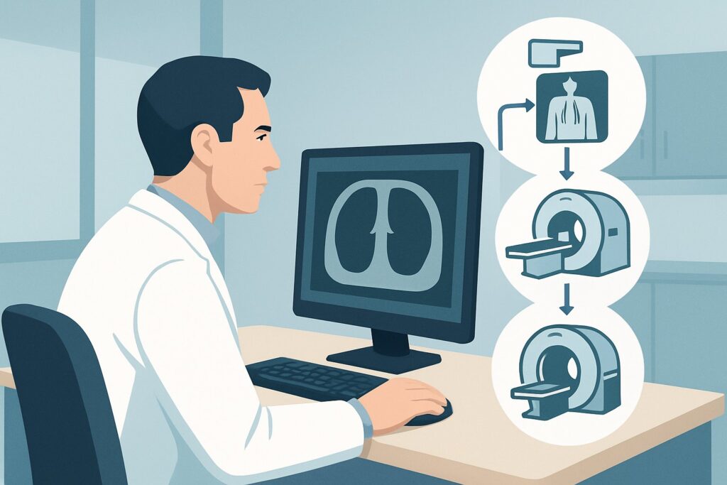
The key to a successful diagnosis is the imaging protocol. A standard, inspiratory-only chest CT is insufficient and will likely miss the diagnosis. The order must specifically request a dynamic expiratory or paired end-inspiration/end-expiration protocol. This technique captures images of the airways when they are fully distended (inspiration) and when they are under the most pressure to collapse (forced expiration). By comparing the airway caliber between these two phases, radiologists can directly measure the degree of luminal narrowing and confirm the diagnosis of TBM or EDAC.
Intravenous contrast is not required because the diagnostic information is based entirely on the air-filled lumen and the airway wall morphology. Adding contrast does not improve the visualization of airway collapse and introduces risks such as allergic reaction and contrast-induced nephropathy. It also increases the radiation dose, particularly if multiple phases are acquired. Therefore, a non-contrast study is the most direct, safest, and most effective approach.
Alternative studies are rated lower for specific reasons:
- Radiography chest is rated May be appropriate (Disagreement). While a chest X-ray might reveal a “saber-sheath” trachea (coronal narrowing with sagittal widening), it is a static image with very low sensitivity for a dynamic process. It cannot quantify collapse and serves primarily to exclude other gross pathologies.
- CT chest with IV contrast is rated Usually not appropriate. As noted, contrast adds risk without benefit for the primary question of airway malacia. It would only become appropriate if the clinical suspicion shifted toward a vascular ring, mediastinal mass, or other pathology requiring vascular assessment, which represents a different clinical scenario.
The radiation dose for a dynamic chest CT is moderate (ACR RRL ☢☢☢, 1-10 mSv), but this is justified by its definitive diagnostic capability, which often prevents the need for more invasive procedures like bronchoscopy.
Once you’ve decided on this study, our protocol guide covers the essential technical details. For more on the technique, contrast, and reading principles, see our complete guide: CT Chest Without Contrast.
What Are the Next Steps After a Dynamic Chest CT?
The results of the dynamic chest CT will guide the subsequent clinical workflow, which typically involves a multidisciplinary discussion between pulmonology, thoracic surgery, and the patient.
- If the CT is positive for significant TBM/EDAC: A finding of >50% expiratory luminal collapse confirms the diagnosis. The next step is to correlate the imaging findings with the severity of the patient’s symptoms and pulmonary function tests. Management is typically stepwise, starting with conservative measures like positive airway pressure therapy (CPAP), pulmonary rehabilitation, and mucolytics. For severe, refractory cases, more invasive options like airway stenting (as a trial) or surgical tracheobronchoplasty may be considered.
- If the CT is negative: A negative dynamic CT, showing no significant expiratory collapse, effectively rules out moderate-to-severe TBM/EDAC as the cause of the patient’s symptoms. The clinical workup should then pivot to investigate other potential causes. This may involve further pulmonary function testing, laryngoscopy to evaluate for vocal cord dysfunction, or esophageal studies to assess for GERD and aspiration, which can mimic respiratory symptoms.
- If the CT is indeterminate or shows borderline findings: In cases where the collapse is mild or borderline (e.g., 40-50% collapse), the clinical significance can be uncertain. In these situations, the next step is often a direct dynamic evaluation with flexible bronchoscopy. Bronchoscopy is considered the gold standard as it allows for direct visualization of the airway during tidal breathing and forced expiratory maneuvers, providing a real-time assessment of airway dynamics.
Pitfalls to Avoid (and When to Get Help)
When working up suspected tracheomalacia, several common pitfalls can lead to diagnostic delays or errors.
1. Ordering a Standard CT: The most frequent error is ordering a routine inspiratory-only chest CT. This will almost always miss the diagnosis. Always specify “paired inspiratory/expiratory” or “dynamic expiratory for tracheomalacia” on the imaging request.
2. Misattributing Symptoms to COPD: Given the high prevalence of co-existing COPD, it is easy to dismiss a patient’s barking cough or severe dyspnea as simply a feature of their underlying lung disease. Maintain a high index of suspicion for TBM in COPD patients with refractory or atypical symptoms.
3. Ignoring Patient Effort: The quality of a dynamic expiratory CT depends on the patient’s ability to perform a forceful exhalation. If the patient cannot cooperate effectively, the degree of collapse may be underestimated. If the imaging is technically limited, this should prompt consideration of bronchoscopy.
If a patient presents with acute respiratory distress, stridor, or signs of impending respiratory failure, this constitutes a medical emergency. The diagnostic workup should be expedited, and you should escalate care immediately to a pulmonologist or critical care specialist.
Related ACR Topics and Tools
This article focuses on a single clinical scenario. For a comprehensive overview of imaging for all related presentations, from stenosis to bronchiectasis, please consult our parent topic guide.
- For breadth across all scenarios in Tracheobronchial Disease, see our parent guide: Tracheobronchial Disease: ACR Appropriateness Decoded.
- To explore imaging guidelines for other clinical problems, use the ACR Appropriateness Criteria Lookup.
- To review technical specifications for hundreds of imaging studies, visit the Imaging Protocol Library.
- For discussions about cumulative radiation exposure with patients, our Radiation Dose Calculator can help frame the conversation.
Frequently Asked Questions
Why is bronchoscopy not the first-line test for suspected tracheomalacia?
While bronchoscopy is considered the gold standard for visualizing dynamic airway collapse, it is an invasive procedure with risks of sedation, laryngospasm, and respiratory compromise. A non-invasive dynamic expiratory CT is highly accurate and is therefore recommended as the initial imaging test. Bronchoscopy is typically reserved for cases where CT is indeterminate, technically limited, or when a therapeutic intervention like stenting is being considered.
Can an MRI be used to diagnose tracheomalacia?
No, MRI is rated as ‘Usually not appropriate’ by the ACR for this indication. While MRI avoids ionizing radiation, it has lower spatial resolution for the airways compared to CT and is more susceptible to motion artifact. More importantly, dynamic ‘real-time’ MRI sequences are not as standardized or widely available as dynamic CT protocols, making CT the superior modality for assessing airway collapse.
What is the specific definition of a ‘positive’ CT for tracheomalacia?
A dynamic expiratory CT is generally considered positive for tracheomalacia or excessive dynamic airway collapse (EDAC) if there is a reduction of 50% or more in the cross-sectional area of the tracheal or main bronchial lumen when comparing the end-inspiratory and dynamic end-expiratory images.
Does the presence of a ‘saber-sheath’ trachea on a chest X-ray confirm tracheomalacia?
No. A saber-sheath trachea, which is a marked coronal narrowing and sagittal widening of the intrathoracic trachea, is a static finding often associated with COPD. While it can be a clue, it does not confirm the dynamic collapse characteristic of tracheomalacia. Many patients with TBM have a normal-appearing trachea on a standard chest radiograph.
If the CT shows external compression on the trachea, is it still considered tracheomalacia?
If an external structure (e.g., a thyroid mass, vascular anomaly, or mediastinal tumor) is compressing the airway and causing it to collapse, this is considered secondary tracheomalacia. The CT is crucial in these cases not only for diagnosing the collapse but also for identifying the underlying extrinsic cause, which then becomes the primary target for treatment.
Reviewed by Pouyan Golshani, MD, Interventional Radiologist — May 30, 2026