What Imaging Should You Order for a New Positive TB Test in an Asymptomatic Patient?
A 34-year-old nurse undergoes routine occupational health screening. Her annual Purified Protein Derivative (PPD) skin test, which has always been negative, is now positive with 12 mm of induration. She feels perfectly well, reporting no cough, fever, night sweats, or weight loss. As the primary care physician managing her follow-up, you must determine if this represents latent infection or subclinical active disease. This decision dictates a vastly different management path, from prophylactic medication to full isolation and multi-drug therapy. The critical first step is imaging.
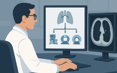
This article provides a detailed clinical workflow for this specific scenario—a newly positive PPD or Interferon-Gamma Release Assay (IGRA), or a positive test with unknown prior status in an asymptomatic patient. Based on the American College of Radiology (ACR) Appropriateness Criteria, the initial imaging study, Radiography chest, is rated Usually Appropriate.
Who Fits This Clinical Scenario?
This guidance is tailored for a very specific and common clinical situation: the asymptomatic patient with new laboratory evidence of exposure to Mycobacterium tuberculosis. The key inclusion criteria are:
- A newly positive PPD skin test or IGRA blood test in a patient with previously documented negative results.
- A positive PPD or IGRA in a patient whose prior testing history is unknown.
- A complete absence of clinical signs or symptoms of active tuberculosis (e.g., no productive cough, hemoptysis, fever, chills, night sweats, or unintentional weight loss).
It is crucial to distinguish this scenario from similar but distinct clinical presentations that require a different diagnostic approach. This workflow does not apply if:
- The patient has symptoms. A patient with a positive TB test and symptoms like a persistent cough or fever falls into the “Suspect active tuberculosis” scenario, which has a different imaging workup and urgency.
- Screening is for facility placement without a test result. An asymptomatic patient requiring screening for placement in a group home or skilled nursing facility, where a PPD test is not available or feasible, represents a separate ACR variant.
Correctly identifying your patient within this specific context ensures the most appropriate, highest-yield, and lowest-risk imaging is ordered first.
What Diagnoses Are You Working Up in This Scenario?
When an asymptomatic patient has a positive PPD or IGRA, the primary goal of imaging is to differentiate between latent infection and subclinical active disease. This distinction is the central branch point for all subsequent management.
Latent Tuberculosis Infection (LTBI)
This is the most common and expected finding. In LTBI, the patient’s immune system has successfully contained the mycobacteria, preventing it from causing active disease. The patient is not infectious. Imaging is often completely normal. In some cases, it may reveal signs of old, healed infection, such as a calcified parenchymal granuloma (Ghon focus) or calcified hilar lymph nodes, sometimes together forming a Ranke complex. These findings confirm prior exposure but do not indicate active disease.
Subclinical Active Pulmonary Tuberculosis
This is the critical diagnosis to exclude. A minority of patients with new positive tests may have early, active disease without yet developing symptoms. Identifying this is paramount to prevent disease progression and community transmission. Imaging findings suggestive of active TB include apical or upper lobe infiltrates (often with a cavitary lesion), pleural effusions, or miliary (diffuse, small nodular) patterns. The presence of any of these findings shifts the patient’s diagnosis from LTBI to active TB, requiring immediate public health reporting and multi-drug treatment.
Non-tuberculous Mycobacterial (NTM) Infection
Less commonly, pulmonary disease from NTM can produce imaging findings that mimic TB, such as nodules, bronchiectasis, or cavities. While NTM infection can also cause a positive IGRA in some cases, the chest radiograph helps identify parenchymal abnormalities that may warrant further investigation with sputum cultures to differentiate NTM from M. tuberculosis.
Why Is a Chest Radiograph the Recommended Study for a New Positive TB Test?
The ACR designates a standard two-view chest radiograph as Usually Appropriate for this scenario because it expertly balances diagnostic yield with patient safety. It is a fast, low-cost, and low-radiation tool that is highly effective at answering the primary clinical question: is there evidence of active pulmonary disease?
A chest radiograph provides excellent visualization of the lung parenchyma, pleura, and mediastinum, making it well-suited to detect the classic signs of active TB, such as consolidation, cavitation, and effusions. For the vast majority of patients who have LTBI, a normal chest radiograph provides the necessary reassurance to proceed with treatment for latent infection, which is a much simpler and shorter regimen than that for active disease.
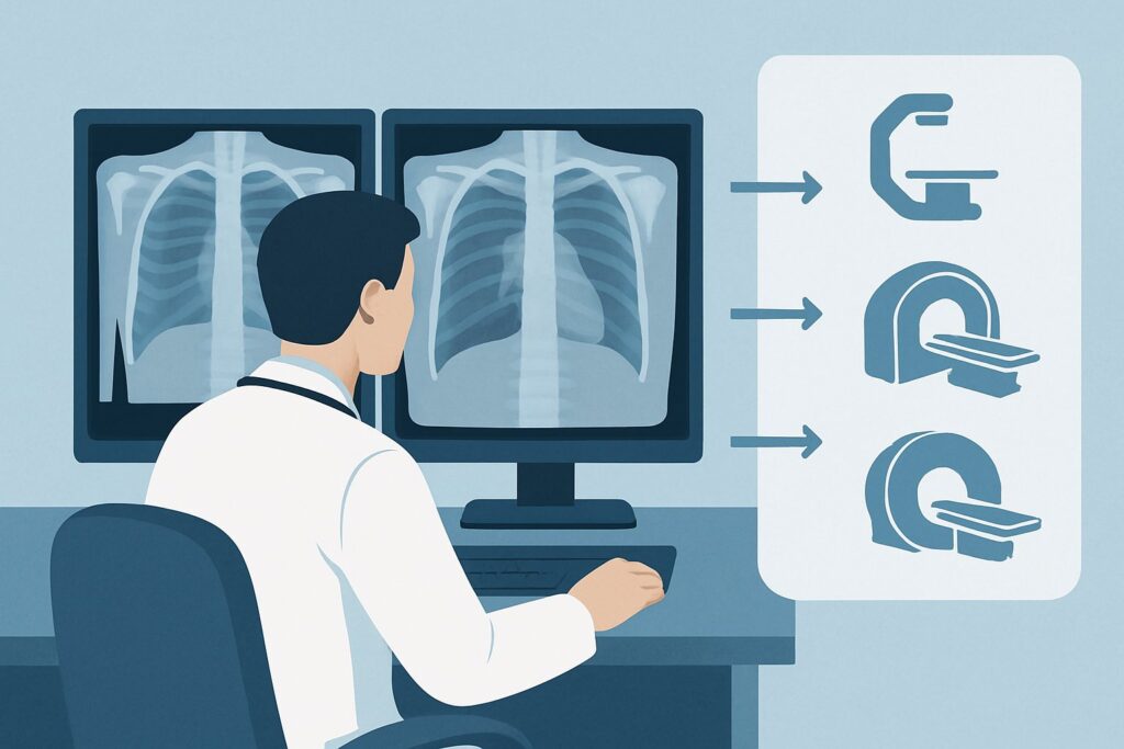
Alternative imaging modalities are rated lower for specific, important reasons in this initial screening context:
- CT chest without IV contrast is rated Usually not appropriate as a first-line test. While CT is more sensitive than radiography for subtle nodules or early infiltrates, this increased sensitivity comes at the cost of a significantly higher radiation dose (☢☢☢ 1-10 mSv for CT versus ☢ <0.1 mSv for radiography). In an asymptomatic screening population, its routine use would lead to the over-investigation of incidental and clinically insignificant findings, increasing patient anxiety and healthcare costs without a clear benefit over initial radiography.
- MRI chest without and with IV contrast is also rated Usually not appropriate. MRI offers poor spatial resolution of the lung parenchyma and is insensitive for detecting the calcifications and fine parenchymal details crucial for evaluating possible TB. It has no established role in the initial evaluation of a positive TB test.
The choice of a chest radiograph is a deliberate one, prioritizing a high-value, low-risk examination to stratify patients effectively. The minimal radiation exposure (adult relative radiation level ☢ <0.1 mSv; pediatric ☢ <0.03 mSv) makes it a safe and repeatable screening tool.
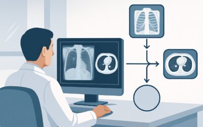
What’s Next After the Chest Radiograph? Downstream Workflow
The results of the chest radiograph directly guide the next steps in patient management and public health intervention. The workflow branches into three main paths.
If the Radiograph is Negative
A normal chest radiograph in an asymptomatic patient with a positive PPD or IGRA confirms the diagnosis of Latent Tuberculosis Infection (LTBI). The patient is not considered to have active, infectious disease. The next step is clinical consultation to discuss treatment options for LTBI. This typically involves a shorter, simpler prophylactic antibiotic regimen (e.g., isoniazid, rifampin, or combination therapy) to reduce the lifetime risk of the latent infection reactivating into active disease. No further imaging is typically required unless the patient develops symptoms.
If the Radiograph is Positive for Active TB
If the radiograph shows findings suggestive of active pulmonary tuberculosis (e.g., apical infiltrate, cavity, effusion), the patient is presumed to have active TB until proven otherwise. This is a medical and public health priority. The immediate next steps include:
- Initiating respiratory isolation to prevent transmission.
- Collecting multiple sputum samples for acid-fast bacilli (AFB) smear, culture, and nucleic acid amplification testing (NAAT) to confirm the diagnosis and determine drug susceptibility.
- Reporting the case to the local public health department for contact tracing.
- Starting a multi-drug treatment regimen for active TB.
Further imaging, such as a CT scan, may be considered to better define the extent of the disease, but it should not delay the initiation of these critical steps.
If the Radiograph is Indeterminate or Equivocal
Occasionally, the findings may be unclear—for example, stable-appearing apical scarring or a solitary pulmonary nodule of uncertain significance. In these cases, the next step is to obtain any prior chest imaging for comparison. If no prior images exist or if comparison fails to resolve the uncertainty, a CT chest (often without contrast) may be warranted to better characterize the findings and assess for subtle signs of activity. This is the situation where a CT scan, rated May be appropriate by the ACR, becomes a valuable second-line tool.
Pitfalls to Avoid (and When to Get Help)
Navigating this workflow requires attention to several potential pitfalls to ensure accurate diagnosis and appropriate management.
- Ignoring a “normal” read in a high-risk patient: In severely immunocompromised patients (e.g., advanced HIV, on TNF-alpha inhibitors), active TB can present with subtle or atypical radiographic findings. A low threshold for obtaining sputum samples and considering a follow-up CT is wise if clinical suspicion remains high despite a normal radiograph.
- Dismissing apical scarring as “old disease”: While often benign, apical scarring can also be a sign of active or reactivating TB. Always compare with prior imaging. If no priors are available, consider the finding indeterminate and proceed with sputum collection and potentially a CT scan.
- Delaying public health notification: If the chest radiograph is suspicious for active TB, do not wait for sputum culture results (which can take weeks) to report the case. Presumptive active TB is a reportable condition.
- Forgetting pediatric considerations: Children with TB may present with different radiographic patterns, most commonly hilar or mediastinal lymphadenopathy, which can be subtle on a chest radiograph. Ensure the interpreting radiologist is aware of the patient’s age.
If the imaging findings are complex or the clinical picture is incongruous with the radiographic results, consultation with a pulmonologist or infectious disease specialist is the appropriate next step.
Related ACR Topics and Tools
For a comprehensive overview of all clinical scenarios related to tuberculosis imaging, from initial suspicion to treatment follow-up, please see our parent guide. It provides a breadth of information that complements this in-depth article.
- For breadth across all scenarios in Imaging of Possible Tuberculosis, see our parent guide: Imaging of Possible Tuberculosis: ACR Appropriateness Decoded.
- To explore other clinical situations, use the Imaging Appropriateness Selector.
- For details on imaging techniques, consult the Imaging Protocol Library.
- To discuss radiation exposure with your patients, the Radiation Dose Calculator can help frame the conversation.
Frequently Asked Questions
Does a positive IGRA test require a different imaging workup than a positive PPD test?
No, for the purposes of initial imaging in an asymptomatic patient, a positive IGRA (like QuantiFERON-TB Gold or T-SPOT.TB) is treated the same as a positive PPD. Both indicate an immune response to M. tuberculosis, and the next step is a chest radiograph to rule out active pulmonary disease.
If the patient had a BCG vaccine, is a chest radiograph still necessary after a positive PPD?
Yes. While a history of BCG vaccination can cause a false-positive PPD skin test, it does not protect completely against TB infection. Therefore, a positive PPD in a BCG-vaccinated individual still requires a chest radiograph to rule out active disease. Alternatively, an IGRA test can be used, as it is not affected by prior BCG vaccination.
How soon after a positive TB test should the chest radiograph be performed?
The chest radiograph should be performed promptly, typically within days to a week of the positive test result. While the situation is not a medical emergency in an asymptomatic patient, timely evaluation is important to confirm a diagnosis of LTBI and initiate prophylactic therapy, or to identify and treat subclinical active disease to prevent progression and transmission.
What if the patient is pregnant and has a new positive TB test?
A chest radiograph is still the recommended next step. With proper abdominal shielding, the radiation dose to the fetus is negligible and far below the threshold known to cause harm. The risk of missing active TB in a pregnant patient, which can have severe consequences for both the mother and the fetus, greatly outweighs the minimal risk of the radiograph.
Is a follow-up chest radiograph needed after completing treatment for LTBI?
Generally, no. For patients with LTBI who had a normal baseline chest radiograph and successfully complete prophylactic treatment, routine follow-up imaging is not recommended unless they develop symptoms suggestive of active TB in the future.
Reviewed by Pouyan Golshani, MD, Interventional Radiologist — May 26, 2026