What Is the Right Supplemental Breast Screening for Average-Risk Women with Nondense Breasts?
It’s a busy afternoon in your primary care clinic. Your next patient is a 52-year-old woman here for her annual wellness visit. She has no new complaints and is diligent about her health screenings. Her mammogram report from last year is on your screen; it was negative and noted “scattered areas of fibroglandular density,” or BI-RADS category B. She asks if the standard mammogram is enough, or if she should be getting any other tests. For this specific clinical scenario—an average-risk woman with nondense breasts—the decision for supplemental screening requires a clear understanding of risk versus benefit.
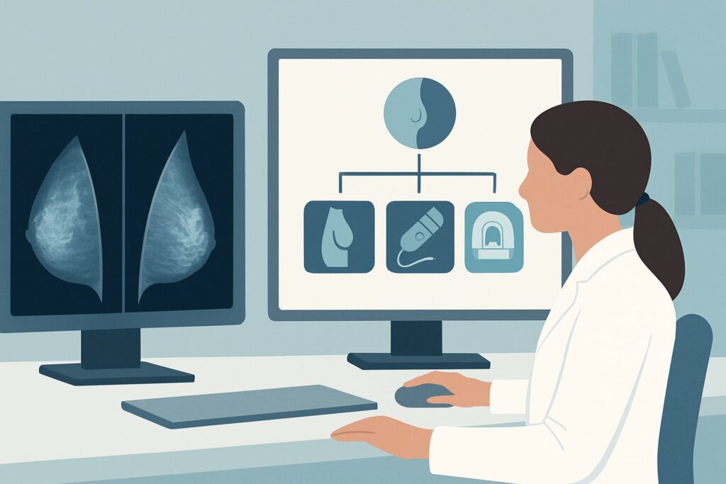
This article provides a focused workflow for this exact presentation, explaining why the American College of Radiology (ACR) Appropriateness Criteria designates Digital breast tomosynthesis screening as “Usually appropriate” while other modalities are not recommended for routine supplemental screening in this population.
Who Fits This Clinical Scenario?
This guidance applies specifically to adult women who meet a precise set of criteria. Correctly identifying your patient’s category is the critical first step to ordering the right imaging study and avoiding unnecessary tests.
Inclusion Criteria for This Workflow:
- Risk Level: The patient must be at average risk for breast cancer. This is defined as a lifetime risk of <15% and no significant risk factors such as a known pathogenic mutation (e.g., BRCA1/2), a strong family history suggestive of one, or a history of chest mantle radiation at a young age.
- Breast Density: The patient must have nondense breasts, as determined on a prior mammogram. This corresponds to Breast Imaging Reporting and Data System (BI-RADS) density categories A (“almost entirely fatty”) or B (“scattered areas of fibroglandular density”).
- Clinical Context: The patient is asymptomatic, and the imaging is being ordered for routine, supplemental screening, not for a diagnostic workup of a new sign or symptom (e.g., a palpable lump, skin changes, or nipple discharge).
Exclusion Criteria (Who This Does NOT Apply To):
It is crucial to distinguish this scenario from similar but distinct clinical presentations. This guidance should not be applied to patients with dense breasts (BI-RADS C or D), as the masking effect of dense tissue fundamentally changes the screening paradigm. Similarly, women with an intermediate risk (e.g., personal history of breast cancer, lobular carcinoma in situ) or high risk (e.g., known genetic mutation) follow different screening algorithms, often involving breast MRI, regardless of their breast density.
What Diagnoses Are You Working Up in This Scenario?
In any screening context, the goal is to detect disease before it becomes clinically apparent. For an average-risk woman with nondense breasts, the primary target of screening is the early detection of occult breast cancer. The “differential” is less about distinguishing between multiple active pathologies and more about identifying the earliest signs of malignancy that could be missed even with standard imaging.
Occult Invasive Breast Cancer: The most important target is detecting small, invasive carcinomas, most commonly invasive ductal carcinoma (IDC), while they are still confined to the breast. In nondense tissue, mammography is highly effective, but very small or subtle architectural distortions representing an early cancer can still be obscured by the limited overlapping fibroglandular tissue. The goal is to find these cancers before they become palpable or metastasize.
Ductal Carcinoma In Situ (DCIS): Screening also aims to identify DCIS, a non-invasive precursor to invasive cancer. DCIS often presents as microcalcifications on mammography. While typically visible in nondense breasts, some forms of non-calcified or subtle DCIS can be challenging to detect on standard 2D imaging. Improving detection of high-grade DCIS is a key objective of advanced screening techniques.
While not the primary goal of screening, advanced imaging can also help better characterize benign or indeterminate findings from prior studies, potentially reducing the need for short-term follow-up or unnecessary biopsies. However, the central purpose remains the detection of otherwise invisible malignant disease.
Why Is Digital Breast Tomosynthesis Screening the Recommended Study for This Presentation?
For an average-risk woman with nondense breasts, the ACR designates Digital breast tomosynthesis (DBT) screening as “Usually appropriate.” This recommendation is based on a favorable balance of improved cancer detection, reduced false positives, and a low radiation dose profile.
DBT, often called 3D mammography, acquires multiple low-dose X-ray images of the breast from different angles. A computer then reconstructs these images into thin, one-millimeter “slices.” This technique effectively minimizes the effect of overlapping breast tissue, which is the primary reason lesions are missed on conventional 2D mammography. Even in nondense breasts with scattered fibroglandular tissue, this overlap can obscure the subtle architectural distortion or spiculations of an early cancer. By “scrolling” through the breast tissue slice by slice, the radiologist has a clearer view, which studies have shown increases the cancer detection rate and, just as importantly, decreases the recall rate for non-cancerous findings.
In contrast, other powerful imaging modalities are rated “Usually not appropriate” for supplemental screening in this specific patient group:
- Breast Ultrasound (US): While useful for evaluating dense breasts and for diagnostic workups, whole-breast screening US is not recommended for average-risk women with nondense breasts. Without the masking effect of dense tissue, the incremental cancer detection benefit is minimal, while the rate of false-positive findings remains high, leading to unnecessary anxiety and biopsies.
- Breast MRI with contrast: This is the most sensitive test for detecting breast cancer. However, for an average-risk woman, its use for screening is “Usually not appropriate.” The rationale is multifactorial: it has a much higher rate of false positives compared to mammography, requires an intravenous contrast agent (gadolinium), is significantly more expensive, and has limited availability. Its high sensitivity is reserved for high-risk screening, where the pre-test probability of finding a cancer is much greater.
The radiation exposure from DBT (ACR Relative Radiation Level ☢☢, 0.1-1 mSv) is considered low and is comparable to that of a standard 2D mammogram. Given its proven benefits in reducing recalls and improving cancer detection, it has become the standard of care for screening in most practices. For detailed procedural considerations, refer to our guide on the Screening Mammography (with DBT) protocol.
What’s Next After Digital Breast Tomosynthesis Screening? Downstream Workflow
The result of the screening DBT, reported using the BI-RADS classification, dictates the next steps in the patient’s care. A clear understanding of this downstream workflow is essential for counseling patients and ensuring timely follow-up.
Negative or Benign Result (BI-RADS 1 or 2): If the study is interpreted as negative (BI-RADS 1) or shows a clearly benign finding (BI-RADS 2), the workflow is simple. The patient should be advised to continue with her routine annual screening mammography. No further immediate action is needed.
Incomplete or Indeterminate Result (BI-RADS 0 or 3):
- A BI-RADS 0 (Incomplete) assessment means the radiologist needs additional imaging to make a final determination. This is not a definitive positive or negative result. The patient will be called back for a diagnostic workup, which typically includes diagnostic mammographic views (e.g., spot compression, magnification) and/or a targeted breast ultrasound of the area in question.
- A BI-RADS 3 (Probably Benign) finding has a very low (<2%) likelihood of malignancy. The standard recommendation is short-interval follow-up, typically with a unilateral diagnostic mammogram or ultrasound in six months, to ensure stability.
Suspicious or Highly Suggestive of Malignancy (BI-RADS 4 or 5):
- A BI-RADS 4 (Suspicious) or BI-RADS 5 (Highly Suggestive of Malignancy) finding requires tissue sampling for a definitive diagnosis. The patient should be referred for an image-guided biopsy, most commonly performed under ultrasound or stereotactic (mammographic) guidance. Prompt referral to a breast surgeon or radiologist for biopsy is the critical next step.
Pitfalls to Avoid (and When to Get Help)
Navigating supplemental screening for this scenario requires careful attention to detail. Here are several common pitfalls to avoid:
- Ordering Supplemental Screening Without a Recent Mammogram: Breast density can change over time. The decision to forgo supplemental imaging should be based on a recent mammogram confirming nondense (BI-RADS A or B) tissue.
- Misclassifying Patient Risk: Overlooking elements of a patient’s personal or family history can lead to under-screening. A patient who seems average-risk may actually meet criteria for intermediate or high-risk screening, which follows a different algorithm. Use a formal risk assessment model if there is any uncertainty.
- Confusing Screening with Diagnostic Workup: If a patient presents with a new symptom, such as a palpable lump or focal pain, a screening mammogram is inappropriate. The correct order is a diagnostic mammogram and targeted ultrasound, regardless of her risk status or breast density.
If a screening study returns a suspicious finding (BI-RADS 4 or 5), or if the clinical picture is complex, immediate escalation to a breast imaging specialist or breast surgeon is warranted for coordination of further diagnostic workup and biopsy.
Related ACR Topics and Tools
The ACR Appropriateness Criteria are extensive, covering thousands of clinical scenarios. For a broader overview of how breast density and risk level interact to guide imaging choices, please see our parent topic hub article. Additionally, several GigHz tools can help streamline your clinical workflow.
- For breadth across all scenarios in Supplemental Breast Cancer Screening Based on Breast Density, see our parent guide: Supplemental Breast Cancer Screening Based on Breast Density: ACR Appropriateness Decoded.
- ACR Appropriateness Criteria Lookup — for adjacent scenarios
- Imaging Protocol Library — for technique on the recommended study
- Radiation Dose Calculator — for cumulative dose conversations
Frequently Asked Questions
Is a 3D mammogram (DBT) really necessary every year for an average-risk woman with nondense breasts?
Yes, the American College of Radiology and Society of Breast Imaging recommend annual screening with digital breast tomosynthesis (DBT) for all average-risk women starting at age 40. Even in nondense breasts, DBT has been shown to improve cancer detection and reduce false-positive recalls compared to 2D mammography alone.
If my patient with nondense breasts has a family history of breast cancer, does she still fit this ‘average-risk’ scenario?
Not necessarily. A single second-degree relative with postmenopausal breast cancer may not elevate her risk significantly. However, a first-degree relative (mother, sister, daughter) with breast cancer, or multiple relatives on the same side of the family, may push her into the intermediate- or high-risk category, which requires a different screening protocol. A formal risk assessment (e.g., using the Tyrer-Cuzick model) is recommended to clarify her status.
Why is screening breast ultrasound ‘Usually not appropriate’ for this patient group when it’s recommended for women with dense breasts?
The primary value of screening ultrasound is to find cancers that are ‘masked’ or hidden by dense fibroglandular tissue on a mammogram. In women with nondense (fatty) breasts, there is very little tissue to cause masking, so mammography is highly effective on its own. In this context, adding ultrasound provides minimal benefit in cancer detection while significantly increasing the rate of false positives, leading to unnecessary biopsies and patient anxiety.
Does a BI-RADS B (‘scattered density’) classification require a different approach than a BI-RADS A (‘almost entirely fatty’)?
No, for the purposes of screening guidelines, both BI-RADS A and B are categorized as ‘nondense.’ The workflow and recommendations described in this article apply equally to both groups. The need for supplemental screening with modalities like ultrasound or MRI is triggered by dense breast classifications (BI-RADS C or D).
If a patient is average-risk with nondense breasts but has breast implants, does that change the recommendation?
Yes, this is a special consideration. While the patient is still considered to have nondense native breast tissue, the presence of implants can obscure tissue on standard mammographic views. Patients with implants require additional ‘implant-displaced’ (Eklund) views during their mammogram. Digital breast tomosynthesis is still the recommended primary screening modality, but the technique is modified.
Reviewed by Pouyan Golshani, MD, Interventional Radiologist — May 29, 2026