What Is the Next Step for a Suspicious Breast Mass in a Woman Under 30?
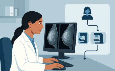
A 27-year-old graduate student presents to your clinic with a palpable lump in her right breast she noticed a month ago. The initial targeted ultrasound is complete, and the report is concerning: a 1.5 cm irregular, hypoechoic mass with indistinct margins, classified as BI-RADS 4 (Suspicious). You know that breast cancer is uncommon in this age group, but this finding cannot be ignored. The immediate question is which imaging study to order next to properly characterize the lesion and guide the definitive next step, which will be a biopsy. This article details the clinical workflow for this specific scenario, guided by the American College of Radiology (ACR) Appropriateness Criteria, which rates a diagnostic digital breast tomosynthesis as Usually Appropriate.
## Who Fits This Clinical Scenario?
This guidance applies to a very specific patient presentation: an adult female, younger than 30 years of age, who has a palpable breast mass that has already been evaluated with ultrasound and found to be suspicious or highly suggestive of malignancy (BI-RADS 4 or 5).
The key inclusion criteria are:
- Age: Younger than 30 years.
- Presentation: A palpable breast mass.
- Prior Imaging: An initial ultrasound has already been performed.
- Ultrasound Result: Findings are categorized as BI-RADS 4 (Suspicious), 4A (Low suspicion), 4B (Moderate suspicion), 4C (High suspicion), or 5 (Highly suggestive of malignancy).
This workflow is not appropriate for several similar-sounding but distinct clinical situations. For instance, this guidance does not apply to the initial imaging workup of a palpable mass in this age group; that scenario begins with ultrasound. It also does not apply to women over 30 or 40, where the pre-test probability of malignancy is higher and mammography often plays an earlier role. Finally, if the ultrasound findings were probably benign (BI-RADS 3) or benign (BI-RADS 2), the management pathway would be entirely different, typically involving short-interval follow-up or routine care, respectively.
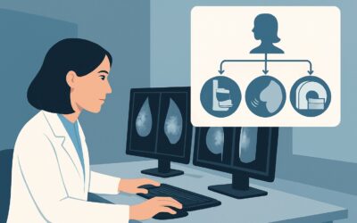
## What Diagnoses Are You Working Up in This Scenario?
With a BI-RADS 4 or 5 finding on ultrasound, the differential diagnosis narrows significantly, and the primary goal is to exclude malignancy. While benign entities can sometimes have suspicious features, the imaging characteristics in this scenario compel a workup for cancer until proven otherwise.
Invasive Breast Carcinoma: Although breast cancer accounts for a small fraction of cancers in women under 30, it is the most consequential diagnosis to exclude. A suspicious palpable mass is the most common presentation for these rare cases. The goal of further imaging is to define the extent of the primary lesion and identify any other occult findings, such as suspicious microcalcifications, that would influence surgical planning.
Phyllodes Tumor: These are rare fibroepithelial tumors that can be benign, borderline, or malignant. They often present as large, palpable, rapidly growing masses and can be indistinguishable from fibroadenomas on ultrasound. However, suspicious features warrant a full workup, as malignant phyllodes tumors require wide local excision.
Other Malignancies: Extremely rare possibilities in this age group include primary breast lymphoma or metastasis from another primary cancer (e.g., melanoma, sarcoma). While not the leading considerations, the aggressive workup prompted by a BI-RADS 4 or 5 finding will inherently evaluate for these possibilities.
Benign Mimics: Certain benign conditions can rarely present with suspicious imaging features. These include complex fibroadenomas with atypical features, diabetic mastopathy, or granulomatous mastitis. However, given the BI-RADS 4 or 5 classification, these are diagnoses of exclusion made only after a biopsy has ruled out malignancy.
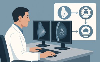
## Why Is Diagnostic Digital Breast Tomosynthesis the Recommended Study for This Presentation?
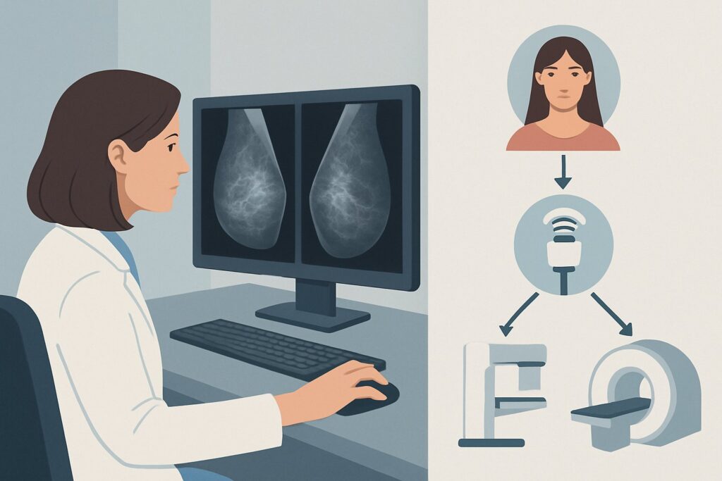
The ACR Appropriateness Criteria rate Digital breast tomosynthesis diagnostic and Mammography diagnostic as Usually Appropriate for this clinical scenario. The immediate next step after a suspicious ultrasound is not just to get tissue, but to fully evaluate the breast for the extent of the disease before the biopsy.
Diagnostic mammography, particularly with tomosynthesis (DBT or “3D mammography”), is crucial for several reasons. In younger women, who typically have dense breast tissue, DBT excels at reducing the effect of overlapping tissue. This allows for better delineation of the mass’s margins and can reveal subtle associated findings like architectural distortion or suspicious microcalcifications that may not be visible on ultrasound. Identifying the full extent of a lesion, especially any associated ductal carcinoma in situ (DCIS) component which often manifests as calcifications, is critical for accurate surgical planning.
The radiation dose for a diagnostic DBT is low (ACR relative radiation level ☢☢, corresponding to 0.1-1 mSv). While any radiation exposure in a young patient is considered carefully, the potential benefit of accurately characterizing a highly suspicious lesion and planning for a potential cancer diagnosis far outweighs this minimal risk.
It is also important to note that Image-guided core biopsy breast is rated Usually Appropriate. The diagnostic mammogram is not a replacement for biopsy; it is the prerequisite step that informs the biopsy. It ensures the correct lesion is targeted and provides a complete imaging baseline.
Why are other studies not recommended at this stage?
- MRI breast without and with IV contrast is rated Usually not appropriate. While breast MRI is extremely sensitive, its role is primarily in staging after a biopsy has confirmed cancer. Using it before a tissue diagnosis can introduce false positives due to lower specificity, potentially leading to unnecessary additional biopsies and patient anxiety.
- Screening mammography is also rated Usually not appropriate. This is a diagnostic problem, not a screening one. A diagnostic exam is supervised by a radiologist and includes tailored, problem-solving views (e.g., spot compression, magnification) that are essential for characterizing a known abnormality. A standard screening study is inadequate for this purpose.
## What’s Next After Diagnostic Digital Breast Tomosynthesis? Downstream Workflow
The results of the diagnostic mammogram or DBT directly inform the subsequent steps, which almost invariably lead to a biopsy.
- If DBT confirms the ultrasound finding and shows no other suspicious lesions: The patient proceeds to an image-guided core needle biopsy, typically under ultrasound guidance for the known mass. The mammographic findings serve as the baseline for post-biopsy and pre-surgical evaluation.
- If DBT reveals additional suspicious findings (e.g., microcalcifications): The biopsy plan must be adjusted. This may involve a stereotactic (mammogram-guided) biopsy of the calcifications in addition to the ultrasound-guided biopsy of the mass. This ensures all suspicious areas are sampled to get a complete pathologic picture.
- After the biopsy results are available:
- Malignant: The patient is referred to a multidisciplinary breast cancer team, including surgical, medical, and radiation oncology. Further staging workup, which now often includes a breast MRI and systemic imaging, will be initiated.
- Benign, but discordant: If the pathology report returns a benign finding (e.g., fibroadenoma) for a lesion that was highly suspicious on imaging (BI-RADS 4C or 5), this is considered a discordant result. An imaging-pathology correlation review is mandatory, and this situation typically requires a surgical excisional biopsy to resolve the discrepancy.
- Benign and concordant: If the benign pathology is consistent with the level of imaging suspicion (e.g., for a BI-RADS 4A lesion), management may revert to short-term imaging follow-up.
- High-Risk Lesion (e.g., ADH, LCIS): These findings often prompt a recommendation for surgical excision due to the risk of associated or future malignancy.
## Pitfalls to Avoid (and When to Get Help)
Navigating this high-stakes scenario requires avoiding several common missteps to ensure optimal patient care.
1. Skipping the Diagnostic Mammogram: A common pitfall is proceeding directly from a suspicious ultrasound to biopsy without obtaining a mammogram. This risks missing the true extent of the disease, particularly an associated calcific component, which can lead to an incomplete initial surgery.
2. Ordering a Screening Study: Requesting a “screening mammogram” instead of a “diagnostic mammogram” will result in an inadequate examination. The order must specify a diagnostic study to ensure the radiologist performs the necessary problem-solving views.
3. Prematurely Ordering an MRI: Ordering a breast MRI before a tissue diagnosis is established can complicate the clinical picture with false positives and is not the standard of care. Reserve MRI for post-biopsy staging.
If there is any discordance between the imaging findings and the pathology results from the core biopsy, it is critical to escalate. This should be discussed immediately with the interpreting radiologist and referred for multidisciplinary review or surgical consultation.
## Related ACR Topics and Tools
This article covers one specific variant within the broader topic of palpable breast masses. The clinical context, such as patient age or prior imaging results, significantly alters the recommended imaging pathway.
For breadth across all scenarios in Palpable Breast Masses, see our parent guide: Palpable Breast Masses: ACR Appropriateness Decoded.
For additional decision support and technical details, the following GigHz tools are available:
- ACR Appropriateness Criteria Lookup — for exploring adjacent clinical scenarios
- Imaging Protocol Library — for technique on the recommended study
- Radiation Dose Calculator — for discussing cumulative dose with patients
Frequently Asked Questions
Why not go straight to biopsy after a suspicious ultrasound in a young woman?
A pre-biopsy diagnostic mammogram or digital breast tomosynthesis (DBT) is essential to evaluate the entire breast. It can reveal the full extent of the mass and identify other subtle findings, like suspicious microcalcifications, that ultrasound may miss. This information is critical for accurate biopsy targeting and planning for potential surgery.
Is breast cancer common in women under 30?
No, it is rare, representing a very small percentage of all breast cancer diagnoses. However, because it can occur and may be more aggressive in younger women, any finding classified as BI-RADS 4 (Suspicious) or 5 (Highly suggestive of malignancy) on ultrasound must be fully worked up to definitively exclude cancer.
Why is breast MRI rated ‘Usually not appropriate’ at this specific stage?
While breast MRI is the most sensitive imaging test for breast cancer, its lower specificity can lead to false positives. At this pre-biopsy stage, a false positive could lead to unnecessary additional biopsies. MRI’s primary role is for staging—to assess the extent of disease in the breast and check the other breast—after a cancer diagnosis has already been confirmed by biopsy.
What is the difference between a screening and a diagnostic mammogram?
A screening mammogram consists of standard, routine views and is performed on asymptomatic women to detect early cancer. A diagnostic mammogram is a problem-solving exam tailored to a specific clinical finding, like a palpable mass or an abnormal screening result. It is directly supervised by a radiologist and includes additional, specialized views like spot compression and magnification to fully characterize the area of concern.
Does the radiation from a diagnostic mammogram pose a significant risk to a young patient?
The radiation dose from a modern diagnostic mammogram or DBT is very low (ACR level ☢☢, 0.1-1 mSv), roughly equivalent to a few months of natural background radiation. The clinical benefit of accurately diagnosing a potentially life-threatening condition like breast cancer far outweighs this minimal theoretical risk.
Reviewed by Pouyan Golshani, MD, Interventional Radiologist — May 30, 2026